. 1990 Aug 10;62(3):469-80.
doi: 10.1016/0092-8674(90)90012-4.

The three postblastoderm cell cycles of Drosophila embryogenesis are regulated in G2 by string

Affiliations

The three postblastoderm cell cycles of Drosophila embryogenesis are regulated in G2 by string

B A Edgar et al. Cell. .

Abstract

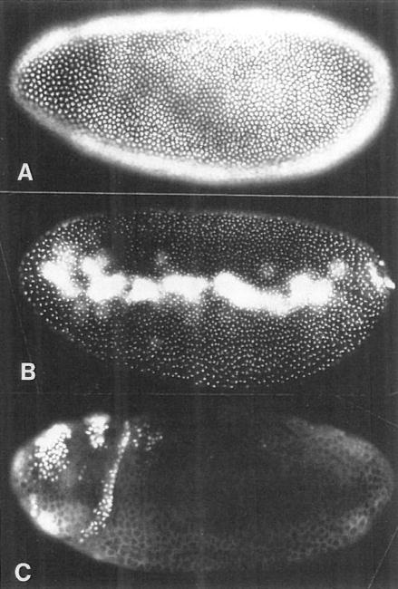
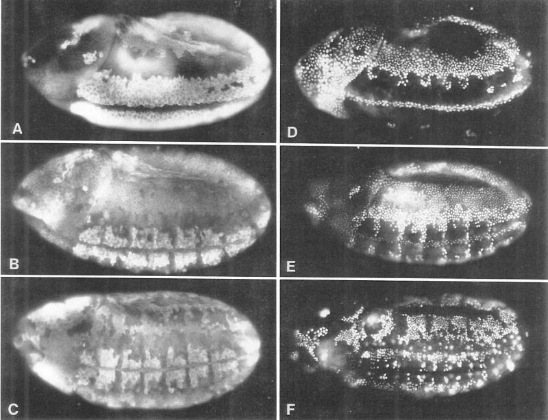
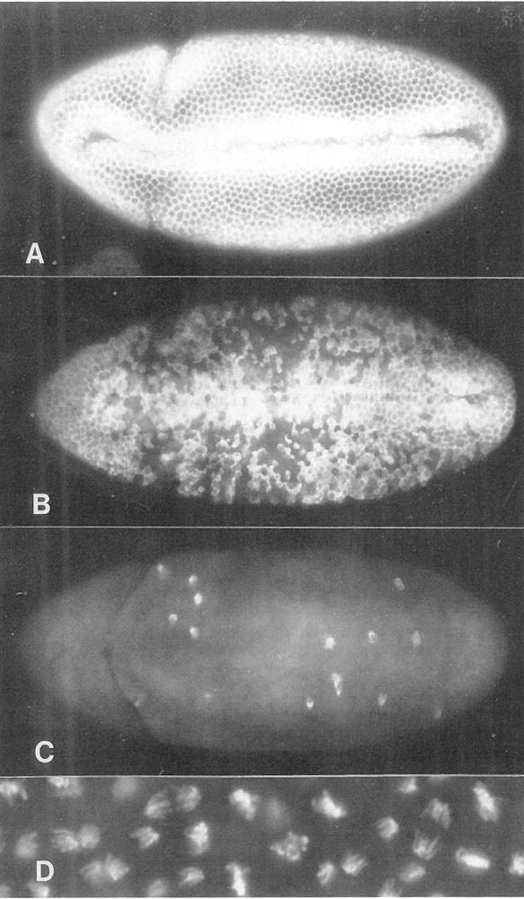
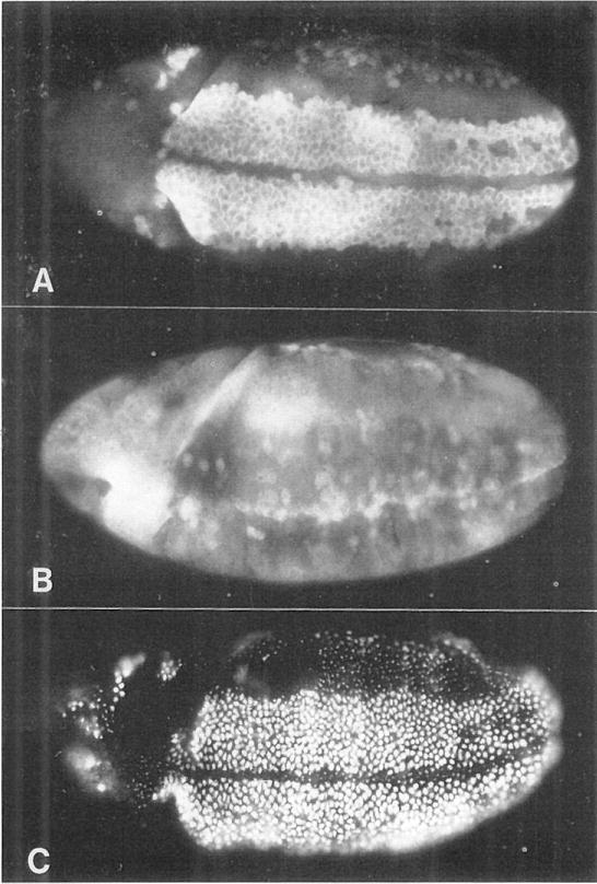
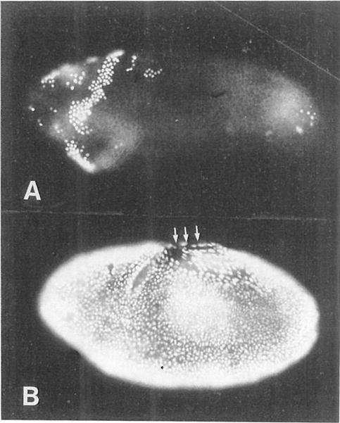
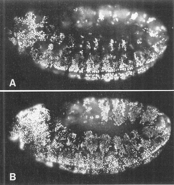
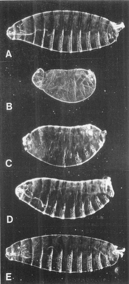
The string (stg) locus of Drosophila encodes a factor that is thought to trigger mitosis by activating the p34cdc2 protein kinase. stg is required for mitosis early in development and is transcribed in a dynamic pattern that anticipates the pattern of embryonic cell divisions. Here we show that differential cell cycle regulation during postblastoderm development (cell cycles 14-16) occurs in G2. We demonstrate that stg mRNA expressed from a heat shock promotor triggers mitosis, and an associated S phase, in G2 cells during these cycles. Hence, differential cell cycle timing at this developmental stage is controlled by stg. Finally, we use heat-induced stg expression to alter the normal pattern of embryonic mitoses. Surprisingly, the complex mitotic pattern evident during normal development is not essential for many features of pattern formation or for viability.


Figures

Figure 1
Figure 1. S Phase 14 Begins and Ends Synchronously without a G1 Period
We show three embryos pulse labeled with BrdU for 20 min at progressively later stages of interphase 14. The BrdU is detected by immunoflourescence, and labeled nuclei appear white. (A) An embryo labeled for the first 20 min of interphase 14 (130–150 min after egg deposition [AED]) shows all blastoderm nuclei heavily and uniformly labeled. (B) An embryo labeled at about 160–180 min AED shows spots of late replicating centromeric heterochromatin in each blastoderm nucleus. Note that nuclei in all regions of the blastoderm are similarly labeled, indicating that S phase completion is not spatially patterned. The yolk nuclei and pole cell nuclei are heavily labeled in this embryo, indicating that they replicate their DNA later than the blastoderm nuclei. (C) An embryo labeled at about 180–200 min AED shows most of the blastoderm nuclei unlabeled, indicating a true G2 period. Patches of labeled nuclei in the anterior are those that have already progressed through mitosis 14 and entered S phase 15. This photo is overexposed to show cytoplasmic background staining.
Figure 2
Figure 2. S Phase 15 Follows Directly after Mitosis 14
(A), (B), and (C) show cycle 14 and early cycle 15 embryos of increasing age stained with antibodies to cyclin A. The cyclin staining patterns indicate cell cycle states; cells in late interphase are brightly labeled, whereas dark cells have just passed through mitosis. (D), (E), and (F) show embryos pulse labeled with BrdU for 20 min immediately prior to fixation. Embryos in (D), (E), and (F) are the same age as embryos in (A), (B), and (C), respectively. The cyclin and BrdU patterns are almost exactly inverse to each other. This indicates that cyclin degradation, which occurs at metaphase (Lehner and O’Farrell, 1989), occurs a constant number of minutes before DNA replication. From comparing distributions of metaphase figures and BrdU-labeled nuclei in single embryos (not shown) and using the map of mitotic times (Foe, 1989), we estimate that the metaphase to S phase time interval is <5 min. All embryos show a 3/4 ventrolateral view; anteriors are to the left and dorsal surfaces uppermost.
Figure 3
Figure 3. stg Expression in G2 of Cycle 14 Induces Mitosis
(A), (B), and (C) show ventral views of hs-stg embryos that were fixed and stained with anti-cyclin A antibodies 0, 5, and 15 min after a 25 min heat pulse (37°C), initiated at 175 min AED. Cyclin degradation, which occurs at metaphase, occurs in a stochastic pattern that encompasses virtually all of the embryonic cells within 15 min of the heat pulse. See Figures 2A, 2B, and 2C for normal cyclin A expression patterns during cycle 14. (D) shows anaphase chromosomes in a hs-stg–induced mitosis. Note the absence of abnormalities such as chromosome bridges.
Figure 4
Figure 4. stg Induces Division Only in G2 Cells
(A) The cyclin A pattern in a normal embryo at about 230 min AED. Brightly labeled cells on the ventral surface are in G2 of cycle 14. The other, darker cells have divided and are in S phase 15. (B) Cyclin A pattern in a hs-stg embryo heat pulsed between 210 and 240 min AED and fixed at about 250 min AED. The G2 cells along the ventral surface have divided and degraded their cyclin A. Cells in other regions have not been induced to divide, as evidenced by their relatively high levels of cyclin accumulation. (C) A hs-stg embryo treated as in (B), but labeled with BrdU for 20 min after the heat shock. Cells in the ventral region, which have been induced to divide, have entered S phase 15 and are heavily labeled. Cells in other regions show only spots of late replicating heterochromatin, indicating that they were in mid-S phase during the heat shock and in late S phase during the labeling period that followed. A normal, substantially different BrdU labeling pattern for this stage can be seen in Figure 2E. All embryos are 3/4, ventrolateral views, with anterior to the left and dorsal uppermost.
Figure 5
Figure 5. Mitosis 14 Triggers S Phase 15
(A) A wild-type embryo that was heat pulsed between 165 and 190 min AED (during G2 of interphase 14) and then labeled for 20 min with BrdU. The labeling pattern is normal; nuclei in mitotic domains 1–4 have progressed through mitosis and into S phase 15. (B) A hs-stg embryo that was similarly treated. All of the nuclei have been induced to divide and have progressed into S phase 15 prematurely. Note the labeled nuclei in the amnioserosa (upper middle region, arrows), which is normally nondividing and nonreplicating at this time. Both embryos are 3/4, ventrolateral views, with anterior to the left and dorsal uppermost.
Figure 6
Figure 6. The Decision to Enter G1 of Cycle 17 Is Executed in G2 of Cycle 16
(A) A wild-type embryo heat pulsed from 350–380 min AED and then labeled with BrdU for 30 min. This pattern is identical to that seen in wild-type embryos that were not heat pulsed. Ventral cells are completing S phase 16 and are heavily labeled. Dorsolateral cells have progressed through mitosis 16, but only a few of them (one small cluster per segment) have entered S phase 17 and become labeled. The majority of these dorsolateral cells do not label with BrdU after mitosis 16, but instead enter their first G1 period. (B) A hs-stg embryo that was similarly heat pulsed and BrdU labeled. In this embryo, the dorsolateral cells have been induced to go through mitosis 16 prematurely, and many of them have become heavily labeled with BrdU. More than the normal number of labeled cells can also be seen in the head, to the left. Thus, premature entry into mitosis 16 triggers an unscheduled S phase 17. Mitotic and replication patterns in the ventral region are essentially unaffected. Both embryos are lateral views, with anterior to the left. These are germ-band extended embryos, so the dorsal-most tissue (the amnioserosa) is the region in the center of the embryo, and strips of dorsolateral tissue curl around it. Ventral tissues are at the periphery of the embryo, on both the top and the bottom.
Figure 7
Figure 7. The Normal Cell Division Pattern Is Not Essential for Pattern Formation in the Epidermis
In this experiment, we attempted to rescue stg embryos using cell divisions driven by the hs-stg gene. (A) Cuticle preparation of a wild-type embryo just before hatching (about 23 hr AED). (B) Cuticle of a stg7B/stg7B embryo. Note the lack of head involution and denticles. (C–E) stg7B/stg7B embryos carrying the hs-stg gene, which were heat pulsed 0, 1, or 2 times, respectively. The hs-stg gene we used in this experiment (RK-STG2) is leaky, and induces one postblastoderm division in the abdomen without a heat pulse. This accounts for the partial rescue of the abdomen without a heat shock (C). The cuticle of the embryo heat pulsed once (D) has the proper number and polarity of segments, but each segment has fewer denticles than normal (typically 4 rows rather than the normal 6 or 7). Embryos heat pulsed two (E) or three times (not shown) developed essentially normal cuticles. Unhatched embryos (about 36 hr AED) were dechorionated, devitellinized in methanol/heptane, rehydrated, mounted in 1:1 lactic acid: Hoyer’s mountant, and baked at 60°C for 12 hr. Heads are to the left and dorsal surfaces uppermost.

Similar articles

Cited by

References

    1. Bier E, Ackerman L, Barbel S, Jan L, Jan YN. Identification and characterization of a neuron specific antigen in Drosophila. Science. 1988;240:913–916. - PubMed
    1. Blow JJ, Laskey RA. A role for the nuclear envelope in controlling DNA replication within the cell cycle. Nature. 1988;332:546–548. - PubMed
    1. Blumenthal AB, Kriegstein HJ, Hogness DS. The units of DNA replication in Drosophila melanogaster chromosomes. Cold Spring Harbor Symp Quant Biol. 1974;38:205–223. - PubMed
    1. Bodmer R, Carretto R, Jan YN. Neurogenesis of the peripheral nervous system in Drosophila embryos: DNA replication patterns and cell lineages. Neuron. 1989;3:21–32. - PubMed
    1. Ducommun B, Dreatta G, Young P, Beach D. Fission yeast cdc25 is a cell-cycle regulated protein. Biochem Biophys Res Commun. 1990;167:301–309. - PubMed

Publication types

LinkOut - more resources