. 2012 Jan;40(2):847-60.
doi: 10.1093/nar/gkr788. Epub 2011 Sep 29.

Creating highly specific nucleases by fusion of active restriction endonucleases and catalytically inactive homing endonucleases

Affiliations

Creating highly specific nucleases by fusion of active restriction endonucleases and catalytically inactive homing endonucleases

Ines Fonfara et al. Nucleic Acids Res. 2012 Jan.

Abstract

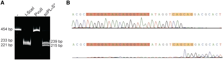
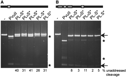
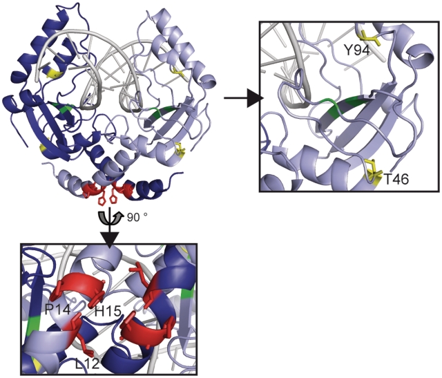
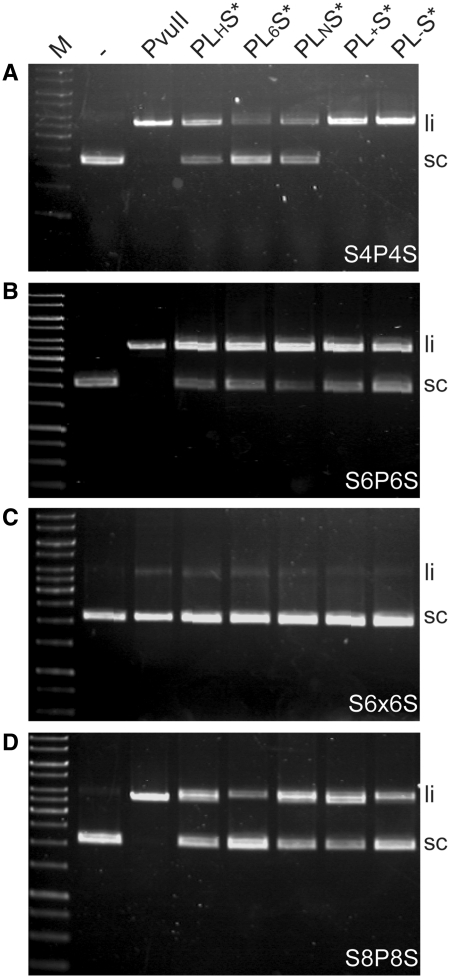
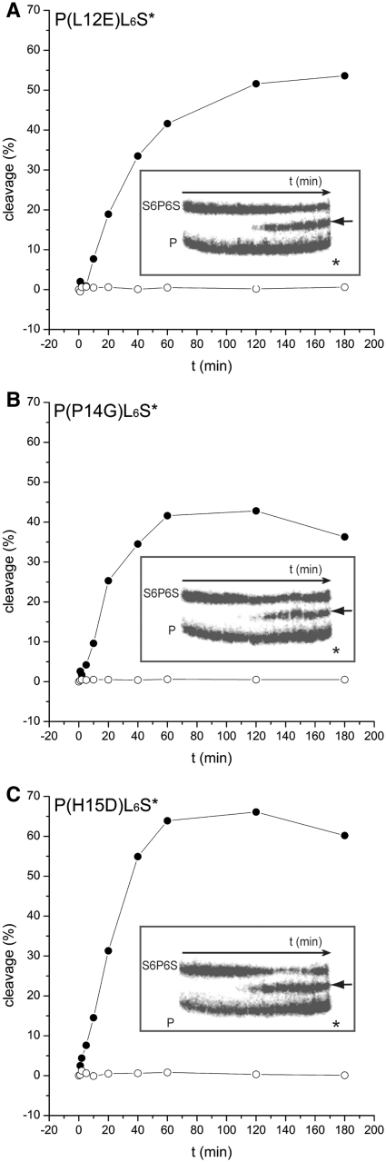
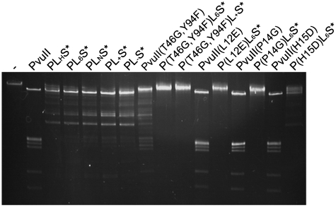
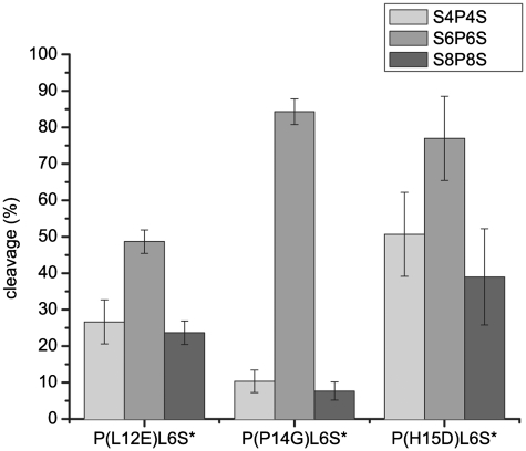
Zinc-finger nucleases and TALE nucleases are produced by combining a specific DNA-binding module and a non-specific DNA-cleavage module, resulting in nucleases able to cleave DNA at a unique sequence. Here a new approach for creating highly specific nucleases was pursued by fusing a catalytically inactive variant of the homing endonuclease I-SceI, as DNA binding-module, to the type IIP restriction enzyme PvuII, as cleavage module. The fusion enzymes were designed to recognize a composite site comprising the recognition site of PvuII flanked by the recognition site of I-SceI. In order to reduce activity on PvuII sites lacking the flanking I-SceI sites, the enzymes were optimized so that the binding of I-SceI to its sites positions PvuII for cleavage of the composite site. This was achieved by optimization of the linker and by introducing amino acid substitutions in PvuII which decrease its activity or disturb its dimer interface. The most specific variant showed a more than 1000-fold preference for the addressed composite site over an unaddressed PvuII site. These results indicate that using a specific restriction enzyme, such as PvuII, as cleavage module, offers an alternative to the otherwise often used catalytic domain of FokI, which by itself does not contribute to the specificity of the engineered nuclease.


Figures

Figure 1.
Figure 1.
(A) Cartoon representation of the fusion enzyme. The C-terminus of one subunit of a dimeric restriction enzyme (PvuII; blue) is fused via a peptide linker (gray) to the N-terminus of a catalytically inactive homing endonuclease (I-SceI; green). This fusion enzyme binds as a homodimer to a tripartite recognition site consisting of the PvuII recognition site (orange) flanked by two I-SceI sites (red) separated by an un-specified DNA (gray). The very C-terminal Strep-tag used for affinity purification is depicted in yellow. (B) Model of the engineered fusion enzyme. One subunit of the fusion enzyme consists of one I-SceI [green; pdb 1r7 m (40)] and one subunit of the PvuII dimer [blue, pdb 1pvi (47)]. This model was built by aligning the recognition sites from the crystal structures of the individual proteins on a DNA composed of the PvuII recognition site (orange) and two I-SceI sites (red) separated by 6 bp up- and downstream. The C-terminus of PvuII and the N-terminus of I-SceI, separated by 2.6 nm, are indicated by red spheres; this distance must be covered by a peptide linker of suitable length.
Figure 2.
Figure 2.
(A) Cleavage of 50 nM of a 454 bp PCR fragment containing an I-SceI recognition site. Lane 1 (-): uncleaved DNA fragment; Lane 2: cleavage products obtained by incubation of the 454 bp PCR fragment with 5 U I-SceI (Fermentas) in Buffer Tango; Lanes 3 and 4: cleavage products obtained by incubation of the 454 bp PCR fragment with 100 nM PvuII and 100 nM scPLHS, respectively, in Buffer Green. (B) Sequencing results of the cleavage product of scPLHS* from (A). The fragments were gelpurified and sequenced in forward (upper panel) and reverse (lower panel) direction. The I-SceI recognition sequence is indicated by a red bar and the cleavage site, which corresponds to a PvuII ‘star’ site by an orange bar. These two sites are separated by 6 bp.
Figure 3.
Figure 3.
Cleavage activity of fusion enzymes with different linkers on linearized DNA. The DNA substrate contains either (A) a single PvuII site (light gray bar) or (B) the tripartite recognition site consisting of a PvuII site (light gray bar) flanked by two I-SceI sites (dark gray bars) each 6 bp away, and an additional unaddressed PvuII site. The experiment was performed under optimized conditions for the corresponding enzymes, namely Buffer Green for PvuII and optimized KGB for the fusion enzymes. The products of addressed cleavage at the tripartite recognition site are indicated by arrows and the products of unaddressed cleavage at PvuII sites by asterisks. The percentage of unaddressed cleavage is shown at the bottom.
Figure 4.
Figure 4.
Sedimentation coefficient distributions in an analytical ultracentrifugation experiment with PL6S* at different salt concentrations. PL6S* at concentrations of 3.3 or 3.5 µM was analyzed in sedimentation velocity experiments at 500 mM NaCl (solid line) or 100 mM NaCl (dashed line), respectively. c(s) analysis using the program SEDFIT yielded a single sedimenting species with s20,W = 4.4 S under high salt and s20,W = 4.6 S under low salt conditions, indicating that PL6S* is a stable homodimer. The purity and stability of the protein analyzed had been determined before by SDS-PAGE of 1.7 µg protein diluted in either low or high salt buffer (see insert). The theoretical molecular weight of one subunit of PL6S* is 46.7 kDa.
Figure 5.
Figure 5.
Structural details of PvuII (pdb 1pvi). The catalytic centre composed of the residues D58, E68 and K70 is highlighted in green. The positions of substitutions introduced into PvuII are: T46G (yellow) increases the fidelity of PvuII; Y94F (yellow) has a reduced Mg2+ binding ability (50); L12E, P14G and H15D (red) are likely to weaken the dimer interface.
Figure 6.
Figure 6.
Competition cleavage experiments with fusion enzyme variants. The internally 32P labeled DNA fragments contain either the tripartite recognition sequence S6P6S (a) or a single PvuII site (u), at equimolar concentrations of 10 nM of both substrates. The two substrates were incubated with (A) PL6S*, (B) P(T46G, Y94F)L6S*, (C) PL+S* and (D) P(T46G,Y94F)L+S* for 3 h at 37°C; after defined time intervals samples were withdrawn from the reaction mixture and analyzed by gel electrophoresis. The cleavage product obtained by cleavage at the addressed tripartite recognition site is indicated by an arrow and the position of the expected cleavage product obtained by cleavage at the unaddressed PvuII site by an asterisk. (E) The autoradiograms (A–D) were quantified and the percentage of cleavage was plotted against time. Filled symbols show the addressed cleavage and open ones the unaddressed cleavage.
Figure 7.
Figure 7.
DNA cleavage experiments with all linker variants and the different substrates at equimolar concentrations (8 nM). (A) S4P4S, (B) S6P6S, (C) S6x6S and (D) S8P8S were incubated with PvuII, PLHS*, PL6S*, PLNS*, PL+S* and PL-S*, respectively, for 3 h under optimized conditions (see main text).
Figure 8.
Figure 8.
Competition cleavage experiments with the fusion enzyme variants containing amino acid substitutions in the dimer interface and internally 32P labeled DNA fragments containing the addressed tripartite recognition site (S6P6S) or the unaddressed PvuII site alone (P). The experiment was performed as described in Figure 5. The autoradiogram is shown in the insert, the result of the quantification is shown as a reaction progress curve (percent cleavage vs. time): (A) P(L12E)L6S*, (B) P(P14G)L6S* and (C) P(H15D)L6S*. The arrow indicates the cleavage product obtained by addressed cleavage (closed circle) and the asterisk the position of the expected cleavage product of unaddressed cleavage (open circle).
Figure 9.
Figure 9.
DNA cleavage experiments with all fusion enzymes and all PvuII variants and bacteriophage λ-DNA, which harbors 15 PvuII sites. DNA and enzyme concentrations were chosen such, that the molar ratio between enzyme and PvuII sites is 1:1.
Figure 10.
Figure 10.
Evaluation of DNA cleavage assays with three different plasmid substrates containing the sites S4P4S (light gray), S6P6S (gray) and S8P8S (dark gray). The variants P(L12E)L6S*, P(P14G)L6S* and P(H15D)L6S* were tested for cleavage of these substrates in equimolar concentrations of DNA and protein for 3 h at 37°C under optimized conditions. The amount of linear DNA was estimated by densitometry of the ethidium bromide stained agarose gel and the mean and standard deviation were calculated and plotted (n = 3).

Similar articles

Cited by

References

    1. Smih F, Rouet P, Romanienko PJ, Jasin M. Double-strand breaks at the target locus stimulate gene targeting in embryonic stem cells. Nucleic Acids Res. 1995;23:5012–5019. - PMC - PubMed
    1. Choulika A, Perrin A, Dujon B, Nicolas JF. Induction of homologous Recombination in mammalian chromosomes by using the I-SceI system of Saccharomyces cerevisiae. Mol. Cell. Biol. 1995;15:1968–1973. - PMC - PubMed
    1. Pingoud A, Wende W. Generation of novel nucleases with extended specificity by rational and combinatorial strategies. ChemBioChem. 2011;12:1495–1500. - PubMed
    1. Wu J, Kandavelou K, Chandrasegaran S. Custom-designed zinc finger nucleases: what is next? Cell. Mol. Life Sci. 2007;64:2933–2944. - PMC - PubMed
    1. Carroll D. Progress and prospects: zinc-finger nucleases as gene therapy agents. Gene Ther. 2008;15:1463–1468. - PMC - PubMed

Publication types

MeSH terms

Substances