

Dietary restriction ameliorates diabetic nephropathy through anti-inflammatory effects and regulation of the autophagy via restoration of Sirt1 in diabetic Wistar fatty (fa/fa) rats: a model of type 2 diabetes
- PMID: 21949662
- PMCID: PMC3178150
- DOI: 10.1155/2011/908185
Dietary restriction ameliorates diabetic nephropathy through anti-inflammatory effects and regulation of the autophagy via restoration of Sirt1 in diabetic Wistar fatty (fa/fa) rats: a model of type 2 diabetes
Abstract
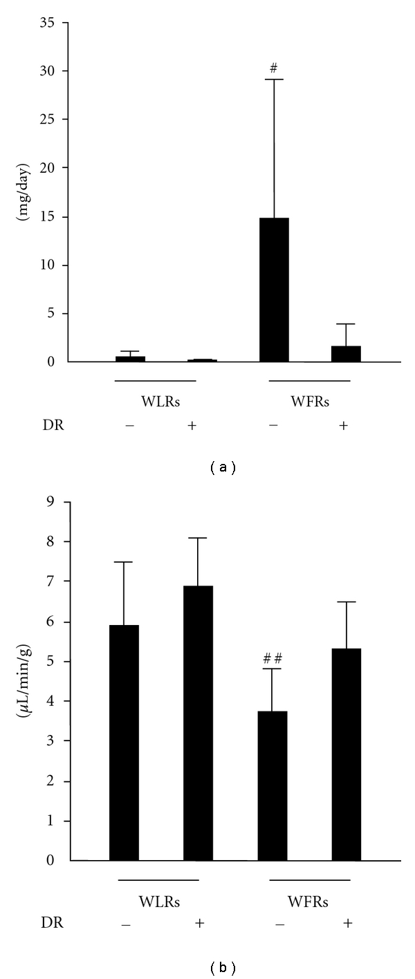
Aim: Despite the beneficial effects of dietary restriction (DR) on lifespan, age-related diseases, including diabetes and cardiovascular diseases, its effects on type 2 diabetic nephropathy remain unknown. This study examined the renoprotective effects of DR in Wistar fatty (fa/fa) rats (WFRs).
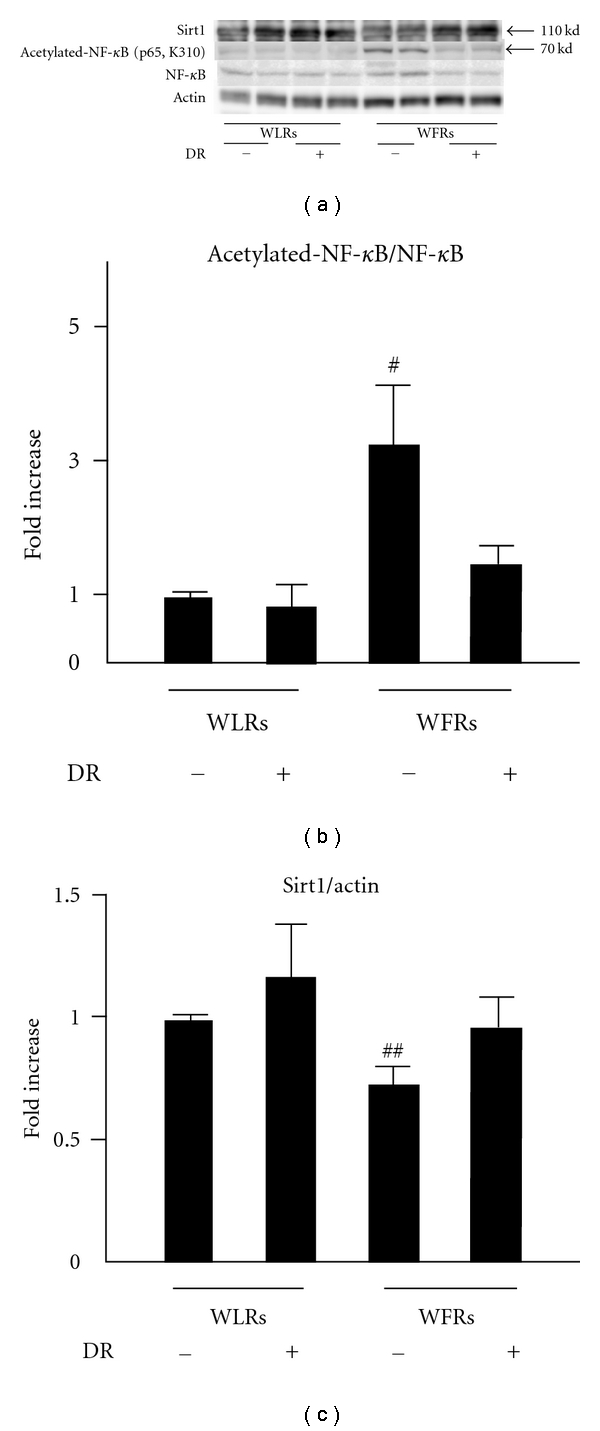
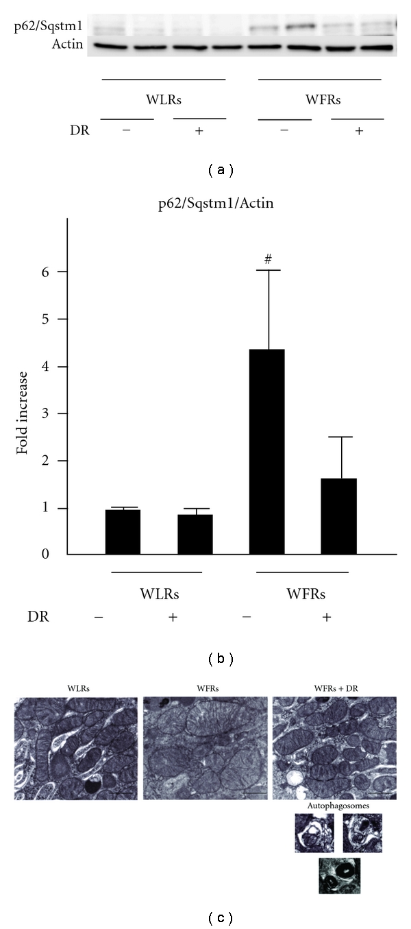
Methods: WFRs were treated with DR (40% restriction) for 24 weeks. Urinary albumin excretion, creatinine clearance, renal histologies, acetylated-NF-κB (p65), Sirt1 protein expression, and p62/Sqstm 1 accumulation in the renal cortex, as well as electron microscopic observation of mitochondrial morphology and autophagosomes in proximal tubular cells were estimated.
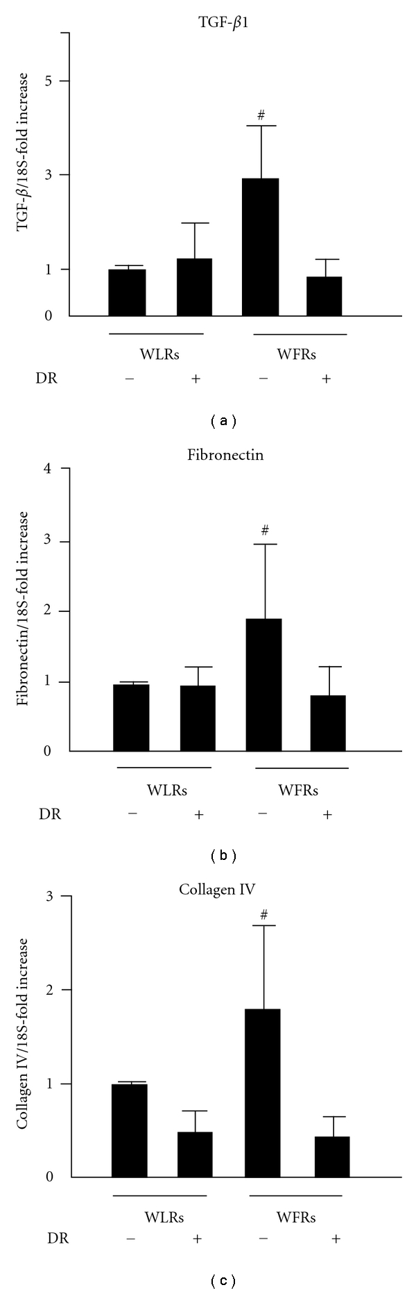
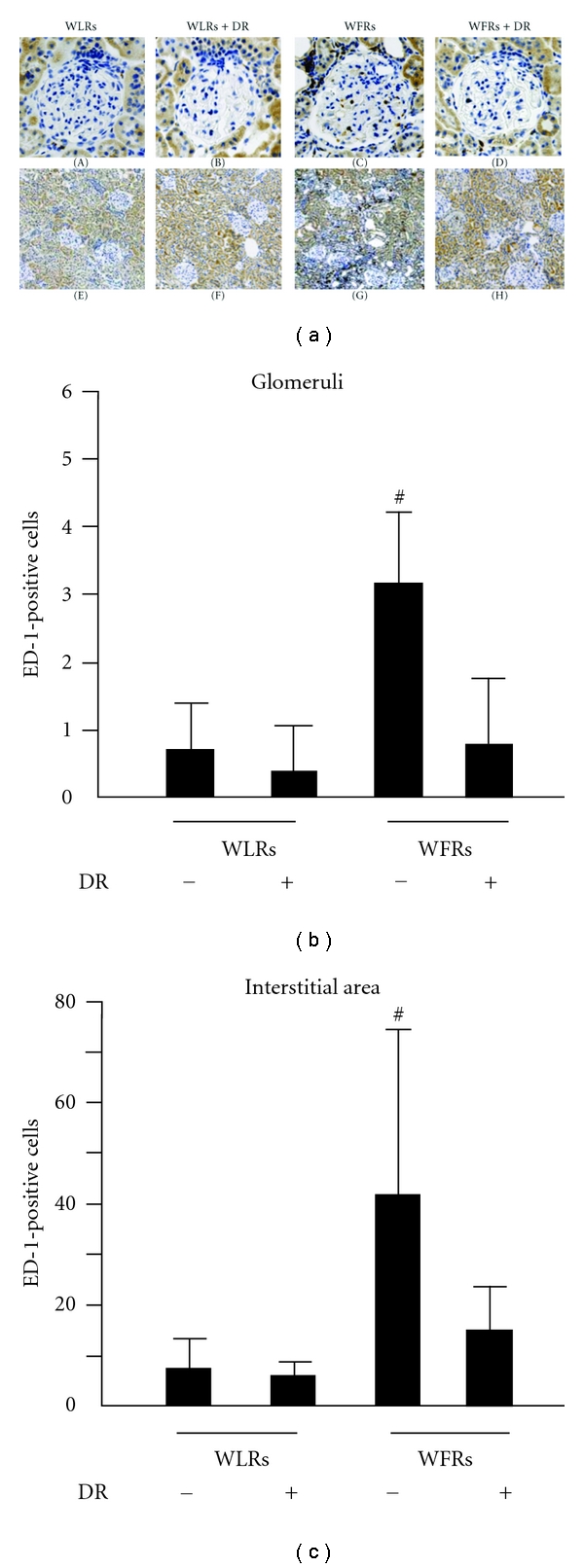
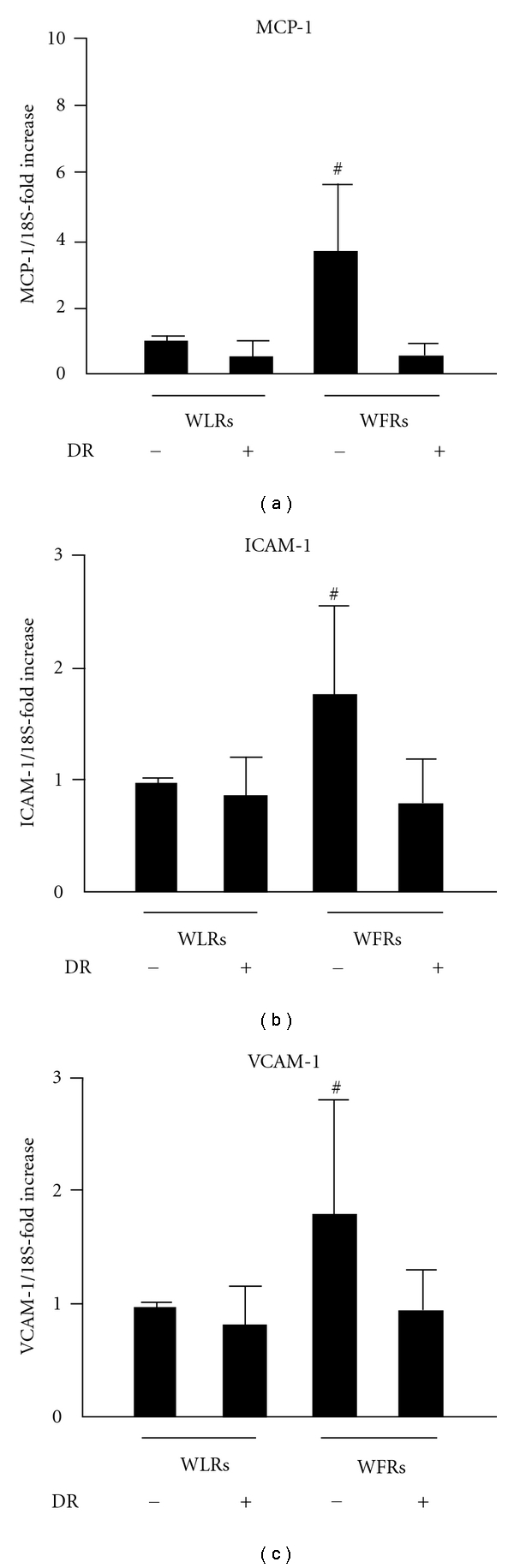
Results: DR ameliorated renal abnormalities including inflammation in WFRs. The decrease in Sirt1 levels, increase in acetylated-NF-κB, and impaired autophagy in WFRs were improved by DR.
Conclusions: DR exerted anti-inflammatory effects and improved the dysregulation of autophagy through the restoration of Sirt1 in the kidneys of WFRs, which resulted in the amelioration of renal injuries in type 2 diabetes.
Copyright © 2011 Munehiro Kitada et al.
Figures

Similar articles
-
A very-low-protein diet ameliorates advanced diabetic nephropathy through autophagy induction by suppression of the mTORC1 pathway in Wistar fatty rats, an animal model of type 2 diabetes and obesity.Diabetologia. 2016 Jun;59(6):1307-17. doi: 10.1007/s00125-016-3925-4. Epub 2016 Mar 28. Diabetologia. 2016. PMID: 27020449
-
Cyclic and intermittent very low-protein diet can have beneficial effects against advanced diabetic nephropathy in Wistar fatty (fa/fa) rats, an animal model of type 2 diabetes and obesity.Nephrology (Carlton). 2017 Dec;22(12):1030-1034. doi: 10.1111/nep.13152. Nephrology (Carlton). 2017. PMID: 28802091
-
A low-protein diet exerts a beneficial effect on diabetic status and prevents diabetic nephropathy in Wistar fatty rats, an animal model of type 2 diabetes and obesity.Nutr Metab (Lond). 2018 Mar 2;15:20. doi: 10.1186/s12986-018-0255-1. eCollection 2018. Nutr Metab (Lond). 2018. PMID: 29507597 Free PMC article.
-
Role of Impaired Nutrient and Oxygen Deprivation Signaling and Deficient Autophagic Flux in Diabetic CKD Development: Implications for Understanding the Effects of Sodium-Glucose Cotransporter 2-Inhibitors.J Am Soc Nephrol. 2020 May;31(5):907-919. doi: 10.1681/ASN.2020010010. Epub 2020 Apr 10. J Am Soc Nephrol. 2020. PMID: 32276962 Free PMC article. Review.
-
Interplay of adenosine monophosphate-activated protein kinase/sirtuin-1 activation and sodium influx inhibition mediates the renal benefits of sodium-glucose co-transporter-2 inhibitors in type 2 diabetes: A novel conceptual framework.Diabetes Obes Metab. 2020 May;22(5):734-742. doi: 10.1111/dom.13961. Epub 2020 Feb 20. Diabetes Obes Metab. 2020. PMID: 31916329 Review.
Cited by
-
The Effect of Allograft Inflammatory Factor-1 on Inflammation, Oxidative Stress, and Autophagy via miR-34a/ATG4B Pathway in Diabetic Kidney Disease.Oxid Med Cell Longev. 2022 Oct 29;2022:1668000. doi: 10.1155/2022/1668000. eCollection 2022. Oxid Med Cell Longev. 2022. PMID: 36345369 Free PMC article.
-
Sirtuin 1: A Target for Kidney Diseases.Mol Med. 2015 Jan 12;21(1):87-97. doi: 10.2119/molmed.2014.00211. Mol Med. 2015. PMID: 25587857 Free PMC article. Review.
-
Ursolic acid attenuates diabetic mesangial cell injury through the up-regulation of autophagy via miRNA-21/PTEN/Akt/mTOR suppression.PLoS One. 2015 Feb 17;10(2):e0117400. doi: 10.1371/journal.pone.0117400. eCollection 2015. PLoS One. 2015. PMID: 25689721 Free PMC article.
-
Nutrient sensing, autophagy, and diabetic nephropathy.Diabetes. 2012 Jan;61(1):23-9. doi: 10.2337/db11-0555. Diabetes. 2012. PMID: 22187371 Free PMC article. Review. No abstract available.
-
Calorie Restriction Protects against Contrast-Induced Nephropathy via SIRT1/GPX4 Activation.Oxid Med Cell Longev. 2021 Oct 19;2021:2999296. doi: 10.1155/2021/2999296. eCollection 2021. Oxid Med Cell Longev. 2021. PMID: 34712381 Free PMC article.
References
-
- International Diabetes Foundation. 2009, http://www.diabetesatlas.org/content/foreward.
-
- Navarro-González JF, Mora-Fernández C, De Fuentes MM, García-Pérez J. Inflammatory molecules and pathways in the pathogenesis of diabetic nephropathy. Nature Reviews Nephrology. 2011;7(6):327–340. - PubMed
-
- Lin SJ, Defossez PA, Guarente L. Requirement of NAD and SIR2 for life-span extension by calorie restriction in saccharomyces cerevisiae. Science. 2000;289(5487):2126–2128. - PubMed
Publication types
MeSH terms
Substances
LinkOut - more resources
Full Text Sources
Medical
