. 2012 Jan;40(2):787-800.
doi: 10.1093/nar/gkr783. Epub 2011 Sep 24.

RNA-binding protein HuR autoregulates its expression by promoting alternative polyadenylation site usage

Affiliations

RNA-binding protein HuR autoregulates its expression by promoting alternative polyadenylation site usage

Weijun Dai et al. Nucleic Acids Res. 2012 Jan.

Abstract

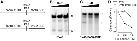
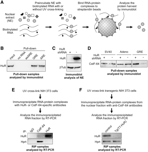
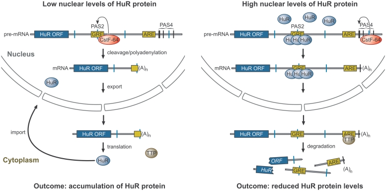
RNA-binding protein HuR modulates the stability and translational efficiency of messenger RNAs (mRNAs) encoding essential components of the cellular proliferation, growth and survival pathways. Consistent with these functions, HuR levels are often elevated in cancer cells and reduced in senescent and quiescent cells. However, the molecular mechanisms that control HuR expression are poorly understood. Here we show that HuR protein autoregulates its abundance through a negative feedback loop that involves interaction of the nuclear HuR protein with a GU-rich element (GRE) overlapping with the HuR major polyadenylation signal (PAS2). An increase in the cellular HuR protein levels stimulates the expression of long HuR mRNA species containing an AU-rich element (ARE) that destabilizes the mRNAs and thus reduces the protein production output. The PAS2 read-through occurs due to a reduced recruitment of the CstF-64 subunit of the pre-mRNA cleavage stimulation factor in the presence of the GRE-bound HuR. We propose that this mechanism maintains HuR homeostasis in proliferating cells. Since only the nuclear HuR is expected to contribute to the auto-regulation, our model may explain the longstanding observation that the increase in the total HuR expression in cancer cells often correlates with the accumulation of its substantial fraction in the cytoplasm.


Figures

Figure 1.
Figure 1.
Endogenous HuR protein levels are reduced in cells constitutively expressing a HuR transgene. (A) Diagram outlining the HILO-RMCE procedure used to obtain P19 and NIH 3T3 cells constitutively expressing T7-tagged HuR transgene. (B) Immunofluorescence analysis of the T7-HuR expression in a transgenic P19 population confirming that virtually all cells express this protein at comparable levels. Note that the T7-HuR protein is concentrated in the nucleus and undetectable in the cytoplasm in a majority of the interphase cells (e.g. the cell indicated with the white arrowhead). In a fraction of interphase cells, T7-HuR is predominantely nuclear but also detected in the cytoplasm (yellow arrowhead), consistent with the nucleocytoplasmic HuR shuttling during the cell cycle (16,27,33). Finally, T7-HuR occupies the entire volume of mitotic and prometaphase cells (orange arrowhead) due to the breakdown of the nuclear envelope at these stages. (C) HuR expression in the parental P19 cells (parent) and two independently generated T7-HuR transgenic cell populations (Tg1 and Tg2) was analyzed by immunoblot. Note that the expression of the endogenous HuR protein is reduced in the presence of the transgenic T7-HuR. (D) Immunoblot analysis of the HuR expression in the NIH 3T3 cell population expressing the T7-HuR transgene (Tg) and control transgenic cells lacking the T7-HuR (control). Similar to (C), the expression of the endogenous HuR protein is reduced in the presence of T7-HuR. (C and D) βTub, β-tubulin loading control.
Figure 2.
Figure 2.
HuR 3′-UTR is involved in the HuR auto-regulation. (A) Diagram of the mouse HuR pre-mRNA. Positions of the PASs containing the AUUAAA hexamer are indicated by vertical blue lines. PASs containing the AAUAAA sequence are shown by vertical black lines. Yellow rectangles represent the GRE and ARE sequences. Positions of the primer pairs used in mRNA expression analyses are marked by gray dashed lines. The plot at the bottom shows phastCons probability values reflecting the likelihood of sequence conservation across placental mammalian species (81). (B) Graph showing the number of sequences in the cDNA clone database that support the utilization of the corresponding PASs. (C) HuR 3′-UTR reduces the RLuc expression in the presence of T7-HuR. HEK293T cells were co-transfected with RLuc-3′HuR-wt or RLuc control plasmids and pBOS-T7-HuR or pEF-BOS vector and the RLuc expression was estimated by luciferase activity assay. (D) Over-expression of T7-HuR differentially regulates the expression of the recombinant RLuc mRNA forms terminated at different PASs. The transfection experiment described in (C) was repeated and the RLuc-3HuR mRNA levels analyzed by RT–qPCR using the primer pairs depicted in (A). (E) Northern blot demonstrating that the three mouse cell lines used in this study (P19, NIH 3T3 and L929) express predominantly PAS2-terminated HuR mRNA species. Expected positions of the four HuR mRNA forms are marked on the left. (F and G) RT–qPCR analyses showing that the expression of the transgenic T7-HuR protein in P19 and NIH 3T3 cells leads to a downregulation of the total HuR mRNA levels and a simultaneous increase in the PAS4-terminated mRNA fraction. Parental P19 cells are used as a control in (F), whereas NIH 3T3 cells containing a transgenic vector sequence are used as a control in (G). Data in (C and D) and (F and G) are averaged from three experiments ±SD.
Figure 3.
Figure 3.
The PAS4-terminated form of the mouse HuR 3′-UTR represses gene expression through an ARE-dependent mechanism. (A) Diagrams of the RLuc reporter constructs containing the wild-type and mutant HuR 3′-UTR. The PAS2 mutation (PAS2mut) converts the ATTAAA sequence to ATGGAA. ΔARE corresponds to the deletion of a 66 nucleotide sequence containing the ARE sequence (Supplementary Figure S1A). (B) PAS2mut changes the mRNA isoform pattern. HEK293T cells were transfected with either the RLuc-3′HuR-wt or the RLuc-3′HuR-PAS2mut constructs and the RNA extracted from these samples analyzed by northern blotting using an RLuc coding sequence-specific probe. Note that the PAS2 inactivation leads to the disappearance of both the PAS2 and the PAS3-terminated mRNA species and that the combined RLuc-specific signal intensities are significantly lower in RLuc-3′HuR-PAS2mut compared to the RLuc-3′HuR-wt. (C) Estimation of the apparent mRNA half-lives in HEK293T cells transfected with either RLuc-3′HuR-wt or the RLuc-3′HuR-PAS2mut plasmids. (D) The transfection experiment in (B) was repeated and followed by the luciferase activity assay. (E) Deletion of the ARE sequence rescues the expression of the RLuc-3′HuR reporters. HEK293T cells were transfected with the four constructs depicted in (A) and the luciferase activity was measured 24 h post-transfection. (F and, G) The effect of TTP knockdown on the expression of RLuc-3′HuR reporters. (F) HEK293T cells were transfected with either TTP-specific siRNA (siTTP) or non-targeting control siRNA (siControl) and the expression levels of the TTP mRNA were analyzed 48 h post-transfection using RT–qPCR. (G) HEK293T cells pre-treated with siRNAs as described in (F) were transfected with RLuc-3′HuR-PAS2mut reporters with (PAS2mut) or without the ARE sequence (PAS2mut/ARE) and the RLuc activity measured 24 h after transfection.
Figure 4.
Figure 4.
HuR represses the PAS2 utilization in vivo. (A) Bicistronic constructs containing dTomato and EGFP coding sequences separated with either the wild-type PAS2/GRE spacer (Bic-PAS2wt) or a similar spacer containing mutated PAS2 sequence (Bic-PAS2mut). (B and C) Knocking down the endogenous HuR protein stimulates the pre-mRNA processing at the PAS2 sequence. (B) The bicistronic constructs were integrated into the P19 cell genome using RMCE and the corresponding stable cell populations transfected with either a HuR-specific siRNA (siHuR) or a non-targeting control siRNA (siControl). Total RNA samples were extracted 48 h post-transfection and the efficiency of HuR knockdown was analyzed by RT–qPCR (C) RNA samples from (B) were further analyzed by RT–qPCR to determine the ratio between the EGFP and dTomato expression levels as a measure of the pre-mRNA processing efficiency at the PAS2.
Figure 5.
Figure 5.
HuR inhibits pre-mRNA cleavage in vitro in a PAS2 DSE-dependent manner. (A) Diagram of the RNA substrates used in the cleavage assay. The top RNA is from the late SV40 PAS and the bottom RNA contains the SV40-specific 5′ part and the HuR PAS2-specific DSE. (B and C) In vitro cleavage reactions carried out with 32P-labeled SV40 (B) and SV40-PAS2-DSE substrates in the presence of increasing amounts of purified N-terminally His-tagged HuR protein. (D) Quantitation of the relative cleavage efficiencies from (B and C) as a function of the purified HuR concentration added to the reaction mixtures.
Figure 6.
Figure 6.
HuR interacts with the PAS2 GRE and hinders CstF-64 binding to this sequence. (A) Diagram of the RNA pull-down assays. (B) Nuclear extract prepared from mouse L929 cells was incubated with biotinylated RNA probes corresponding to the GRE or ARE sequences from the mouse HuR mRNA followed by capturing the RNA–protein complexes on strepavidin beads. Control pull-down samples contained either no RNA or the ARE antisense sequence (asARE). The analysis of the RNA-bound protein fraction by immunoblotting with anti-HuR antibodies shows that HuR interacts with both GRE and ARE sequences in vitro. (C and D) To examine the effect of HuR knockdown on the association of CstF-64 with the HuR GRE sequence, nuclear extracts were prepared from HEK293T cells encoding Dox-inducible shRNA against human HuR, which were cultured either with (HuR knockdown) or without Dox (normal HuR protein levels). The pull-down experiments were then carried out using the two nuclear extracts and the interaction of CstF-64 protein with different DSE-containing RNAs was assessed by immunoblotting. (C) Immunoblot analysis of total nuclear extracts confirming an efficient HuR knockdown in cells expressing HuR-specific shRNA (Dox+) as compared to the Dox-negative control. (D) Immunoblot analysis of the pull-down fractions showing that the CstF-64 binding to the GRE sequence is enhanced in the absence of HuR. On the other hand, CstF-64 interaction with the SV40- and adenovirus-specific probes is not affected by the change in the HuR abundance. (E) RNA immunoprecipitation experiment confirming the interaction of the HuR and CstF-64 proteins with the endogenous HuR mRNA in NIH 3T3 cells. Note that the Hprt mRNA used here as a control efficiently recruits CstF-64 but not HuR (F) RNA immunoprecpitation carried out with the nuclear fraction from transgenic NIH 3T3 cells expressing either vector sequences (control) or T7-HuR [Tg(T7-HuR)] shows dramatically diminished interaction of CstF-64 with the HuR mRNA in the presence of T7-HuR. On the other hand, the T7-HuR expression has no effect on the CstF-64 interaction with the Hprt mRNA.
Figure 7.
Figure 7.
Model for the HuR homeostatic auto-regulation.

Similar articles

Cited by

References

    1. Moore MJ. From birth to death: the complex lives of eukaryotic mRNAs. Science. 2005;309:1514–1518. - PubMed
    1. Moore MJ, Proudfoot NJ. Pre-mRNA processing reaches back to transcription and ahead to translation. Cell. 2009;136:688–700. - PubMed
    1. Glisovic T, Bachorik JL, Yong J, Dreyfuss G. RNA-binding proteins and post-transcriptional gene regulation. FEBS Lett. 2008;582:1977–1986. - PMC - PubMed
    1. Sanchez-Diaz P, Penalva LO. Post-transcription meets post-genomic: the saga of RNA binding proteins in a new era. RNA Biol. 2006;3:101–109. - PubMed
    1. McKee AE, Minet E, Stern C, Riahi S, Stiles CD, Silver PA. A genome-wide in situ hybridization map of RNA-binding proteins reveals anatomically restricted expression in the developing mouse brain. BMC Dev. Biol. 2005;5:14. - PMC - PubMed

Publication types