

Exploring weak, transient protein--protein interactions in crowded in vivo environments by in-cell nuclear magnetic resonance spectroscopy
- PMID: 21942871
- PMCID: PMC3202675
- DOI: 10.1021/bi201287e
Exploring weak, transient protein--protein interactions in crowded in vivo environments by in-cell nuclear magnetic resonance spectroscopy
Abstract
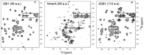
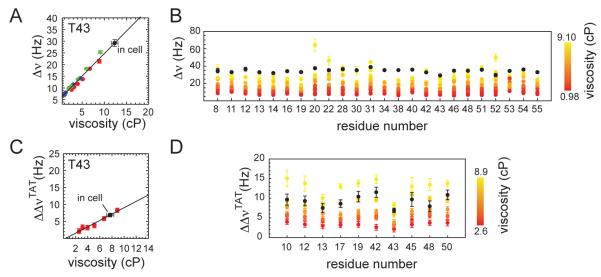
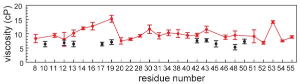
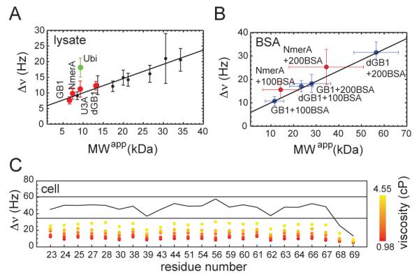
Biology relies on functional interplay of proteins in the crowded and heterogeneous environment inside cells, and functional protein interactions are often weak and transient. Thus, methods that preserve these interactions and provide information about them are needed. In-cell nuclear magnetic resonance (NMR) spectroscopy is an attractive method for studying a protein's behavior in cells because it may provide residue-level structural and dynamic information, yet several factors limit the feasibility of protein NMR spectroscopy in cells; among them, slow rotational diffusion has emerged as the most important. In this paper, we seek to elucidate the causes of the dramatically slow protein tumbling in cells and in so doing to gain insight into how the intracellular viscosity and weak, transient interactions modulate protein mobility. To address these questions, we characterized the rotational diffusion of three model globular proteins in Escherichia coli cells using two-dimensional heteronuclear NMR spectroscopy. These proteins have a similar molecular size and globular fold but very different surface properties, and indeed, they show very different rotational diffusion in the E. coli intracellular environment. Our data are consistent with an intracellular viscosity approximately 8 times that of water, too low to be a limiting factor for observation of small globular proteins by in-cell NMR spectroscopy. Thus, we conclude that transient interactions with cytoplasmic components significantly and differentially affect the mobility of proteins and therefore their NMR detectability. Moreover, we suggest that an intricate interplay of total protein charge and hydrophobic interactions plays a key role in regulating these weak intermolecular interactions in cells.
Figures
Similar articles
-
(19) F NMR spectroscopy as a probe of cytoplasmic viscosity and weak protein interactions in living cells.Chemistry. 2013 Sep 16;19(38):12705-10. doi: 10.1002/chem.201301657. Epub 2013 Aug 6. Chemistry. 2013. PMID: 23922149
-
Quantification of size effect on protein rotational mobility in cells by 19F NMR spectroscopy.Anal Bioanal Chem. 2018 Jan;410(3):869-874. doi: 10.1007/s00216-017-0745-4. Epub 2017 Nov 28. Anal Bioanal Chem. 2018. PMID: 29184995
-
In-cell NMR spectroscopy in Escherichia coli.Methods Mol Biol. 2012;831:261-77. doi: 10.1007/978-1-61779-480-3_15. Methods Mol Biol. 2012. PMID: 22167679 Review.
-
Intracellular pH modulates quinary structure.Protein Sci. 2015 Nov;24(11):1748-55. doi: 10.1002/pro.2765. Epub 2015 Aug 30. Protein Sci. 2015. PMID: 26257390 Free PMC article.
-
Studying protein stability in crowded environments by NMR.Prog Nucl Magn Reson Spectrosc. 2024 Apr-Jun;140-141:42-48. doi: 10.1016/j.pnmrs.2024.01.001. Epub 2024 Feb 8. Prog Nucl Magn Reson Spectrosc. 2024. PMID: 38705635 Review.
Cited by
-
Diversity in the origins of proteostasis networks--a driver for protein function in evolution.Nat Rev Mol Cell Biol. 2013 Apr;14(4):237-48. doi: 10.1038/nrm3542. Epub 2013 Mar 6. Nat Rev Mol Cell Biol. 2013. PMID: 23463216 Free PMC article. Review.
-
Analysis of Fluorescent Proteins for Observing Single Gene Locus in a Live and Fixed Escherichia coli Cell.J Phys Chem B. 2024 Jul 18;128(28):6730-6741. doi: 10.1021/acs.jpcb.4c01816. Epub 2024 Jul 5. J Phys Chem B. 2024. PMID: 38968413 Free PMC article.
-
Exposing the Moving Parts of Proteins with NMR Spectroscopy.J Phys Chem Lett. 2012 Apr 19;3(8):1039-1051. doi: 10.1021/jz3002103. Epub 2012 Apr 2. J Phys Chem Lett. 2012. PMID: 22545175 Free PMC article.
-
Radio Signals from Live Cells: The Coming of Age of In-Cell Solution NMR.Chem Rev. 2022 May 25;122(10):9267-9306. doi: 10.1021/acs.chemrev.1c00790. Epub 2022 Jan 21. Chem Rev. 2022. PMID: 35061391 Free PMC article. Review.
-
Real-time monitoring of New Delhi metallo-β-lactamase activity in living bacterial cells by 1H NMR spectroscopy.Angew Chem Int Ed Engl. 2014 Feb 17;53(8):2130-3. doi: 10.1002/anie.201308636. Epub 2014 Jan 23. Angew Chem Int Ed Engl. 2014. PMID: 24458501 Free PMC article.
References
Publication types
MeSH terms
Substances
Grants and funding
LinkOut - more resources
Full Text Sources
