

E-cadherin promotes accumulation of a unique memory CD8 T-cell population in murine salivary glands
- PMID: 21930933
- PMCID: PMC3189029
- DOI: 10.1073/pnas.1107200108
E-cadherin promotes accumulation of a unique memory CD8 T-cell population in murine salivary glands
Abstract
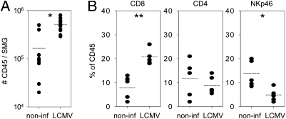
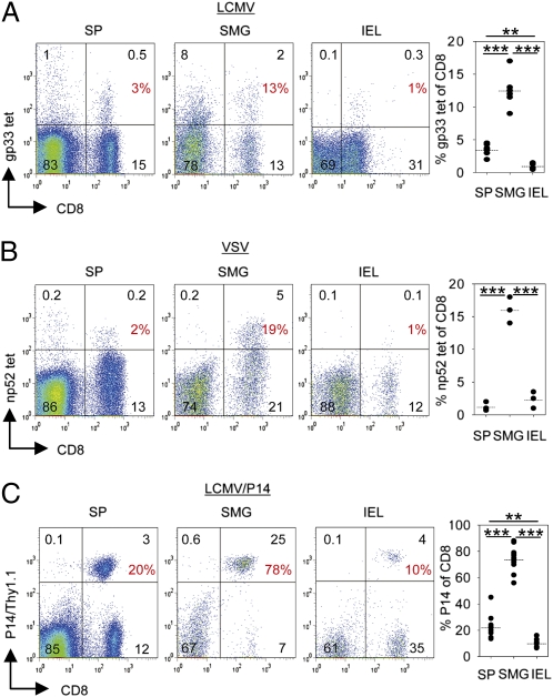
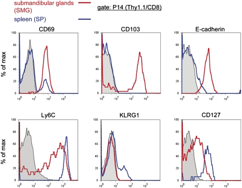
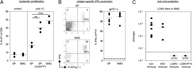
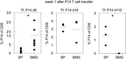
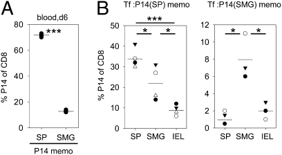
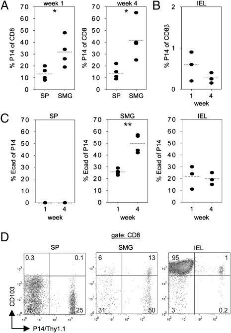
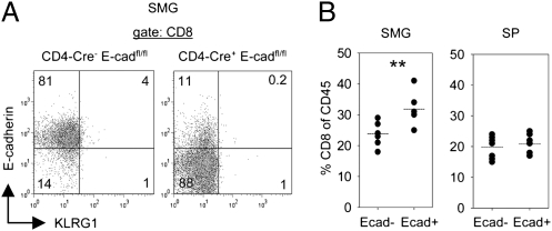
The salivary glands are important effector sites for IgA-mediated humoral immunity to protect oral surfaces. Within murine submandibular glands (SMG), we identified a memory CD8 T-cell population that exhibited a unique cell-surface phenotype distinct from memory CD8 T cells in spleen but similar to memory T cells resident in the intraepithelial lymphocyte compartment of the intestinal mucosa. In mice immune to lymphocytic choriomeningitis virus (LCMV) or vesicular stomatitis virus(VSV), virus-specific memory CD8 T cells with this unusual phenotype were present in SMG at remarkably high frequencies. LCMV-specific memory CD8 T cells in SMG showed potent functional activities in vivo, including cytokine-induced bystander proliferation, antigen-triggered IFNγ production, and viral clearance. Adoptive transfer experiments further revealed that the capacity to accumulate in SMG decreased during CD8 T-cell differentiation and that SMG CD8 T cells were poorly replenished from the circulation, indicating that they were tissue-resident. Moreover, they preferentially relocalized within their tissue of origin after adoptive transfer and antigen rechallenge, thus revealing an imprinted differentiation status. Accumulation of memory CD8 T cells within SMG did not require local antigen presentation but was promoted by the epithelial differentiation molecule E-cadherin intrinsically expressed by these CD8 T cells. This finding extends the epithelial-restricted function of E-cadherin to an impact on lymphocyte accumulation within epithelial tissues.
Conflict of interest statement
The authors declare no conflict of interest.
Figures
Similar articles
-
Thymus-resident memory CD8+ T cells mediate local immunity.Eur J Immunol. 2013 Sep;43(9):2295-304. doi: 10.1002/eji.201343519. Epub 2013 Jun 26. Eur J Immunol. 2013. PMID: 23715993
-
α4 β1 integrin promotes accumulation of tissue-resident memory CD8+ T cells in salivary glands.Eur J Immunol. 2017 Feb;47(2):244-250. doi: 10.1002/eji.201646722. Epub 2016 Dec 7. Eur J Immunol. 2017. PMID: 27861803
-
Circulating memory CD8+ T cells are limited in forming CD103+ tissue-resident memory T cells at mucosal sites after reinfection.Eur J Immunol. 2021 Jan;51(1):151-166. doi: 10.1002/eji.202048737. Epub 2020 Aug 31. Eur J Immunol. 2021. PMID: 32762051
-
Bone marrow contains virus-specific cytotoxic T lymphocytes.Blood. 1997 Sep 1;90(5):2103-8. Blood. 1997. PMID: 9292550
-
Differences in the transduction of canonical Wnt signals demarcate effector and memory CD8 T cells with distinct recall proliferation capacity.J Immunol. 2014 Sep 15;193(6):2784-91. doi: 10.4049/jimmunol.1400465. Epub 2014 Aug 15. J Immunol. 2014. PMID: 25127860
Cited by
-
Tissue-resident memory T cells.Immunity. 2014 Dec 18;41(6):886-97. doi: 10.1016/j.immuni.2014.12.007. Epub 2014 Dec 6. Immunity. 2014. PMID: 25526304 Free PMC article. Review.
-
The Effects of Acute Neutrophil Depletion on Resolution of Acute Influenza Infection, Establishment of Tissue Resident Memory (TRM), and Heterosubtypic Immunity.PLoS One. 2016 Oct 14;11(10):e0164247. doi: 10.1371/journal.pone.0164247. eCollection 2016. PLoS One. 2016. PMID: 27741316 Free PMC article.
-
Skin-resident memory CD4+ T cells enhance protection against Leishmania major infection.J Exp Med. 2015 Aug 24;212(9):1405-14. doi: 10.1084/jem.20142101. Epub 2015 Jul 27. J Exp Med. 2015. PMID: 26216123 Free PMC article.
-
Antigen-independent differentiation and maintenance of effector-like resident memory T cells in tissues.J Immunol. 2012 May 15;188(10):4866-75. doi: 10.4049/jimmunol.1200402. Epub 2012 Apr 13. J Immunol. 2012. PMID: 22504644 Free PMC article.
-
Organoids in immunological research.Nat Rev Immunol. 2020 May;20(5):279-293. doi: 10.1038/s41577-019-0248-y. Epub 2019 Dec 18. Nat Rev Immunol. 2020. PMID: 31853049 Review.
References
-
- Masopust D, Vezys V, Marzo AL, Lefrançois L. Preferential localization of effector memory cells in nonlymphoid tissue. Science. 2001;291:2413–2417. - PubMed
-
- Sallusto F, Lenig D, Förster R, Lipp M, Lanzavecchia A. Two subsets of memory T lymphocytes with distinct homing potentials and effector functions. Nature. 1999;401:708–712. - PubMed
-
- Klonowski KD, et al. Dynamics of blood-borne CD8 memory T cell migration in vivo. Immunity. 2004;20:551–562. - PubMed
-
- Gebhardt T, et al. Memory T cells in nonlymphoid tissue that provide enhanced local immunity during infection with herpes simplex virus. Nat Immunol. 2009;10:524–530. - PubMed
Publication types
MeSH terms
Substances
LinkOut - more resources
Full Text Sources
Molecular Biology Databases
Research Materials
Miscellaneous
