. 2011 Sep 26;208(10):2113-24.
doi: 10.1084/jem.20110522. Epub 2011 Sep 19.

Airborne lipid antigens mobilize resident intravascular NKT cells to induce allergic airway inflammation

Affiliations

Airborne lipid antigens mobilize resident intravascular NKT cells to induce allergic airway inflammation

Seth T Scanlon et al. J Exp Med. .

Abstract

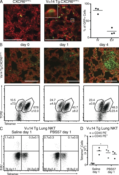
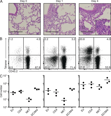
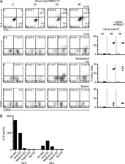
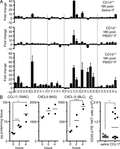
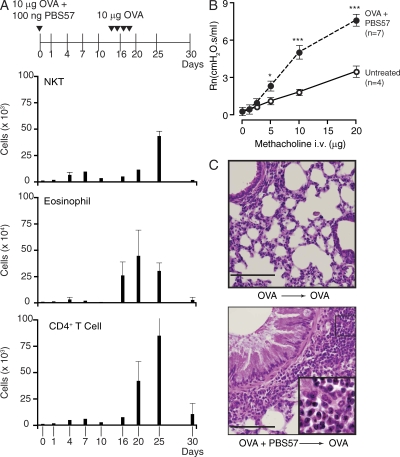
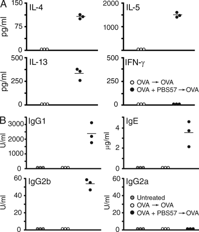
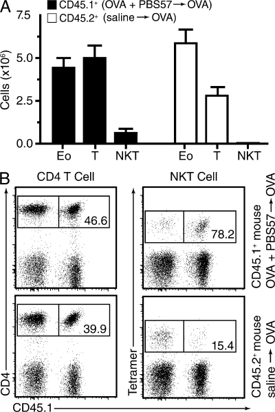
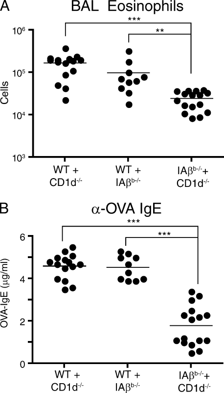
Airborne exposure to microbial cell wall lipids such as lipopolysaccharide triggers innate immune responses that regulate susceptibility to allergic airway inflammation. α-Glycosylceramides represent another widespread class of microbial lipids that directly stimulate innate-like, IL-4- and IL-13-producing, CD1d-restricted NKT cells. In this study, we demonstrate that NKT cells constitutively accumulate and reside in the microvasculature of the mouse lung. After a single airborne exposure to lipid antigen, they promptly extravasate to orchestrate the formation of peribronchiolar and interstitial lymphohistiocytic granulomas containing numerous eosinophils. Concomitant airborne exposure to ovalbumin (OVA) induces the priming of OVA-specific Th2 cells and IgE antibodies by the same dendritic cell coexpressing CD1d and MHC class II. Although NKT cell activation remains confined to the lipid-exposed lung and draining lymph nodes, Th2 cells recirculate and seed the lung of a parabiotic partner, conferring susceptibility to OVA challenge months after the initial exposure, in a manner independent of NKT cells and CD1d. Thus, transient recruitment and activation of lung-resident intravascular NKT cells can trigger long-term susceptibility to allergic airway inflammation.


Figures

Figure 1.
Figure 1.
Intravascular location of lung Vα14 NKT cells. (A, left) Two-photon fluorescence microscopy of lung harvested from CXCR6GFP/+ mice and Vα14tg CXCR6GFP/+ mice (as indicated) injected with DyLight592–tomato lectin to visualize blood vessel endothelia. The inset demonstrates the intravascular location of a GFP+ cell (boxed area). (right) The frequency of intravascular (IV) and extravascular (EV) NKT cells was measured in three independent Vα14tg CXCR6GFP/+ samples counting at least 100 cells per lung. A GFP+ cell was scored as intravascular if it appeared wholly within the structure of a blood vessel within the 50-µm depth of examination. (B, top) Images of rehydrated 10-µm frozen sections of Vα14 transgenic CXCR6GFP/+ lungs at days 0, 1, and 4 after airway administration of 100 ng of the NKT ligand PBS57. (bottom) Characterization of GFP+ cells by TCR-β and CD1d–α-GalCer tetramer staining. Numbers indicate mean percentage and SEM in each gate (n = 3/group). (C) EdU incorporation (3 h after i.v. injection) by Vα14tg lung lymphocytes stained with tetramers (n = 3/group). (D) Anti–CD45-PE label acquisition by CD1d–α-GalCer tetramer-gated lymphocytes obtained from lungs harvested 2 min after i.v. injection of the antibody in Vα14tg mice exposed to airborne saline or PBS57, as indicated (n = 3/group). (A and D) Horizontal bars indicate the mean. *, P < 0.05. Bars, 100 µm.
Figure 2.
Figure 2.
Extravasation of intravascular NKT cells upon airborne exposure to their lipid antigen. BALB/c mice received one intratracheal instillation of 100 ng PBS57 at day 0. (A) H&E staining of lungs (inset shows granuloma, day 4), representative of three mice/group. Bars, 200 µm. (B) Anti–CD45-PE label acquisition by B220TCR-β+ gated lymphocytes obtained from lungs harvested 2 min after i.v. injection of the antibody (n = 3 mice/group). (C) Absolute numbers of eosinophils (Eo), CD4+ T cells (CD4), NKT cells (NKT), and DCs/macrophages (DC/MΦ) harvested from the lungs at the corresponding time points. All experiments are representative of three mice per group. Horizontal bars indicate the mean.
Figure 3.
Figure 3.
Local activation of lung NKT cells after airborne exposure to lipid ligand. (A, top row) Intracellular IL-4 and IFN-γ staining of NKT cells (B220, TCR-β+, CD1d-tetramer+) in the lung of BALB/c mice at the indicated times after intratracheal (IT) instillation of 100 ng PBS57. Quadrant numbers represent percent mean and SEM of three mice per group. (bottom rows) CD25 and CD69 expression by tetramer+ NKT cells at the indicated times in the lung, mediastinal LNs, and spleen. The right column shows the percentage of EdU+ NKT cells at the indicated times after intratracheal instillation of PBS57 or saline in the corresponding tissues. All of the mice in the EdU incorporation experiment also received 10 µg OVA intratracheally. Horizontal bars indicate the mean. (B) Lung macrophages and DCs from lung, mediastinal (Med) and inguinal (Ing) LNs, and spleen (Spl) were FACS sorted 24 and 48 h after intratracheal instillation of 100 ng PBS57 and cultured with the PBS57-specific NKT cell hybridoma DN32.D3 for 1 d before assessing IL-2 released in supernatants. Data are representative of two independent experiments.
Figure 4.
Figure 4.
Local chemokines induce NKT cell extravasation. (A) Chemokine messenger RNA expression profile of total lungs from WT or CD1d−/− mice 18 h after airway exposure to saline or PBS57 as indicated. Data were normalized to control unmanipulated WT mice, with bars showing the mean ± SEM of two independent experiments. Shown are ccl1 (01), ccl2 (02), ccl3 (03), ccl4 (04), ccl5 (05), ccl6 (06), ccl7 (07), ccl8 (08), ccl9 (09), ccl11 (10), ccl12 (11), ccl17 (12), ccl19 (13), ccl20 (14), ccl21 (15), ccl22 (16), ccl24 (17), ccl25 (18), ccl27a (19), ccl28 (20), cxcl1 (21), cxcl2 (22), cxcl3 (23), pf4/cxcl4 (24), cxcl5 (25), ppbp/cxcl7 (26), cxcl9 (27), cxcl10 (28), cxcl11 (29), cxcl12 (30), cxcl13 (31), cxcl14 (32), cxcl15 (33), cxcl16 (34), cxcl17 (35), cx3cl1 (36), and xcl1 (37). IT, intratracheal. (B) Time course of protein expression of CCL17 (TARC), CXCL9 (MIG), and CXCL13 (BLC) in total lung lysates after intratracheal exposure to PBS57. Data are representative of two separate experiments with two to three individual mice analyzed at each time point. (C) NKT cell extravasation 48 h after intratracheal administration of 100 ng CCL17 was measured by the absolute number of lung tetramer+ cells that were protected from a 2-min intravascular anti–CD45-PE labeling. EdU incorporation after a 3-h pulse was <1% in all groups (not depicted). (B and C) Horizontal bars indicate the mean. *, P < 0.05; ***, P < 0.001.
Figure 5.
Figure 5.
Airborne exposure to PBS57 + OVA renders mice susceptible to asthma-like response upon OVA-only challenge. (A) Mice received 10 µg OVA along with 100 ng PBS57 intratracheally at day 0 and were challenged intratracheally with 10 µg OVA at days 14, 15, 17, and 18. Absolute numbers of CD1d–α-GalCer+ NKT cells, eosinophils, and CD4+ T cells in the BAL at the indicated days are shown as mean ± SD of three mice per group. (B) Airway hyperresponsiveness in response to MCh assessed at day 20 (n = 4–7/group). Error bars represent SEM. *, P < 0.05; ***, P < 0.001. (C) Representative H&E micrographs of lungs from mice sensitized with OVA alone or with OVA + PBS57 at day 0 and challenged with OVA alone before harvesting at day 20. Note the mucus hypersecretion and the lymphocytic and eosinophilic infiltration (inset) in mice sensitized with PBS57. Micrographs are representative of four mice per group. Bars, 100 µm.
Figure 6.
Figure 6.
Th2 polarization of OVA-specific T cells in mediastinal LNs. (A) Cytokine release by LN cells extracted at day 20 and cultured with 50 µg/ml OVA for 3 d. Circles represent individual mice. (B) Serum isotypes of anti-OVA antibodies at day 20 in mice untreated or exposed to OVA and PBS57 as indicated. Data are representative of two independent experiments, each with three mice per group. (A and B) Horizontal bars indicate the mean.
Figure 7.
Figure 7.
Airborne priming induces recirculating effectors of allergic airway inflammation. (A) The CD45.1+ member of a parabiotic pair was sensitized with OVA + PBS57, whereas its CD45.2+ partner received saline at day 0. Both mice were then challenged with OVA alone at days 14, 15, 17, and 18, and lungs were harvested at day 20 to measure absolute numbers of eosinophils (Eo), T cells, and NKT cells. Error bars represent SEM. (B) Origin of the CD4 and NKT cells recovered from the lungs of each parabiont at day 20. Data are representative of three parabiotic pairs.
Figure 8.
Figure 8.
Allergic airway inflammation in CD1d1fl/fl mice. (A–C) Cell counts of BAL eosinophils (Eo) and T cells and serum levels of anti-OVA IgE and IgG1 at day 20 in Cd11c-Cd1dΔ/Δ (A), Lyz2-Cd1dΔ/Δ (B), or Cd19-Cd1dΔ/Δ (C) mice sensitized with OVA + PBS57 at day 0 and challenged with OVA on days 14, 15, 17, and 18. Controls were littermates expressing the Cre transgene and CD1d1fl/+ or expressing CD1d1fl/fl in the absence of the Cre transgene. Circles represent individual mice. Horizontal bars indicate the mean. *, P < 0.05; ***, P < 0.001.
Figure 9.
Figure 9.
Mixed bone marrow chimeras demonstrate a requirement for joint expression of CD1d and MHC class II by APCs. (A and B) BAL eosinophil counts (A) and anti-OVA IgE serum levels (B) in individual CD1d−/− mice reconstituted with 1:1 mixtures of bone marrows as indicated. Data are a compilation of three separate experiments in which reconstitution by CD45.1+ and CD45.2+ bone marrow–derived cells was 38.8 ± 4.7% and 61.2 ± 4.7%, respectively. Horizontal bars indicate the mean. **, P < 0.01; ***, P < 0.001.

Similar articles

Cited by

References

    1. Agea E., Russano A., Bistoni O., Mannucci R., Nicoletti I., Corazzi L., Postle A.D., De Libero G., Porcelli S.A., Spinozzi F. 2005. Human CD1-restricted T cell recognition of lipids from pollens. J. Exp. Med. 202:295–308 10.1084/jem.20050773 - DOI - PMC - PubMed
    1. Akbari O., Stock P., Meyer E., Kronenberg M., Sidobre S., Nakayama T., Taniguchi M., Grusby M.J., DeKruyff R.H., Umetsu D.T. 2003. Essential role of NKT cells producing IL-4 and IL-13 in the development of allergen-induced airway hyperreactivity. Nat. Med. 9:582–588 10.1038/nm851 - DOI - PubMed
    1. Akbari O., Faul J.L., Hoyte E.G., Berry G.J., Wahlström J., Kronenberg M., DeKruyff R.H., Umetsu D.T. 2006. CD4+ invariant T-cell-receptor+ natural killer T cells in bronchial asthma. N. Engl. J. Med. 354:1117–1129 10.1056/NEJMoa053614 - DOI - PubMed
    1. Barrett N.A., Austen K.F. 2009. Innate cells and T helper 2 cell immunity in airway inflammation. Immunity. 31:425–437 10.1016/j.immuni.2009.08.014 - DOI - PMC - PubMed
    1. Bendelac A., Savage P.B., Teyton L. 2007. The biology of NKT cells. Annu. Rev. Immunol. 25:297–336 10.1146/annurev.immunol.25.022106.141711 - DOI - PubMed

Publication types

MeSH terms