. 2011 Nov 11;286(45):39328-35.
doi: 10.1074/jbc.M111.264341. Epub 2011 Sep 16.

Waixenicin A inhibits cell proliferation through magnesium-dependent block of transient receptor potential melastatin 7 (TRPM7) channels

Affiliations

Waixenicin A inhibits cell proliferation through magnesium-dependent block of transient receptor potential melastatin 7 (TRPM7) channels

Susanna Zierler et al. J Biol Chem. .

Abstract

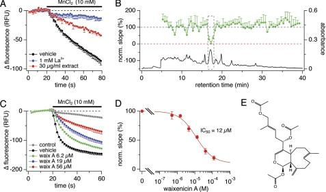
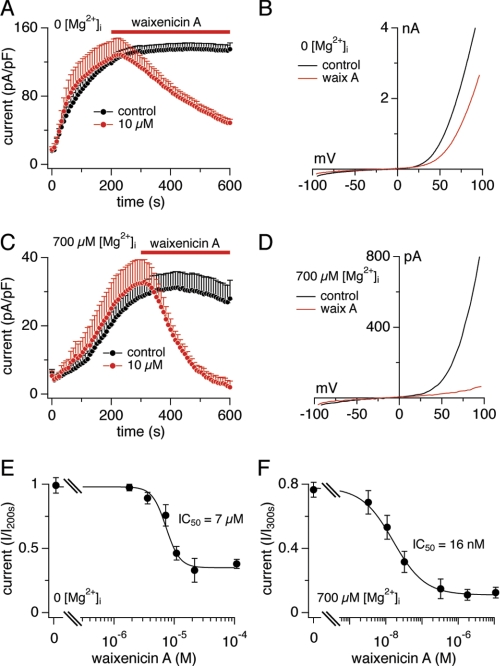
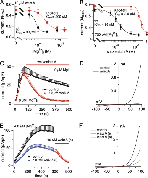
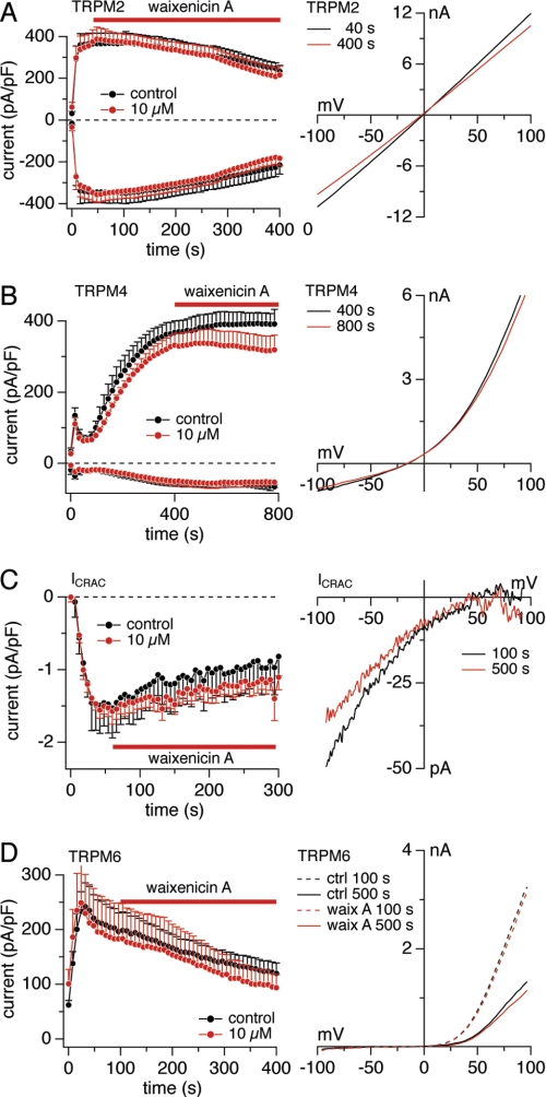
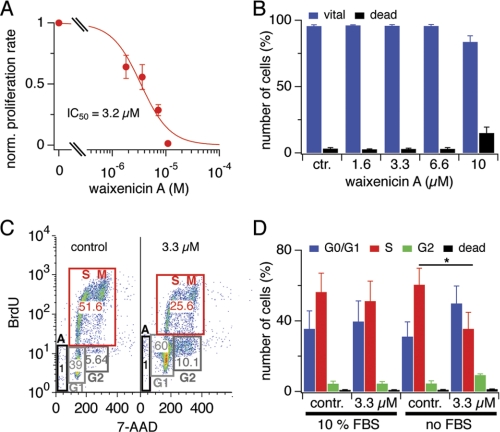
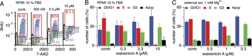
Transient receptor potential melastatin 7 (TRPM7) channels represent the major magnesium-uptake mechanism in mammalian cells and are key regulators of cell growth and proliferation. They are expressed abundantly in a variety of human carcinoma cells controlling survival, growth, and migration. These characteristics are the basis for recent interest in the channel as a target for cancer therapeutics. We screened a chemical library of marine organism-derived extracts and identified waixenicin A from the soft coral Sarcothelia edmondsoni as a strong inhibitor of overexpressed and native TRPM7. Waixenicin A activity was cytosolic and potentiated by intracellular free magnesium (Mg(2+)) concentration. Mutating a Mg(2+) binding site on the TRPM7 kinase domain reduced the potency of the compound, whereas kinase deletion enhanced its efficacy independent of Mg(2+). Waixenicin A failed to inhibit the closely homologous TRPM6 channel and did not significantly affect TRPM2, TRPM4, and Ca(2+) release-activated Ca(2+) current channels. Therefore, waixenicin A represents the first potent and relatively specific inhibitor of TRPM7 ion channels. Consistent with TRPM7 inhibition, the compound blocked cell proliferation in human Jurkat T-cells and rat basophilic leukemia cells. Based on the ability of the compound to inhibit cell proliferation through Mg(2+)-dependent block of TRPM7, waixenicin A, or structural analogs may have cancer-specific therapeutic potential, particularly because certain cancers accumulate cytosolic Mg(2+).


Figures

FIGURE 1.
FIGURE 1.
Screening assay identifies waixenicin A as TRPM7 inhibitor. A, decrease in relative fluorescence units (RFU) following 10 mm MnCl2 application in HEK293-TRPM7. Vehicle was negative control (black, n = 10). La3+ (blue, n = 10) and the extract (red, n = 2) reduced Mn2+-induced fluorescence quench. Error bars represent S.D. B, HPLC chromatogram (UV absorbance at 220–240 nm) of extract fractionation (black) and bioassay profile for the fractions (green) plotted as normalized slopes of fluorescence quench against retention time. Error bars represent S.D. C, HEK293-TRPM7 cells were incubated with waixenicin A for 15 min before 10 mm MnCl2 application: uninduced HEK293 control (gray, n = 8), vehicle (black, n = 8), waixenicin A at 6.2 μm (green, n = 3), 19 μm (blue, n = 3), and 56 μm (red, n = 3). D, maximum slopes of fluorescence quench normalized to vehicle control, plotted against concentration, and approximated by dose-response fit (n = 3–8). Error bars represent S.D. E, chemical structure of waixenicin A (relative configuration shown).
FIGURE 2.
FIGURE 2.
Mg2+ dependence of waixenicin A block. Error bars represent S.E. A, TRPM7 current densities in HEK293-TRPM7 without (black, n = 8) and with 10 μm waixenicin A (red, n = 9). B, I/V relationships are representative currents obtained at 500 s. C, TRPM7 current densities in the presence of 700 μm [Mg2+]i without (black, n = 10) and with waixenicin A (red, n = 8). Same analysis as described in A. D, I/V relationships are representative currents obtained at 600 s. E, waixenicin A was applied as described in A. Currents were extracted at +80 mV at 500 s, normalized to current at 200 s, plotted against concentration, and approximated by dose-response fit (n = 8–10). F, waixenicin A was applied as described in C. Currents were extracted at +80 mV at 600 s, normalized to current at 300 s, and analyzed as described in E (n = 5–13).
FIGURE 3.
FIGURE 3.
Waixenicin A inhibits TRPM7 kinase domain mutants. Error bars represent S.E. A, dose-response curve for [Mg2+]i in waixenicin A-treated HEK293 cells overexpressing TRPM7-wt and TRPM7-K1648R. Current amplitudes at 600 s were normalized to maximal current, averaged, plotted against [Mg2+]i, and approximated by a dose-response fit (n = 6–10). B, data acquisition and analysis as in Fig. 2C. Normalized currents of TRPM7-wt cells (black) taken from Fig. 2F and TRPM7-K1648R cells (red) are plotted against concentration and approximated by dose-response fit (n = 6–9). C, TRPM7-ΔK current densities in zero [Mg2+]i without (black, n = 7) and with 10 μm waixenicin A (waix A; red, n = 8). D, I/V relationships are representative currents obtained at 500 s. E, TRPM7-wt currents in the presence of 700 μm [Mg2+]i without (black, n = 17) and with 10 μm waixenicin A applied first intracellularly (blue/red, n = 14) and then extracellularly (red, n = 14). F, I/V relationships are representative currents obtained at 600 s (black and blue) and 800 s (red).
FIGURE 4.
FIGURE 4.
Waixenicin A is specific for TRPM7. Error bars represent S.E. A, current densities at −80/+80 mV elicited by 100 μm intracellular ADP-ribose in HEK293-TRPM2 cells without (black, n = 6) and with 10 μm waixenicin A (red, n = 8). Corresponding average I/V relationships in response obtained before (black, n = 6) and at the end of waixenicin A (waix A) application (red, n = 8). B, current densities at −80/+80 mV elicited by 3 μm [Ca2+]i in HEK293-TRPM4 cells without (black, n = 6) and with 10 μm waixenicin A (red, n = 7). Corresponding I/V relationships are average currents obtained before (black, n = 6) and at the end of waixenicin A application (red, n = 7). C, native CRAC current densities at −80 mV elicited by 20 μm inositol 1,4,5-trisphosphate in RBL1 cells without (black, n = 6) and with 10 μm waixenicin A (red circles, n = 6). Corresponding I/V relationships are average currents obtained before (black, n = 6) and at the end of waixenicin A application (red, n = 6). D, current densities at +80 mV elicited by Mg2+-free solution in HEK293-TRPM6 cells without (black, n = 11) and with 10 μm waixenicin A application (red, n = 9). E, representative I/V relationships extracted at 100 s (dashed lines) and 500 s (solid lines) from untreated controls (ctrl; black) and waixenicin A-treated cells (red).
FIGURE 5.
FIGURE 5.
Waixenicin A inhibits proliferation of RBL1 cells. Error bars represent S.E. A, normalized proliferation rate of RBL1 cells treated with waixenicin A for 2 days and analyzed via MTT assay (n = 3). The absorbance ratio of untreated controls represents 100% of proliferation rate. B, RBL1 cells were treated as in A, stained with annexin V/phosphatidylinositol, and analyzed via flow cytometry. Bars represent normalized (norm.) cell numbers (n = 3). C, cell cycle analysis of RBL1 cells incubated with or without 10% FBS, 3.3 μm waixenicin A, and BrdU for 6 h. Cells were stained with antiBrdU antibody and 7-AAD and analyzed via flow cytometry. Representative plots of control (contr.) cells and cells treated with 3.3 μm waixenicin A. D, statistical analysis of data set described in E. Bars represent normalized cell numbers (n = 3).
FIGURE 6.
FIGURE 6.
Waixenicin A causes growth arrest in Jurkat T-cells. Error bars represent S.E. A, cell cycle analysis of Jurkat cells incubated in RPMI media with 10% FBS and waixenicin A plus BrdU for 2 h (n = 3). Cells were stained with antiBrdU antibody and 7-AAD and analyzed via flow cytometry. B, statistical analysis of the data set as described in A. Bars represent normalized cell numbers (n = 3). C, Jurkat cells were incubated in standard external solution (sol.) containing 1 mm Mg2+ with no FBS. Different waixenicin A-concentrations were applied for 2 h, and cells were analyzed as described in B. Bars represent average cell numbers (n = 3). Apop, apoptotic cells.

Similar articles

Cited by

  • The Role of TRPM7 in Oncogenesis.
    Köles L, Ribiczey P, Szebeni A, Kádár K, Zelles T, Zsembery Á. Köles L, et al. Int J Mol Sci. 2024 Jan 5;25(2):719. doi: 10.3390/ijms25020719. Int J Mol Sci. 2024. PMID: 38255793 Free PMC article. Review.
  • pH-Channeling in Cancer: How pH-Dependence of Cation Channels Shapes Cancer Pathophysiology.
    Pethő Z, Najder K, Carvalho T, McMorrow R, Todesca LM, Rugi M, Bulk E, Chan A, Löwik CWGM, Reshkin SJ, Schwab A. Pethő Z, et al. Cancers (Basel). 2020 Sep 2;12(9):2484. doi: 10.3390/cancers12092484. Cancers (Basel). 2020. PMID: 32887220 Free PMC article. Review.
  • TRPM7 Ion Channel: Oncogenic Roles and Therapeutic Potential in Breast Cancer.
    Cordier C, Prevarskaya N, Lehen'kyi V. Cordier C, et al. Cancers (Basel). 2021 Dec 16;13(24):6322. doi: 10.3390/cancers13246322. Cancers (Basel). 2021. PMID: 34944940 Free PMC article. Review.
  • The Pathophysiologic Roles of TRPM7 Channel.
    Park HS, Hong C, Kim BJ, So I. Park HS, et al. Korean J Physiol Pharmacol. 2014 Feb;18(1):15-23. doi: 10.4196/kjpp.2014.18.1.15. Epub 2014 Feb 13. Korean J Physiol Pharmacol. 2014. PMID: 24634592 Free PMC article. Review.
  • TRPM7.
    Fleig A, Chubanov V. Fleig A, et al. Handb Exp Pharmacol. 2014;222:521-46. doi: 10.1007/978-3-642-54215-2_21. Handb Exp Pharmacol. 2014. PMID: 24756720 Free PMC article. Review.

References

    1. Li M., Jiang J., Yue L. (2006) J. Gen. Physiol. 127, 525–537 - PMC - PubMed
    1. Nadler M. J., Hermosura M. C., Inabe K., Perraud A. L., Zhu Q., Stokes A. J., Kurosaki T., Kinet J. P., Penner R., Scharenberg A. M., Fleig A. (2001) Nature 411, 590–595 - PubMed
    1. Ryazanova L. V., Dorovkov M. V., Ansari A., Ryazanov A. G. (2004) J. Biol. Chem. 279, 3708–3716 - PubMed
    1. Chubanov V., Gudermann T., Schlingmann K. P. (2005) Pflugers Arch. 451, 228–234 - PubMed
    1. Voets T., Nilius B., Hoefs S., van der Kemp A. W., Droogmans G., Bindels R. J., Hoenderop J. G. (2004) J. Biol. Chem. 279, 19–25 - PubMed

Publication types

LinkOut - more resources