. 2011 Sep 14:10:113.
doi: 10.1186/1476-4598-10-113.

Homeobox gene Dlx-2 is implicated in metabolic stress-induced necrosis

Affiliations

Homeobox gene Dlx-2 is implicated in metabolic stress-induced necrosis

Su Yeon Lee et al. Mol Cancer. .

Abstract

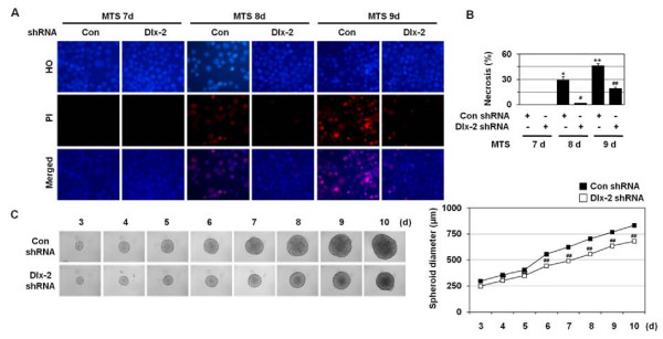
Background: In contrast to tumor-suppressive apoptosis and autophagic cell death, necrosis promotes tumor progression by releasing the pro-inflammatory and tumor-promoting cytokine high mobility group box 1 (HMGB1), and its presence in tumor patients is associated with poor prognosis. Thus, necrosis has important clinical implications in tumor development; however, its molecular mechanism remains poorly understood.

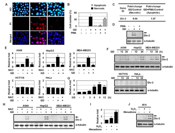
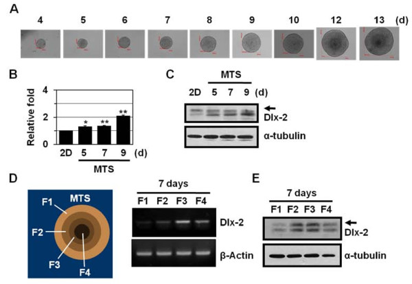
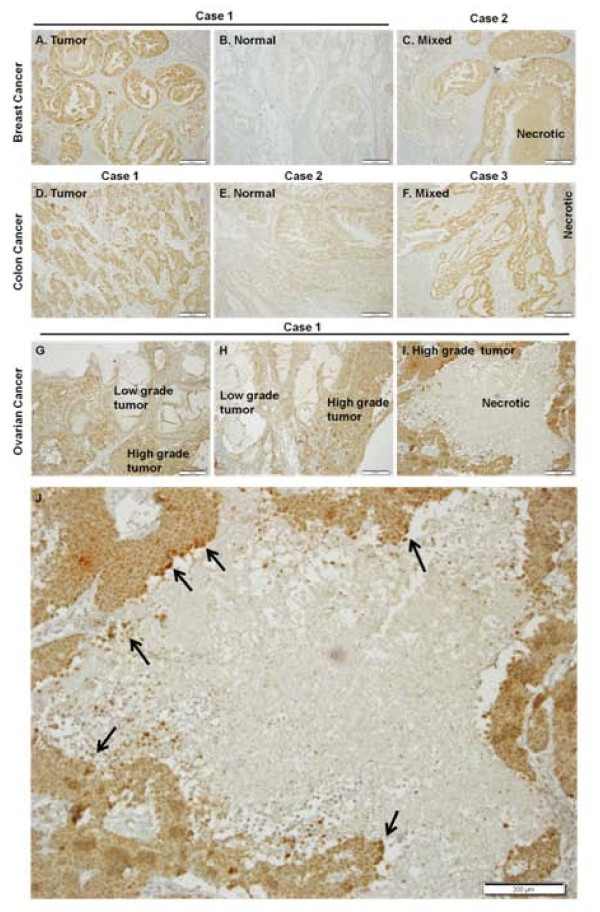
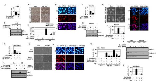
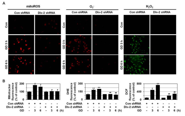
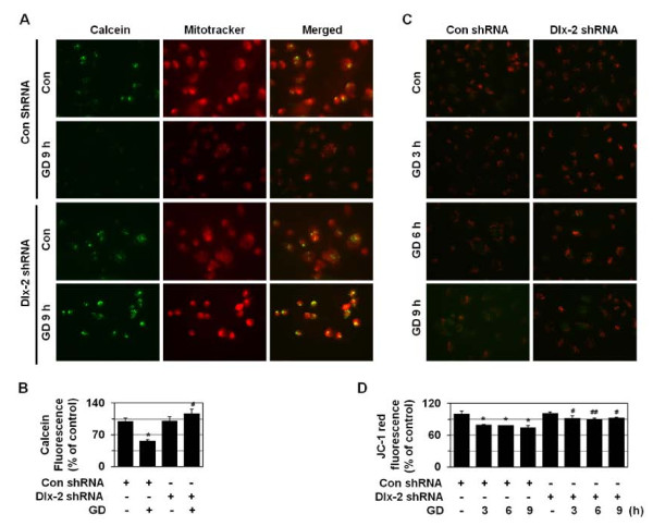
Results: In the present study, we show that Distal-less 2 (Dlx-2), a homeobox gene of the Dlx family that is involved in embryonic development, is induced in cancer cell lines dependently of reactive oxygen species (ROS) in response to glucose deprivation (GD), one of the metabolic stresses occurring in solid tumors. Increased Dlx-2 expression was also detected in the inner regions, which experience metabolic stress, of human tumors and of a multicellular tumor spheroid, an in vitro model of solid tumors. Dlx-2 short hairpin RNA (shRNA) inhibited metabolic stress-induced increase in propidium iodide-positive cell population and HMGB1 and lactate dehydrogenase (LDH) release, indicating the important role(s) of Dlx-2 in metabolic stress-induced necrosis. Dlx-2 shRNA appeared to exert its anti-necrotic effects by preventing metabolic stress-induced increases in mitochondrial ROS, which are responsible for triggering necrosis.

Conclusions: These results suggest that Dlx-2 may be involved in tumor progression via the regulation of metabolic stress-induced necrosis.


Figures

Figure 1
Figure 1
Induction of Dlx-2 during metabolic stress-induced necrosis. (A, B) A549 cells were pretreated with PMA and treated with GD for 18 h, and then stained with HO/PI (A), and apoptotic and necrotic cells were scored (B). (C, D) A549 cells were pretreated with PMA and treated with GD for 12 h and microarray analysis was performed. The numbers mean fold increase in expression as compared with GD-untreated control cells (C). The cells were analyzed using Western blotting (D). (E) Different types of cells were pretreated with NAC and treated with GD for 12 h, and then analyzed by real-time PCR for Dlx-2 expression. (F) Cells were treated with GD and then analyzed using Western blotting. (G) MDA-MB-231 cells were treated with GD, and then analyzed by real-time PCR for Dlx-2 expression. (H) Cells were pretreated with NAC and treated with GD for 12 h, and then analyzed using Western blotting. (I, J) MCF-7 cells were treated with H2O2 and menadione for 48 h, and then analyzed by real-time PCR (I) and Western blotting for Dlx-2 expression (J). The values obtained from HO/PI staining and real-time PCR are expressed as mean ± SE (n = 3). *P < 0.05, **P < 0.01 versus control; #P < 0.05, ##P < 0.01 versus GD-treated cells. Arrow shown in panels D, F, H, and J, a putative modified form of Dlx-2.
Figure 2
Figure 2
Dlx-2 shRNA prevents metabolic stress-induced necrosis in MTS. (A) Formation, growth, and morphology of MTSs derived from MCF-7 cells, which were cultured for up to 13 days. (B-C) MCF-7 spheroids cultured on agarose for the indicated times were analyzed by real-time PCR for Dlx-2 expression (B). The values are expressed as mean ± SE (n = 3). *P < 0.05, **P < 0.01 versus two-dimensional cultured cells. The MCF-7 spheroids were also analyzed using Western blotting with antibodies against Dlx-2 and α-tubulin (C). (D-E) After 7 days of MCF-7 MTS culture, the MTSs were dissociated into subpopulations of cells from different locations in the spheroids, as described in Materials and Methods. The cells isolated from different locations within the MCF-7 spheroids were analyzed by RT-PCR using primers for Dlx-2 and β-actin (D). The cells were also analyzed using Western blotting with antibodies against Dlx-2 and α-tubulin (E). Arrow in panels C and E, a putative modified form of Dlx-2.
Figure 3
Figure 3
Immunohistochemical detection of Dlx-2 in human tumors, including breast, colon, and ovarian cancers. IHC was performed on 4-μm sections of formalin-fixed, paraffin-embedded human tumors, including breast, colon, and ovarian tumor tissues. Sections were incubated with an anti-Dlx-2 antibody and the antibody was visualized with diaminobenzidine chromogen, and sections were counterstained with hematoxylin. Dlx-2, brown staining; nuclei, blue staining (H & E). A-C, breast cancer; D-F, colon cancer; G-I, ovarian cancer, and J, the enlargement image of panel I. Arrows in panel J indicate strong positive Dlx-2 staining in tumor cells adjacent to areas of necrosis and in the cells in the necrotic core. Scale bar, 200 μm.
Figure 4
Figure 4
Dlx-2 plays a role(s) in metabolic stress-induced necrosis. (A-E) A549 cells were stably transfected with Dlx-2 shRNA. The cells were analyzed by real-time PCR for Dlx-2 expression (A), and treated with GD 12 h and analyzed using Western blotting (B). The cells were treated with GD for 18 h (C) and stained with HO/PI (D), and apoptotic and necrotic cells were scored (E). (F-J) HepG2 cells were stably transfected with Dlx-2 shRNA. The cells were analyzed by real-time PCR for Dlx-2 expression (F), and treated with GD for 12 h and analyzed using Western blotting (G). The cells were treated with GD for 18 h (H), stained with HO/PI (I), and apoptotic and necrotic cells were scored (J). (K-Q) MDA-MB-231 cells were stably transfected with 2 different Dlx-2 shRNA (T1 and T2). The cells were analyzed by real-time PCR for Dlx-2 expression (K), and treated with GD for 12 h and analyzed using Western blotting (L). The cells were treated with GD for 18 h (M), stained with HO/PI (N), and apoptotic and necrotic cells were scored (O). The cells were treated with GD for 12 h and analyzed for HMGB1 (P) and LDH release (Q). The values obtained from real-time PCR, HO/PI staining, and LDH release assay are expressed as mean ± SE (n = 3). *P < 0.05, **P < 0.01 versus control; #P < 0.05, ##P < 0.01 versus control shRNA. Arrow in panels B, G, and L, a putative modified form of Dlx-2.
Figure 5
Figure 5
Dlx-2 shRNA prevents metabolic stress-induced necrosis in MTS. (A, B) MCF-7 cells stably transfected with control and Dlx-2 shRNA were seeded into 1.2% agarose-coated 96-well plates at a density of 400 cells per well and cultured for 7, 8, and 9 days. Then the cells were dissociated and stained with HO/PI (A), and apoptotic and necrotic cells were scored. The values are expressed as mean ± SE (n = 3). *P < 0.05, **P < 0.01 versus control; #P < 0.05; ##P < 0.01 versus control shRNA (B). (C) Formation, growth, and morphology of MTSs made using MCF-7 control and Dlx-2 shRNA stable cells. To calculate MTS size, diameters of 5 spheroids were measured every day. Results are expressed as mean ± SE (n = 3). ##P < 0.01 versus control shRNA.
Figure 6
Figure 6
Dlx-2 shRNA prevents metabolic stress-induced ROS production. (A, B) MDA-MB-231 cells were stably transfected with control or Dlx-2 shRNA and treated with GD for 3 or 6 h, and mitochondrial ROS and O2- and intracellular H2O2 production was measured using the MitoTracker Red CM-H2XRos, DHE, and DCFH-DA, respectively, under a fluorescence microscope (X200; Carl Zeiss). Representative images of cells from 3 independent experiments are shown (A). The values are expressed as mean ± SE from approximately 200 cells per treatment group (n = 3) (B). *P < 0.05, **P < 0.01 versus control; #P < 0.05; ##P < 0.01 versus control shRNA.
Figure 7
Figure 7
Dlx-2 shRNA prevents metabolic stress-induced loss of ΔΨm and mPT. (A, B) MDA-MB-231 cells were stably transfected with control or Dlx-2 shRNA and treated with GD for 9 h and loaded with 0.5 μM calcein AM and 5 mM CoCl2 for the final 15 min of the incubation. To detect cytoplasmic mitochondrial distribution, 50 nM MitoTracker CMX-ROS were added during calcein loading. Calcein fluorescence was excited at 488 nm and emitted at 515 nm; MitoTracker Red CMX-ROS was excited at 579 nm, and emitted at 599 nm; and the cells were observed using fluorescence microscopy. Representative images of cells from 3 independent experiments were shown (A). The results are expressed as mean ± SE from 15 to 30 cells per treatment group (n = 3) (B). *P < 0.05 versus control; #P < 0.01 versus control shRNA. (C, D) MDA-MB-231 cells stably transfected with control or Dlx-2 shRNA were treated with GD for the indicated times and then treated with 5 mg/ml JC-1 for 15 min. Representative images of cells from 3 independent experiments are shown (C). The results are expressed as mean ± SE from 50 to 100 cells per group (n = 3) (D). *P < 0.05 versus control; #P < 0.05; ##P < 0.01 versus control shRNA.

Similar articles

Cited by

References

    1. Tomes L, Emberley E, Niu Y, Troup S, Pastorek J, Strange K, Harris A, Watson PH. Necrosis and hypoxia in invasive breast carcinoma. Breast Cancer Res Treat. 2003;81:61–69. doi: 10.1023/A:1025476722493. - DOI - PubMed
    1. Kunz M, Ibrahim SM. Molecular responses to hypoxia in tumor cells. Mol Cancer. 2003;2:23. doi: 10.1186/1476-4598-2-23. - DOI - PMC - PubMed
    1. Gatenby RA, Gillies RJ. Why do cancers have high aerobic glycolysis? Nat Rev Cancer. 2004;4:891–899. doi: 10.1038/nrc1478. - DOI - PubMed
    1. Jin S, DiPaola RS, Mathew R, White E. Metabolic catastrophe as a means to cancer cell death. J Cell Sci. 2007;120:379–383. doi: 10.1242/jcs.03349. - DOI - PMC - PubMed
    1. Jin S, White E. Role of autophagy in cancer: management of metabolic stress. Autophagy. 2007;3:28–31. - PMC - PubMed

Publication types

MeSH terms