. 2011 Nov 4;286(44):38018-38026.
doi: 10.1074/jbc.M111.255844. Epub 2011 Sep 9.

Phosphorylation of the kinase interaction motif in mitogen-activated protein (MAP) kinase phosphatase-4 mediates cross-talk between protein kinase A and MAP kinase signaling pathways

Affiliations

Phosphorylation of the kinase interaction motif in mitogen-activated protein (MAP) kinase phosphatase-4 mediates cross-talk between protein kinase A and MAP kinase signaling pathways

Robin J Dickinson et al. J Biol Chem. .

Abstract

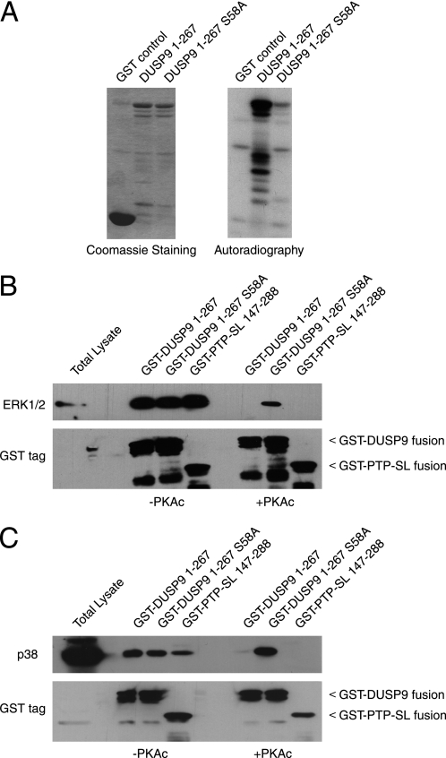
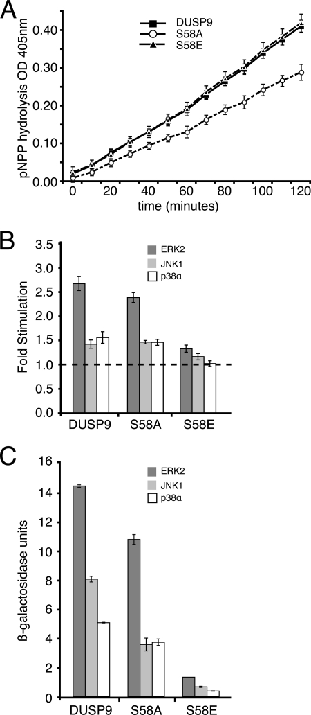
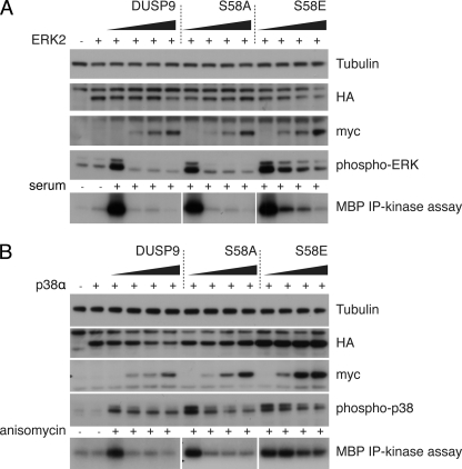
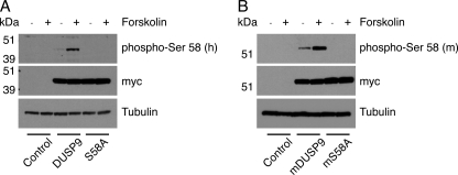
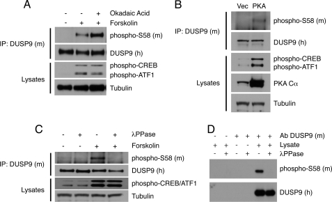
MAP kinase phosphatase 4 (DUSP9/MKP-4) plays an essential role during placental development and is one of a subfamily of three closely related cytoplasmic dual-specificity MAPK phosphatases, which includes the ERK-specific enzymes DUSP6/MKP-3 and DUSP7/MKP-X. However, unlike DUSP6/MKP-3, DUSP9/MKP-4 also inactivates the p38α MAP kinase both in vitro and in vivo. Here we demonstrate that inactivation of both ERK1/2 and p38α by DUSP9/MKP-4 is mediated by a conserved arginine-rich kinase interaction motif located within the amino-terminal non-catalytic domain of the protein. Furthermore, DUSP9/MKP-4 is unique among these cytoplasmic MKPs in containing a conserved PKA consensus phosphorylation site (55)RRXSer-58 immediately adjacent to the kinase interaction motif. DUSP9/MKP-4 is phosphorylated on Ser-58 by PKA in vitro, and phosphorylation abrogates the binding of DUSP9/MKP-4 to both ERK2 and p38α MAP kinases. In addition, although mutation of Ser-58 to either alanine or glutamic acid does not affect the intrinsic catalytic activity of DUSP9/MKP-4, phospho-mimetic (Ser-58 to Glu) substitution inhibits both the interaction of DUSP9/MKP-4 with ERK2 and p38α in vivo and its ability to dephosphorylate and inactivate these MAP kinases. Finally, the use of a phospho-specific antibody demonstrates that endogenous DUSP9/MKP-4 is phosphorylated on Ser-58 in response to the PKA agonist forskolin and is also modified in placental tissue. We conclude that DUSP9/MKP-4 is a bona fide target of PKA signaling and that attenuation of DUSP9/MKP-4 function can mediate cross-talk between the PKA pathway and MAPK signaling through both ERK1/2 and p38α in vivo.


Figures

FIGURE 1.
FIGURE 1.
A conserved KIM mediates the recognition and inactivation of ERK1/2 and p38α by DUSP9/MKP-4. A, shown is amino acid sequence alignment of the region containing the kinase interaction motif of human (Hs), bovine (Bt), murine (Mm), and rat (Rn) DUSP9/MKP-4 with human (Hs) DUSP6/MKP-3 and DUSP7/MKP-X sequences. Amino acid residues are numbered. Basic residues essential for substrate interaction (KIM) are shaded gray, and the putative PKA consensus motif (RRXS) in DUSP9/MKP-4 is highlighted in bold type. The Ser-58 phospho-acceptor is marked with an asterisk. B, mutation of the KIM does not affect the intrinsic phosphatase activity of DUSP9/MKP-4. Time-dependent hydrolysis of p-NPP by either wild-type (■) or mutant (□) DUSP9/MKP-4 was monitored by measuring the change in optical density (OD) at 405 nm. Assays were performed in triplicate, and mean values with associated errors are shown. C, catalytic activation of DUSP9/MKP-4 is KIM-dependent. Catalytic activation is expressed as -fold increase in the initial rate of p-NPP hydrolysis by 5 μg of recombinant phosphatase toward p-NPP assayed in the presence of 5 μg of the indicated MAP kinase. Assays were performed in triplicate, and mean values with associated errors are shown. D and E, analysis of yeast two-hybrid interactions between DUSP9/MKP-4 or DUSP9/MKP-4 KIM and a panel of mitogen- and stress-activated MAPK isoforms is shown. D, pGBKT7, pGBKT7.DUSP9/MKP-4, and pGBKT7.DUSP9/MKP-4 KIM were transformed into PJ69-4A yeast cells and mated with PJ69-4α yeast cells containing empty pGADT7 or the GAL4 activation domain fusions pGADT7.ERK2, pGADT7.JNK1, or pGADT7.p38α. Yeast diploids expressing both binding domain and activation domain fusions were selected on synthetic dropout media deficient in leucine and tryptophan (−Leu/−Trp). Diploid colonies were resuspended and replated for analysis onto −Leu/−Trp or −Leu/−Trp/−His/−Ade-selective plates. Protein-protein interactions were assessed by growth on this selective medium. E, semiquantitative analysis of the two-hybrid interactions based on the level of induction of the β-galactosidase gene is shown. Assays were performed in triplicate, and mean values with associated errors are shown. F and G, the dephosphorylation of ERK2 and p38α MAPKs by DUSP9/MKP-4 is KIM-dependent. COS-1 cells were transfected with either HA-tagged ERK2 (F) or HA-tagged p38α (G) expression constructs (1 μg of plasmid) together with increasing (0, 100, or 250 ng) amounts of plasmid encoding either Myc-tagged wild-type DUSP9/MKP-4 or DUSP9/MKP-4 KIM mutant. Cells were then either left untreated (−) or stimulated (+) with either serum (F) or anisomycin (G) before lysis and analysis of proteins by SDS-PAGE and Western blotting using the indicated antibodies.
FIGURE 2.
FIGURE 2.
Ser-58 within the amino-terminal domain of DUSP9/MKP-4 is phosphorylated by PKA in vitro, and this prevents binding to both ERK2 and p38α. A, GST alone or GST fusions of either DUSP9/MKP-4 1–267 or DUSP9/MKP-4 1–267 S58A were expressed in bacterial cells, purified from cell lysates, and incubated with recombinant PKA and [γ-32P]ATP in vitro. Proteins were then analyzed by SDS-PAGE and Coomassie Blue staining (left panel) or by autoradiography to show 32P incorporation (right panel). B and C, Ser-58 phosphorylation abolishes interaction with MAP kinases in vitro. GST-DUSP9/MKP-4 1–267, GST-DUSP9/MKP-4 1–267 S58A, or GST-PTP-SL-(147–288) fusion proteins were incubated either in the absence (−PKAc) or presence (+PKAc) of PKA. HEK293 cell lysates were then added and incubated for 2 h, and GST fusion proteins were then pulled down using glutathione-Sepharose before analysis by SDS-PAGE and Western blotting. MAP kinases were detected using antibodies against either ERK1/2 (B) or p38α (C). The presence of GST fusion proteins was verified using an antibody against GST (B and C, lower panels). In the first of each panel, total lysate (20 μg) was loaded.
FIGURE 3.
FIGURE 3.
Phospho-mimetic substitution of Ser-58 blocks the catalytic activation and binding of full-length DUSP9/MKP-4 to ERK2 and p38α. A, mutation of Ser-58 does not affect the intrinsic phosphatase activity of DUSP9/MKP-4. Time-dependent hydrolysis of p-NPP by either wild-type (■), mutant S58A (○), or mutant S58E (▴) DUSP9/MKP-4 was monitored by measuring the change in optical density (OD) at 405 nm. Assays were performed in triplicate, and mean values with the associated errors are shown. B, catalytic activation of DUSP9/MKP-4 is substantially reduced by substitution of Ser-58 for glutamic acid. Catalytic activation is expressed as the -fold increase in the initial rate of p-NPP hydrolysis by 5 μg of recombinant phosphatase toward p-NPP assayed in the presence of 5 μg of the indicated MAP kinase. Assays were performed in triplicate, and mean values with associated errors are shown. C, the binding of DUSP9/MKP-4 to MAPKs is substantially reduced by substitution of Ser-58 for glutamic acid. pGBKT7, pGBKT7.DUSP9/MKP-4, pGBKT7.DUSP9/MKP-4 S58A, or pGBKT7.DUSP9/MKP-4 S58E were transformed into PJ69-4A yeast cells and mated with PJ69-4α yeast cells containing empty pGADT7 or the GAL4 activation domain fusions pGADT7.ERK2, pGADT7.JNK1, or pGADT7.p38α. Semiquantitative analysis of the two-hybrid interactions was based on the level of induction of the β-galactosidase gene. Assays were performed in triplicate, and mean values with associated errors are shown.
FIGURE 4.
FIGURE 4.
Phospho-mimetic substitution of Ser-58 greatly reduces the activity of DUSP9/MKP-4 toward MAPK substrates in COS-1 cells. COS-1 cells were transfected with expression constructs (1 μg of plasmid) encoding either HA-tagged ERK2 (A) or HA-tagged p38α (B) together with increasing (0, 50, 100, or 250 ng) amounts of plasmid encoding either Myc-tagged DUSP9/MKP-4, DUSP9/MKP-4 S58A, or DUSP9/MKP-4 S58E. Cells were then either left untreated (−) or stimulated (+) with either serum (A) or anisomycin (B) before lysis. Proteins were then either analyzed by SDS-PAGE and Western blotting using the indicated antibodies (upper panels), or HA-tagged MAPKs were immunoprecipitated (IP), and kinase activities assayed in the presence of [γ-32P]ATP using myelin basic protein (MBP) as the substrate followed by SDS-PAGE and autoradiography (bottom panels).
FIGURE 5.
FIGURE 5.
Both human and murine DUSP9/MKP-4 are phosphorylated on Ser-58 in response to forskolin in COS-1 cells. COS-1 cells were transfected with expression constructs (500 ng of plasmid) encoding either myc-tagged wild-type human DUSP9/MKP-4 or DUSP9/MKP-4 S58A (A) or wild-type murine DUSP9/MKP-4 or DUSP9/MKP-4 S58A (B). Cells were either left untreated (−) or stimulated with forskolin (+) before lysis and analysis of proteins by SDS-PAGE and Western blotting using antisera raised against either human (A) or murine-specific Ser-58 (B) phosphopeptides. An antibody against tubulin was used as a loading control, whereas ectopic expression of wild-type and mutant DUSP9/MKP-4 was detected using an anti-myc monoclonal antibody.
FIGURE 6.
FIGURE 6.
Endogenous DUSP9/MKP-4 is phosphorylated on Ser-58 in both MEFs and murine placental tissue. A, immortalized MEFs were either left untreated or exposed to forskolin (10 μm) either in the absence or presence of the phosphatase inhibitor okadaic acid (1 μm). Cells were then lysed, and endogenous DUSP9/MKP-4 was immunoprecipitated (IP) using a polyclonal antibody (#302) against murine DUSP9/MKP-4 followed by SDS-PAGE and Western blotting using a polyclonal antibody (#657) raised against human DUSP9/MKP-4 and an antibody raised against a murine-specific Ser-58 phosphopeptide (upper two panels). Input lysates were also analyzed by SDS-PAGE and Western blotting using antibodies against phospho-CREB/ATF1, or as a loading control, tubulin (lower two panels). B, Ser-58 phosphorylation is enhanced by expression of a constitutively active mutant of PKA. Immortalized MEFs were transfected with either empty vector (Vec) or an expression vector encoding a constitutively active PKA Cα subunit. Cells were then lysed, and endogenous DUSP9/MKP-4 was immunoprecipitated using a polyclonal antibody (#302) against murine DUSP9/MKP-4 followed by SDS-PAGE and Western blotting using a polyclonal antibody (#657) raised against human DUSP9/MKP-4 and an antibody raised against a murine-specific Ser-58 phosphopeptide (upper two panels). Input lysates were also analyzed by SDS-PAGE and Western blotting using antibodies against phospo-CREB/ATF1, PKA Cα, or as a loading control, tubulin (lower three panels). C, immortalized MEFs were either left untreated or exposed to forskolin (10 μm). Cells were then lysed, and endogenous DUSP9/MKP-4 was immunoprecipitated using a polyclonal antibody (#302) against murine DUSP9/MKP-4. Immunoprecipitates were then incubated either in the absence (−) or presence (+) of λ-phosphatase (λPPase) before analysis by SDS-PAGE and Western blotting using a polyclonal antibody (#657) raised against human DUSP9/MKP-4 and an antibody raised against the murine Ser-58 phosphopeptide (upper two panels). Input lysates were also analyzed by SDS-PAGE and Western blotting using antibodies against phospo-CREB/ATF1 and, as a loading control, tubulin (lower two panels). D, endogenous murine DUSP9/MKP-4 was immunoprecipitated from placental lysates using a polyclonal antibody (Ab, #302) against murine DUSP9/MKP-4 with appropriate controls (no antibody and antibody but no lysate), and the samples were then divided into two aliquots and incubated either in the absence (−) or presence (+) of λ-phosphatase (λPPase). Proteins were then analyzed by SDS-PAGE and Western blotting using a polyclonal antibody (#657) raised against recombinant human DUSP9/MKP-4 and an antibody raised against the murine Ser-58 phosphopeptide as indicated.

Similar articles

Cited by

References

    1. Owens D. M., Keyse S. M. (2007) Oncogene 26, 3203–3213 - PubMed
    1. Camps M., Nichols A., Arkinstall S. (2000) FASEB J. 14, 6–16 - PubMed
    1. Dickinson R. J., Keyse S. M. (2006) J. Cell Sci. 119, 4607–4615 - PubMed
    1. Groom L. A., Sneddon A. A., Alessi D. R., Dowd S., Keyse S. M. (1996) EMBO J. 15, 3621–3632 - PMC - PubMed
    1. Muda M., Theodosiou A., Rodrigues N., Boschert U., Camps M., Gillieron C., Davies K., Ashworth A., Arkinstall S. (1996) J. Biol. Chem. 271, 27205–27208 - PubMed

Publication types

Substances