. 2011 Sep 1;25(17):1847-58.
doi: 10.1101/gad.17020911.

Regulation of the Fanconi anemia pathway by a SUMO-like delivery network

Affiliations

Regulation of the Fanconi anemia pathway by a SUMO-like delivery network

Kailin Yang et al. Genes Dev. .

Abstract

The USP1/UAF1 complex deubiquitinates the Fanconi anemia protein FANCD2, thereby promoting homologous recombination and DNA cross-link repair. How USP1/UAF1 is targeted to the FANCD2/FANCI heterodimer has remained unknown. Here we show that UAF1 contains a tandem repeat of SUMO-like domains in its C terminus (SLD1 and SLD2). SLD2 binds directly to a SUMO-like domain-interacting motif (SIM) on FANCI. Deletion of the SLD2 sequence of UAF1 or mutation of the SIM on FANCI disrupts UAF1/FANCI binding and inhibits FANCD2 deubiquitination and DNA repair. The USP1/UAF1 complex also deubiquitinates PCNA-Ub, and deubiquitination requires the PCNA-binding protein hELG1. The SLD2 sequence of UAF1 binds to a SIM on hELG1, thus targeting the USP1/UAF1 complex to its PCNA-Ub substrate. We propose that the regulated targeting of USP1/UAF1 to its DNA repair substrates, FANCD2-Ub and PCNA-Ub, by SLD-SIM interactions coordinates homologous recombination and translesion DNA synthesis.


Figures

Figure 1.
Figure 1.
The C-terminal region of UAF1 is dispensable for the activation of USP1 activity in vitro. (A) Schematic representation of the domain structure of UAF1-WT and two deletion mutants: ΔWD2 and ΔC. (B) Coomassie stain of purified wild-type and C-terminal truncated human UAF1 proteins from Sf9 insect cells. (C) The indicated wild-type or mutant human UAF1 proteins were mixed with purified USP1, and deubiquitination enzyme activity was monitored using Ub-AMC as the substrate.
Figure 2.
Figure 2.
The C-terminal region of UAF1 is required for DNA repair in vivo. (A) UAF1-ΔC failed to correct the increased FancD2-Ub and PCNA-Ub levels in UAF1−/−/− chicken DT40 cells. Plasmids expressing human UAF1-WT, UAF1-ΔC, or UAF1-ΔWD2 were stably transfected into UAF1−/−/− DT40 cells. Whole-cell lysates were prepared and immunoprecipitated using anti-Flag agarose beads. The protein level of UAF1, USP1, FANCD2, PCNA, and Actin were measured by Western blot. (B) UAF1-ΔC failed to correct the MMC sensitivity of UAF1−/−/− chicken DT40 cells. MMC clonogenic assays were performed on the stable correction clones from A. (C) To investigate the involvement of UAF1 in HR-mediated repair, we integrated the SCneo substrate into the Ovalbumin locus of the UAF1-deficient cells and measured the efficiency of I-SceI-induced gene conversion. Wild-type human UAF1 rescued the HR defect in the transfected cells, but UAF1-ΔC and UAF1-ΔWD2 mutants failed to rescue. The difference between the single-asterisk (*) cells and the wild-type DT40 cells (WT + Vector) was statistically significant (P < 0.01). The difference between the double-asterisk (**) cells and the wild-type DT40 cells (WT + Vector) was also statistically significant (P < 0.001).
Figure 3.
Figure 3.
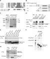
The C-terminal SLD2 of UAF1 binds to a conserved SIM sequence on FANCI. (A) Identification of a conserved SIM in the primary amino acid sequence of human FANCI, aligned with the homologous regions of FANCI from other species: Homo sapiens (hs), Mus musculus (mm), Bos taurus (bt), Gallus gallus (gg), and Danio rerio (dr). (B) Schematic representation of FANCI-WT and two deletion mutants. FANCI-WT and C700 both contain the SIM region, while the SIM was absent in the C500 mutant. The N-terminal Flag epitope was indicated. (C) GST-SLD2 pulled down FANCI-WT and C700, but not C500. Plasmids expressing the indicated deletion mutants of human FANCI were transiently transfected into HEK293T cells. Cell lysates were used for the pull-down experiment with the indicated GST or GST-SLD2 fusion protein. Western blot was then performed using anti-Flag antibody. (D) FANCI interacted with full-length UAF1 in vivo. Flag-tagged wild-type FANCI and a mutant form of full-length FANCI (ΔVIPL) were transfected into HEK293T cells and immunoprecipitated using anti-Flag agarose beads. Wild-type FANCI specifically interacted with UAF1 after MMC treatment, and this interaction was impaired in the SIM mutant (ΔVIPL). (E) GST fusion proteins containing the indicated SUMO or SLD protein sequences were prepared and used to pull down wild-type and the ΔVIPL mutant form of full-length FANCI overexpressed in HEK293T cells. Only SLD2, but not SLD1 or SUMO1/2/3, pulled down wild-type FANCI, and this interaction was dependent on the SIM. By densitometry, the relative band intensities of lanes 6 and 7 were 1.00 and 0.57, respectively. (F) GST fusion proteins containing either SLD2 or SUMO1 were prepared and used to pull down wild-type Flag-FANCI or wild-type Flag-Pias1 protein, which were overexpressed in HEK293T cells. GST-SLD2 specifically pulled down FANCI and GST-SUMO1 specifically pulled down Pias1. (G) GST-SLD2 pull-down of wild-type FANCI but not of FANCI-ΔSIM (another FANCI mutant with a deletion of the entire SIM sequence).
Figure 4.
Figure 4.
Deletion of the SIM domain of FANCI blocks deubiquitination by USP1/UAF1 and disrupts the FA pathway. (A) FANCD2 ubiquitination was impaired in shFANCI cells. HeLa cells were stably transfected with an shRNA specific for the FANCI gene product. Cell lysates were immunoblotted with the indicated antibodies. (B) Both WT and ΔVIPL mutant FANCI rescued FANCD2 monoubiquitination. Cells with stable FANCI knockdown were transfected with an shRNA-resistant cDNA encoding the indicated Flag-tagged wild-type or mutant FANCI proteins. Lysates from stably transfected cells were immunoblotted with the indicated antibodies. The ratio of FANCD2-Ub to FANCD2 (L/S ratio) was indicated. (C) The indicated HeLa cell transfectants were analyzed by the G2/M accumulation assay. The percentage of cells in different phases of the cell cycle was determined by FACS analysis.
Figure 5.
Figure 5.
The SLD2 region of UAF1 is required for binding to a SIM on hELG1. (A) SLD2 is required for UAF1/hELG1 interaction. HEK293T cells were transfected with the cDNA encoding either empty vector (lanes 1,4), Flag-UAF1-WT (lanes 2,5), or Flag-UAF1-ΔC (lanes 3,6). As indicated, immunoprecipitation with anti-Flag agarose beads was performed, and proteins were immunoblotted with the indicated antibodies. (B) Schematic representation of hELG1 mutants. The N400 deletion contains only the first 400 residues of hELG1 protein. The ΔSIM mutation was described previously (Lee et al. 2010). (C) Sequence alignment of the SIM region of hELG1 from multiple species (Homo sapiens [hs], Mus musculus [mm], Bos taurus [bt], Gallus gallus [gg], and Danio rerio [dr]), showing a high level of sequence conservation. The sequence of ΔSIM mutant was also shown. (D) SIM is required for UAF1/hELG1 interaction. HEK293T cells were cotransfected with the cDNAs encoding Flag-tagged wild-type (or ΔC mutant) UAF1 and/or Myc-tagged wild-type hELG1 or a ΔSIM mutant (sequence shown in C), which contains a deletion of 11 residues in the SIM region (Lee et al. 2010). For the experiment, we examined the 1- to 400-amino-acid fragment of hELG1. An anti-Flag immunoprecipitation was performed (lanes 7–12), and proteins were immunoblotted with the indicated antibodies. The asterisk (*) denotes a nonspecific band.
Figure 6.
Figure 6.
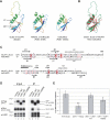
Structural similarity between the SLD2 of UAF1 and SUMO2. (A) Predicted model structure of SLD2 domain of hsUAF1. The structures of the RAWUL domain of hsRing1B (PDB: 3H8H) and hsSUMO2 (PDB: 1WM3) were also shown for comparison. (B) The superposed view of the model structure of the SLD2 domain from hsUAF1 (green) and the structure of hsSUMO2 (red). (C) Structure-based sequence alignment of the SLD2 domain from hsUAF1 and hsSUMO2. The major secondary structures were well conserved, especially the β2/α1 region. The 23-residue sequence of SLD2 (amino acids 616–638), which is not conserved in SUMO2, contains an inverted SIM sequence. This sequence is shown as the long loop in A and B. The asterisk indicates the conserved lysine (K) residue targeted by mutagenesis. (D) The Flag-UAF1-K595E mutant protein binds weakly to the SIM sequence on hELG1. HEK293T cells were cotransfected with the cDNAs encoding Flag-tagged wild-type, ΔC mutant, or K595E mutant UAF1 and Myc-tagged wild-type hELG1 (the 1- to 400-amino-acid fragment of hELG1). Cells were treated with UV (60J/m2, 6 h) before being harvested (Lanes 5–8). An anti-Flag immunoprecipitation was performed, and proteins were immunoblotted with the indicated antibodies. (E) The gene conversion assay was performed as described in Figure 2C. The difference between the single-asterisk (*) cells and the wild-type DT40 cells (WT + Vector) was statistically significant (P < 0.05). The difference between the double-asterisk (**) cells and the wild-type cells (WT + Vector) was also statistically significant (P < 0.001).
Figure 7.
Figure 7.
The USP1/UAF1 complex regulates the ubiquitination level of FANCD2/FANCI and PCNA. (A) The USP1/UAF1 complex regulates the ubiquitination level of both FANCD2/FANCI and PCNA, while hELG1 specifically regulates the ubiquitination level of PCNA. Retroviral shRNAs targeting the genes as indicated were used to stably infect HeLa cells, and the effects on FANCD2/FANCI and PCNA ubiquitination were examined by Western blot. By densitometry, the relative band intensities for the PCNA-Ub band for lanes 14 were 1.00, 4.92, 4.63, and 2.53, respectively. (B) Schematic interaction between the SLD2 region of UAF1 and the SIM sequences on hELG1 and FANCI. Most intracellular USP1 is constitutively bound to UAF1. UAF1 is a more abundant protein than USP1, and it has multiple binding partners. The WD40 domain of UAF1 binds and stimulates USP1. The SLD2 region of UAF1 binds the SIM sequences of hELG1 and of FANCI.

Comment in

Similar articles

Cited by

References

    1. Al-Hakim A, Escribano-Diaz C, Landry MC, O'Donnell L, Panier S, Szilard RK, Durocher D 2010. The ubiquitous role of ubiquitin in the DNA damage response. DNA Repair (Amst) 9: 1229–1240 - PMC - PubMed
    1. Bergink S, Jentsch S 2009. Principles of ubiquitin and SUMO modifications in DNA repair. Nature 458: 461–467 - PubMed
    1. Bezsonova I, Walker JR, Bacik JP, Duan S, Dhe-Paganon S, Arrowsmith CH 2009. Ring1B contains a ubiquitin-like docking module for interaction with Cbx proteins. Biochemistry 48: 10542–10548 - PubMed
    1. Bienko M, Green CM, Sabbioneda S, Crosetto N, Matic I, Hibbert RG, Begovic T, Niimi A, Mann M, Lehmann AR, et al. 2010. Regulation of translesion synthesis DNA polymerase eta by monoubiquitination. Mol Cell 37: 396–407 - PubMed
    1. Cohn MA, Kowal P, Yang K, Haas W, Huang TT, Gygi SP, D'Andrea AD 2007. A UAF1-containing multisubunit protein complex regulates the Fanconi anemia pathway. Mol Cell 28: 786–797 - PubMed

Publication types

MeSH terms

Substances

LinkOut - more resources