. 2011 Aug 31;31(35):12533-42.
doi: 10.1523/JNEUROSCI.2840-11.2011.

Functional recovery after peripheral nerve injury is dependent on the pro-inflammatory cytokines IL-1β and TNF: implications for neuropathic pain

Affiliations

Functional recovery after peripheral nerve injury is dependent on the pro-inflammatory cytokines IL-1β and TNF: implications for neuropathic pain

Sylvain Nadeau et al. J Neurosci. .

Abstract

IL-1β and TNF are potential targets in the management of neuropathic pain after injury. However, the importance of the IL-1 and TNF systems for peripheral nerve regeneration and the mechanisms by which these cytokines mediate effects are to be fully elucidated. Here, we demonstrate that mRNA and protein levels of IL-1β and TNF are rapidly upregulated in the injured mouse sciatic nerve. Mice lacking both IL-1β and TNF, or both IL-1 type 1 receptor (IL-1R1) and TNF type 1 receptor (TNFR1), showed reduced nociceptive sensitivity (mechanical allodynia) compared with wild-type littermates after injury. Microinjecting recombinant IL-1β or TNF at the site of sciatic nerve injury in IL-1β- and TNF-knock-out mice restored mechanical pain thresholds back to levels observed in injured wild-type mice. Importantly, recovery of sciatic nerve function was impaired in IL-1β-, TNF-, and IL-1β/TNF-knock-out mice. Notably, the infiltration of neutrophils was almost completely prevented in the sciatic nerve distal stump of mice lacking both IL-1R1 and TNFR1. Systemic treatment of mice with an anti-Ly6G antibody to deplete neutrophils, cells that play an essential role in the genesis of neuropathic pain, did not affect recovery of neurological function and peripheral axon regeneration. Together, these results suggest that targeting specific IL-1β/TNF-dependent responses, such as neutrophil infiltration, is a better therapeutic strategy for treatment of neuropathic pain after peripheral nerve injury than complete blockage of cytokine production.


Figures
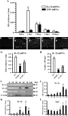
Figure 1.
Figure 1.
Sciatic nerve microcrush lesion rapidly induces production of the bioactive forms of IL-1β and TNF. A, Quantification of in situ hybridization signal for IL-1β and TNF mRNA in the sciatic nerve distal stump at various times after lesion (n = 4 mice/time point). B–F, Representative dark-field photomicrographs showing IL-1β mRNA expression at 0 (B; naive mouse), 1 (C), 3 (D), 7 (E), and 14 (F) d after sciatic nerve lesion in C57BL/6 mice. All photomicrographs were taken at 1 mm distal to the lesion. The arrows in C–F point to the 10–0 suture node indicating the site of lesion. G–H, Quantification of ISH signal for IL-1β mRNA (G) and the number of cells expressing IL-1β mRNA (H) in the sciatic nerve distal stump of MyD88- and IL-1R1-knock-out (−/−) mice and their WT littermates at 1 d after lesion (n = 4 per group). I, Representative immunoblots for IL-1α, IL-1β and TNF performed on cell lysate extracts obtained from the sciatic nerve of naive (N) and injured C57BL/6 mice at various times after lesion (n = 4 mice/time point). J–L, Immunoblot analysis of IL-1α (31 kDa), IL-1β (17 kDa), and TNF (26 kDa) proteins after sciatic nerve injury. The β-actin protein was used as internal standard and control for protein loading. ***p < 0.001, **p < 0.01, and *p < 0.05 compared with naive (A, J–L) or WT (G–H) mice. Scale bar (in F) B–F, 250 μm.
Figure 2.
Figure 2.
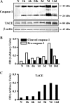
The active forms of the enzymes that mediate the proteolytic cleavage of the pro-forms of IL-1β and TNF into mature (secreted) cytokines are upregulated in the sciatic nerve distal stump after microcrush lesion. A, Representative immunoblots for caspase-1 and TACE performed on cell lysate extracts obtained from the sciatic nerve of naive and injured C57BL/6 mice at various times after lesion. B, C, Quantification of immunoblot analysis of caspase-1 (B) and TACE (C) protein expression revealed that the precursor forms for caspase-1 (45 kDa) and TACE (∼100 kDa) were rapidly converted into the active forms of 26 and ∼80 kDa, respectively, after injury. The β-actin protein was used as internal standard and control for protein loading.
Figure 3.
Figure 3.
IL-1β and TNF are major factors contributing to pain after peripheral nerve injury and are critical for recovery of neurological function. A, Paw withdrawal threshold (in grams) to mechanical stimulation following PSNL or sham-operation (n = 4–6 mice/group). WT mice showed a robust decrease in withdrawal threshold on the hindpaw ipsilateral to nerve injury at day 3 and maintained this hypersensitivity until the end of the testing period. In contrast, mechanical allodynia was almost completely abolished in IL-1R1/TNFR1 double knock-out (−/−) mice. B, Paw withdrawal thresholds (expressed as percentage of baseline) of IL-1β−/−, TNF−/−, and IL-1β/TNF−/− mice compared with WT mice after sciatic nerve injury (n = 11 mice/group). C, Paw withdrawal thresholds (percentage of baseline) of IL-1β−/− and TNF−/−mice following PSNL and injection of recombinant mouse IL-1β (rmIL-1β) or TNF (rmTNF) into the sciatic nerve at 7 d (n = 6–9 mice/group). D, Recovery of sciatic nerve function after microcrush lesion, as determined by the SFI, in IL-1β−/−, TNF−/−, and IL-1β/TNF−/− mice compared with WT mice over a period of 42 d (n = 10–32 mice/group). ***p < 0.001, **p < 0.01, and *p < 0.05 compared with baseline levels in the same group (A), WT (B, D), or sham WT (C).
Figure 4.
Figure 4.
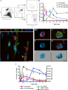
Time course of neutrophil and macrophage influx in the mouse sciatic nerve after microcrush lesion. A, Representative flow cytometry profiles showing the presence of neutrophils (red; CD45+ CD11b+ 7/4+ Ly-6C+ Ly-6G+), proinflammatory M1 macrophages (blue; CD45+ CD11b+ 7/4+ Ly-6Chi Ly-6G), and anti-inflammatory M2 macrophages (green; CD45+ CD11b+ 7/4 Ly-6Clo Ly-6G) in the sciatic nerve distal stump of C57BL/6 mice at 24 h after injury. B, Time course of the presence of neutrophils, M1 macrophages, and M2 macrophages in the sciatic nerve distal stump of C57BL/6 mice during the first week after lesion (n = 4 mice per time point), as revealed by flow cytometry. C–I, Confocal images and 3D reconstructions of Z-stacks showing 7/4+ neutrophils with their characteristic multilobed, segmented nuclei (cells pointed to by red arrows in C and D–F) and proinflammatory M1 macrophages (7/4+ iba1+ cells with monolobed or bilobed nuclei; cell pointed to by the yellow arrow in C and G–I) in the sciatic nerve distal stump at day 1 after lesion. Note that some elongated iba1+ cells did not express the 7/4 marker, suggesting that they could be resident endoneurial macrophages (see the cell pointed to by the green arrow in C). Nuclear counterstaining with DAPI is shown in blue. J, Time course of the presence of neutrophils, M1 macrophages, and M2 macrophages in the sciatic nerve distal stump of C57BL/6 mice during the first week after lesion (n = 4 mice per time point), as revealed by multiple immunofluorescence labeling. Also shown in J is the expression of the neutrophil chemoattractants KC/CXCL1 and MIP-2/CXCL2 and the monocyte chemoattractant protein-1 (MCP-1, also termed CCL2) at the mRNA level. *p < 0.05 compared with naive mice (J). Scale bars: (in C) C, 10 μm; (in I) D–I, 6 μm.
Figure 5.
Figure 5.
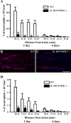
Neutrophils are recruited in an IL-1/TNF-dependent fashion after peripheral nerve injury. A, Quantification of the number of multilobed 7/4+ iba1 neutrophils in the sciatic nerve distal stump of IL-1R1/TNFR1 double-knock-out (−/−) mice and their WT littermates at 1 and 4 d after microcrush lesion (n = 4 mice per group per time point). B, C, Representative photomicrographs showing 7/4 immunostaining in the sciatic nerve distal stump of WT (B) and IL-1R1/TNFR1−/− (C) mice at 1 d after injury. D, Quantification of the number of Ly-6G+ neutrophils in the sciatic nerve distal stump of IL-1R1/TNFR1−/− mice and their WT littermates at 1 and 4 d after lesion (n = 4 per group per time point). *p < 0.05 compared with WT mice. Scale bar (in C) B, C, 150 μm.
Figure 6.
Figure 6.
Neutrophil depletion following peripheral nerve injury decreases pain without interfering with axonal regeneration and functional recovery. A, B, Quantification of the number of neutrophils, characterized by their expression of the Ly-6G (A) and 7/4 (B) antigens and multilobed nucleus, in the sciatic nerve distal stump of mice treated with PBS, isotype control antibody, or anti-Ly-6G antibody and killed at 1 and 4 d after microcrush lesion (n = 4 per group per time point). C, Quantification of the number of M1 macrophages, characterized by their expression of the 7/4 and iba1 antigens and monolobed or bilobed nucleus, in the sciatic nerve distal stump of mice treated with PBS, isotype control antibody, or anti-Ly-6G antibody (n = 4 per group per time point). Note the presence of fewer polymorphonuclear neutrophils, but not M1 macrophages, in the injured sciatic nerve of mice that received treatment with the anti-Ly-6G antibody. D, Paw withdrawal thresholds to mechanical stimulation (von Frey filaments) in partial sciatic nerve-ligated mice treated with either PBS, isotype control antibody, or anti-Ly-6G antibody (n = 4–6 mice/group). E, SFI analysis reveals no significant difference in recovery of locomotor function between neutrophil-depleted mice and control groups over a 42 d period (n = 8–9 mice/group; microcrush lesion model). F, Quantification of the number of YFP-labeled axons at predetermined distances from the proximal host-graft interface following neutrophil depletion at 7 d after grafting (n = 4–6 mice/group). ***p < 0.001, **p < 0.01, and *p < 0.05 compared with PBS-treated mice. †††p < 0.001, ††p < 0.01, and p < 0.05 compared with mice treated with the isotype control antibody.

Similar articles

Cited by

References

    1. Audoy-Rémus J, Richard JF, Soulet D, Zhou H, Kubes P, Vallières L. Rod-shaped monocytes patrol the brain vasculature and give rise to perivascular macrophages under the influence of proinflammatory cytokines and angiopoietin-2. J Neurosci. 2008;28:10187–10199. - PMC - PubMed
    1. Austin PJ, Moalem-Taylor G. The neuro-immune balance in neuropathic pain: involvement of inflammatory immune cells, immune-like glial cells and cytokines. J Neuroimmunol. 2010;229:26–50. - PubMed
    1. Barrette B, Hébert MA, Filali M, Lafortune K, Vallières N, Gowing G, Julien JP, Lacroix S. Requirement of myeloid cells for axon regeneration. J Neurosci. 2008;28:9363–9376. - PMC - PubMed
    1. Boivin A, Pineau I, Barrette B, Filali M, Vallières N, Rivest S, Lacroix S. Toll-like receptor signaling is critical for Wallerian degeneration and functional recovery after peripheral nerve injury. J Neurosci. 2007;27:12565–12576. - PMC - PubMed
    1. Chen CJ, Kono H, Golenbock D, Reed G, Akira S, Rock KL. Identification of a key pathway required for the sterile inflammatory response triggered by dying cells. Nat Med. 2007;13:851–856. - PubMed

Publication types

MeSH terms