. 2011 Dec;39(22):9605-19.
doi: 10.1093/nar/gkr656. Epub 2011 Aug 31.

Ku counteracts mobilization of PARP1 and MRN in chromatin damaged with DNA double-strand breaks

Affiliations

Ku counteracts mobilization of PARP1 and MRN in chromatin damaged with DNA double-strand breaks

Qiao Cheng et al. Nucleic Acids Res. 2011 Dec.

Abstract

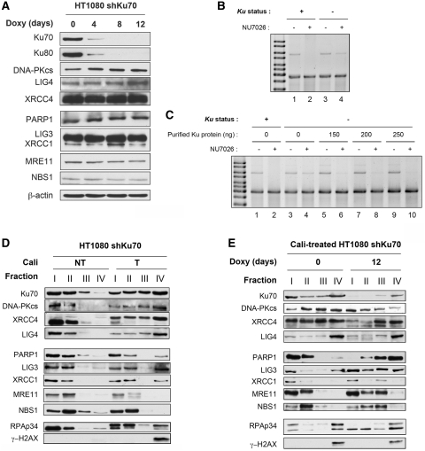
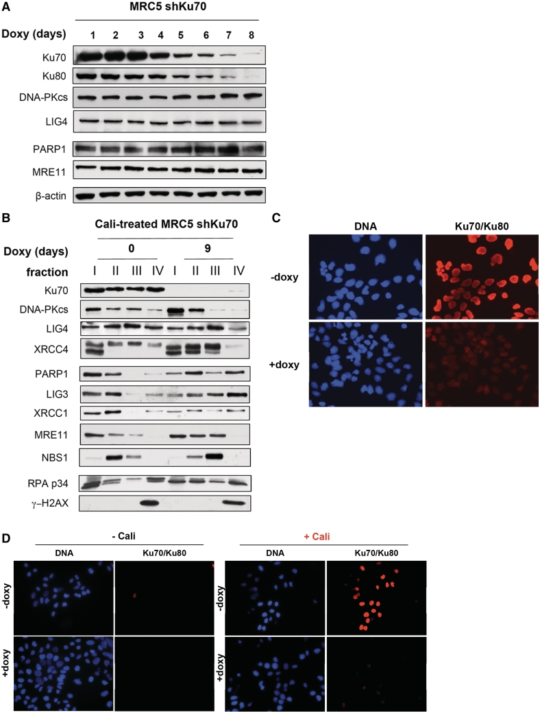
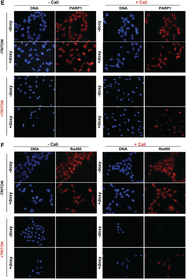
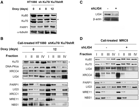
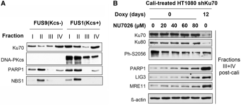
In mammalian cells, the main pathway for DNA double-strand breaks (DSBs) repair is classical non-homologous end joining (C-NHEJ). An alternative or back-up NHEJ (B-NHEJ) pathway has emerged which operates preferentially under C-NHEJ defective conditions. Although B-NHEJ appears particularly relevant to genomic instability associated with cancer, its components and regulation are still largely unknown. To get insights into this pathway, we have knocked-down Ku, the main contributor to C-NHEJ. Thus, models of human cell lines have been engineered in which the expression of Ku70/80 heterodimer can be significantly lowered by the conditional induction of a shRNA against Ku70. On Ku reduction in cells, resulting NHEJ competent protein extracts showed a shift from C- to B-NHEJ that could be reversed by addition of purified Ku protein. Using a cellular fractionation protocol after treatment with a strong DSBs inducer followed by western blotting or immunostaining, we established that, among C-NHEJ factors, Ku is the main counteracting factor against mobilization of PARP1 and the MRN complex to damaged chromatin. In addition, Ku limits PAR synthesis and single-stranded DNA production in response to DSBs. These data support the involvement of PARP1 and the MRN proteins in the B-NHEJ route for the repair of DNA DSBs.


Figures

Figure 1.
Figure 1.
Effect of Ku extinction on the expression and mobilization to damaged chromatin of representative repair proteins and on NHEJ activity in vitro. (A) WCEs of HT1080 shKu70 cells treated with doxycyclin for the indicated time were denatured and separated on 10% SDS–PAGE gel followed by electrotransfer on membrane. The membranes were blotted with the antibodies as indicated. (B) End-joining assay catalyzed under standard reaction conditions with extracts from Ku proficient or deficient cells (treated with doxycylin for 7 days), in the presence or not of DNA-PK specific inhibitor NU7026. DNA ligation products were separated by agarose gel electrophoresis followed by SYBR-Green staining. Ligation efficiency (% of plasmid multimers versus monomer) was 12.4 and 15.1 for lanes 1 and 3 (without NU7026) and 0 and 5.5 for lanes 2 and 4 (with NU7026), respectively. (C) End-joining assay catalyzed under standard reaction conditions with extracts from Ku proficient or deficient cells (treated with doxycylin for 7 days), in the presence or not of the indicated amount of purified Ku protein. DNA ligation products were separated by agarose gel electrophoresis followed by SYBR-Green staining. Ligation efficiency (% of multimers versus monomer) was 13.7, 12.9, 16.6, 16.2 and 15.5 for lanes 1, 3, 5, 7 and 9 (without NU7026) and 0, 6.0, 2.6, 1.7 and 0.9 for lanes 2, 4, 6, 8 and 10 (with NU7026), respectively. (D) HT1080 shKu70 cells were treated (T) or not (NT) with calicheamicin (Cali), collected and fractionated as described in the ‘Materials and Methods’ section, leading to fractions I to IV. Protein samples were denatured and separated on SDS–PAGE gels 10% for standard separation or 15% for γH2AX isolation, followed by electrotransfer and blotting as indicated. (E) HT1080 shKu70 cells were grown for 12 days in the presence or not of doxycyclin, then treated with Cali, fractionated and analyzed as in (D).
Figure 2.
Figure 2.
Effect of Ku extinction on the expression and mobilization to damaged chromatin of representative repair proteins. (A) WCEs of MRC5 shKu70 cells treated with doxycyclin for the indicated time were denatured and separated on 10% SDS–PAGE gel followed by electrotransfer on membrane. The membranes were blotted with the antibodies as indicated. (B) MRC5 shKu70 cells were grown for 9 days in the presence or not of doxycyclin, then treated with Cali, fractionated as described in the ‘Materials and Methods’ section, leading to fractions I–IV. Protein samples were denatured and separated on SDS–PAGE gel, followed by electrotransfer and blotting as indicated. (C) MRC5 shKu70 incubated or not with doxycyclin for 8 days were fixed, permeabilized and immunostained with anti-Ku70/Ku80 heterodimer antibodies followed by incubation with Alexa-Fluor® 594-conjugated goat anti-mouse IgG. DNA was stained with DAPI. (D) MRC5 shKu70 incubated or not with doxycyclin for 8 days were mock-treated or treated with 10 nM Calicheamicin (Cali) for 45 min at 37°C in medium. Cells were permeabilized in buffer containing 0.1% Triton X-100 prior to fixation and immunostaining with anti-Ku70/Ku80 heterodimer antibodies and Alexa-Fluor® 594-conjugated goat anti-mouse antibodies. DNA was stained with DAPI. (E and F) MRC5 shKu70 incubated or not with doxycyclin for 8 days were mock-treated or treated with 10 nM Cali for 45 min at 37°C in medium. Cells were pre-extracted in buffer containing 0.1% Triton X-100 prior to fixation then immunostained with anti-PARP1 antibodies (E) or anti-Rad50 antibodies (F) followed by incubation with Alexa-Fluor® 594-conjugated goat anti-mouse antibodies. DNA was stained with DAPI.
Figure 2.
Figure 2.
Effect of Ku extinction on the expression and mobilization to damaged chromatin of representative repair proteins. (A) WCEs of MRC5 shKu70 cells treated with doxycyclin for the indicated time were denatured and separated on 10% SDS–PAGE gel followed by electrotransfer on membrane. The membranes were blotted with the antibodies as indicated. (B) MRC5 shKu70 cells were grown for 9 days in the presence or not of doxycyclin, then treated with Cali, fractionated as described in the ‘Materials and Methods’ section, leading to fractions I–IV. Protein samples were denatured and separated on SDS–PAGE gel, followed by electrotransfer and blotting as indicated. (C) MRC5 shKu70 incubated or not with doxycyclin for 8 days were fixed, permeabilized and immunostained with anti-Ku70/Ku80 heterodimer antibodies followed by incubation with Alexa-Fluor® 594-conjugated goat anti-mouse IgG. DNA was stained with DAPI. (D) MRC5 shKu70 incubated or not with doxycyclin for 8 days were mock-treated or treated with 10 nM Calicheamicin (Cali) for 45 min at 37°C in medium. Cells were permeabilized in buffer containing 0.1% Triton X-100 prior to fixation and immunostaining with anti-Ku70/Ku80 heterodimer antibodies and Alexa-Fluor® 594-conjugated goat anti-mouse antibodies. DNA was stained with DAPI. (E and F) MRC5 shKu70 incubated or not with doxycyclin for 8 days were mock-treated or treated with 10 nM Cali for 45 min at 37°C in medium. Cells were pre-extracted in buffer containing 0.1% Triton X-100 prior to fixation then immunostained with anti-PARP1 antibodies (E) or anti-Rad50 antibodies (F) followed by incubation with Alexa-Fluor® 594-conjugated goat anti-mouse antibodies. DNA was stained with DAPI.
Figure 3.
Figure 3.
Effect of Ku restoration and LIG4-deficiency on the mobilization to damaged chromatin of representative repair proteins. (A) WCEs of HT1080 shKu70/Ku70shR cells treated with doxycyclin for the indicated time were denatured and separated on 10% SDS–PAGE gel, followed by electrotransfer and blotting as indicated. Asterisk indicates FLAG-Ku70. (B) HT1080 shKu70/Ku70shR cells were grown for 12 days in the presence or not of doxycyclin, then treated with Cali, collected and fractionated as described in the ‘Materials and Methods’ section, leading to fractions I–IV. Protein samples were denatured and separated on 10% SDS–PAGE gel, followed by electrotransfer and blotting as indicated. Asterisk indicates FLAG-Ku70. (C) Western blotting on the WCEs of the MRC5 cells expressing or not shLIG4. (D) MRC5 cells expressing or not shLIG4 were treated with Cali, fractionated and analyzed as in (B).
Figure 4.
Figure 4.
Effect of DNA-PKcs deficiency or inhibition of DNA-PK on the mobilization to damaged chromatin of representative repair proteins. (A) DNA-PKcs deficient (Fus9) and DNA-PKcs complemented (Fus1) glioblastoma cell lines were treated with Cali. collected and fractionated as described in the ‘Materials and Methods’ section, leading to fractions I to IV. Protein samples were denatured and separated on 10% SDS–PAGE gel, followed by electrotransfer and blotting as indicated. (B) HT1080 shKu70 cells were grown for 12 days in the presence or not of doxycyclin, then treated with Cali in the presence of increasing concentrations of the DNA-PKcs inhibitor NU7026 as indicated, fractionated and analyzed as in (A), except that only pooled fractions III and IV were loaded.
Figure 5.
Figure 5.
Consequences of Ku deficiency on PAR synthesis and ssDNA production in damaged chromatin. (A) MRC5 shKu70 incubated or not with doxycyclin for 8 days were mock-treated or treated with 40 µM DPQ for 1 h at 37°C, then 1 mM H2O2 was added to the medium for 15 min Cells were then fixed, permeabilized and immunostained with anti-PAR antibodies followed by incubation with Alexa-Fluor® 594-conjugated goat anti-mouse IgG. DNA was stained with DAPI. (B) MRC5 shKu70 incubated or not with doxycyclin for 8 days were mock-treated or treated with 40 µM DPQ for 1 h at 37°C, then 40 nM Cali was added to the medium for 7 min Cells were fixed, permeabilized and immunostained with anti-PAR antibodies followed by incubation with Alexa-Fluor® 594-conjugated goat anti-mouse IgG. DNA was stained with DAPI. (C and D) After incorporation of 20 µM BrdU, MRC5 shKu70 incubated or not with doxycyclin for 8 days, mock-treated or treated with 10 nM Cali for 45 min and post-incubated for 2 h were subjected to immunofluorescence with anti-BrdU antibodies and Alexa-Fluor® 594-conjugated goat anti-mouse antibodies. In C, a DNA denaturation step was included by incubation in 2N HCl for 10 min prior to immunostaining with anti-BrdU antibodies. DNA was stained with DAPI.
Figure 5.
Figure 5.
Consequences of Ku deficiency on PAR synthesis and ssDNA production in damaged chromatin. (A) MRC5 shKu70 incubated or not with doxycyclin for 8 days were mock-treated or treated with 40 µM DPQ for 1 h at 37°C, then 1 mM H2O2 was added to the medium for 15 min Cells were then fixed, permeabilized and immunostained with anti-PAR antibodies followed by incubation with Alexa-Fluor® 594-conjugated goat anti-mouse IgG. DNA was stained with DAPI. (B) MRC5 shKu70 incubated or not with doxycyclin for 8 days were mock-treated or treated with 40 µM DPQ for 1 h at 37°C, then 40 nM Cali was added to the medium for 7 min Cells were fixed, permeabilized and immunostained with anti-PAR antibodies followed by incubation with Alexa-Fluor® 594-conjugated goat anti-mouse IgG. DNA was stained with DAPI. (C and D) After incorporation of 20 µM BrdU, MRC5 shKu70 incubated or not with doxycyclin for 8 days, mock-treated or treated with 10 nM Cali for 45 min and post-incubated for 2 h were subjected to immunofluorescence with anti-BrdU antibodies and Alexa-Fluor® 594-conjugated goat anti-mouse antibodies. In C, a DNA denaturation step was included by incubation in 2N HCl for 10 min prior to immunostaining with anti-BrdU antibodies. DNA was stained with DAPI.

Similar articles

Cited by

References

    1. O'Driscoll M, Jeggo PA. The role of double-strand break repair - insights from human genetics. Nat. Rev. Genet. 2006;7:45–54. - PubMed
    1. Wyman C, Kanaar R. DNA double-strand break repair: all's well that ends well. Annu. Rev. Genet. 2006;40:363–383. - PubMed
    1. Hartlerode AJ, Scully R. Mechanisms of double-strand break repair in somatic mammalian cells. Biochem. J. 2009;423:157–168. - PMC - PubMed
    1. Pardo B, Gomez-Gonzalez B, Aguilera A. DNA double-strand break repair: how to fix a broken relationship. Cell Mol. Life Sci. 2009;66:1039–1056. - PMC - PubMed
    1. Weterings E, Chen DJ. The endless tale of non-homologous end-joining. Cell Res. 2008;18:114–124. - PubMed

Publication types

MeSH terms