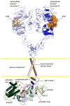
Regulation of the catalytic activity of the EGF receptor
- PMID: 21868214
- PMCID: PMC3232302
- DOI: 10.1016/j.sbi.2011.07.007
Regulation of the catalytic activity of the EGF receptor
Abstract
The epidermal growth factor receptor (EGFR) is a receptor tyrosine kinase involved in cell growth that is often misregulated in cancer. Several recent studies highlight the unique structural mechanisms involved in its regulation. Some elucidate the important role that the juxtamembrane segment and the transmembrane helix play in stabilizing the activating asymmetric kinase dimer, and suggest that its activation mechanism is likely to be conserved among the other human EGFR-related receptors. Other studies provide new explanations for two long observed, but poorly understood phenomena, the apparent heterogeneity in ligand binding and the formation of ligand-independent dimers. New insights into the allosteric mechanisms utilized by intracellular regulators of EGFR provide hope that allosteric sites could be used as targets for drug development.
Copyright © 2011 Elsevier Ltd. All rights reserved.
Conflict of interest statement
Figures


Similar articles
-
Mechanism for activation of the EGF receptor catalytic domain by the juxtamembrane segment.Cell. 2009 Jun 26;137(7):1293-307. doi: 10.1016/j.cell.2009.04.025. Cell. 2009. PMID: 19563760 Free PMC article.
-
Architecture and membrane interactions of the EGF receptor.Cell. 2013 Jan 31;152(3):557-69. doi: 10.1016/j.cell.2012.12.030. Cell. 2013. PMID: 23374350 Free PMC article.
-
Structural basis for negative cooperativity in growth factor binding to an EGF receptor.Cell. 2010 Aug 20;142(4):568-79. doi: 10.1016/j.cell.2010.07.015. Cell. 2010. PMID: 20723758 Free PMC article.
-
Emerging Allosteric Mechanism of EGFR Activation in Physiological and Pathological Contexts.Biophys J. 2019 Jul 9;117(1):5-13. doi: 10.1016/j.bpj.2019.05.021. Epub 2019 May 28. Biophys J. 2019. PMID: 31202480 Free PMC article. Review.
-
Asymmetric tyrosine kinase arrangements in activation or autophosphorylation of receptor tyrosine kinases.Mol Cells. 2010 May;29(5):443-8. doi: 10.1007/s10059-010-0080-5. Epub 2010 Apr 28. Mol Cells. 2010. PMID: 20432069 Review.
Cited by
-
Finding the missing links in EGFR.Nat Struct Mol Biol. 2012 Jan 5;19(1):1-3. doi: 10.1038/nsmb.2221. Nat Struct Mol Biol. 2012. PMID: 22218287 No abstract available.
-
In planta assessment of the role of thioredoxin h proteins in the regulation of S-locus receptor kinase signaling in transgenic Arabidopsis.Plant Physiol. 2013 Nov;163(3):1387-95. doi: 10.1104/pp.113.225672. Epub 2013 Sep 27. Plant Physiol. 2013. PMID: 24077073 Free PMC article.
-
Allosteric targeting of receptor tyrosine kinases.Nat Biotechnol. 2014 Nov;32(11):1113-20. doi: 10.1038/nbt.3028. Nat Biotechnol. 2014. PMID: 25380447
-
Structural basis of the effect of activating mutations on the EGF receptor.Elife. 2021 Jul 28;10:e65824. doi: 10.7554/eLife.65824. Elife. 2021. PMID: 34319231 Free PMC article.
-
Ack1: activation and regulation by allostery.PLoS One. 2013;8(1):e53994. doi: 10.1371/journal.pone.0053994. Epub 2013 Jan 14. PLoS One. 2013. PMID: 23342057 Free PMC article.
References
-
- Cohen S. Isolation of a mouse submaxillary gland protein accelerating incisor eruption and eyelid opening in the new-born animal. J Biol Chem. 1962;237:1555–1562. - PubMed
-
- Carpenter G, Lembach KJ, Morrison MM, Cohen S. Characterization of the binding of 125-I-labeled epidermal growth factor to human fibroblasts. J Biol Chem. 1975;250:4297–4304. - PubMed
-
- Hynes NE, MacDonald G. ErbB receptors and signaling pathways in cancer. Curr Opin Cell Biol. 2009;21:177–184. - PubMed
-
- Yarden Y, Schlessinger J. Epidermal growth factor induces rapid, reversible aggregation of the purified epidermal growth factor receptor. Biochemistry. 1987;26:1443–1451. - PubMed
-
- Yarden Y, Schlessinger J. Self-phosphorylation of epidermal growth factor receptor: evidence for a model of intermolecular allosteric activation. Biochemistry. 1987;26:1434–1442. - PubMed
Publication types
MeSH terms
Substances
Grants and funding
LinkOut - more resources
Full Text Sources
Other Literature Sources
Research Materials
Miscellaneous

