. 2011;6(8):e23185.
doi: 10.1371/journal.pone.0023185. Epub 2011 Aug 16.

IL-1 and IL-23 mediate early IL-17A production in pulmonary inflammation leading to late fibrosis

Affiliations

IL-1 and IL-23 mediate early IL-17A production in pulmonary inflammation leading to late fibrosis

Paméla Gasse et al. PLoS One. 2011.

Abstract

Background: Idiopathic pulmonary fibrosis is a devastating as yet untreatable disease. We demonstrated recently the predominant role of the NLRP3 inflammasome activation and IL-1β expression in the establishment of pulmonary inflammation and fibrosis in mice.

Methods: The contribution of IL-23 or IL-17 in pulmonary inflammation and fibrosis was assessed using the bleomycin model in deficient mice.

Results: We show that bleomycin or IL-1β-induced lung injury leads to increased expression of early IL-23p19, and IL-17A or IL-17F expression. Early IL-23p19 and IL-17A, but not IL-17F, and IL-17RA signaling are required for inflammatory response to BLM as shown with gene deficient mice or mice treated with neutralizing antibodies. Using FACS analysis, we show a very early IL-17A and IL-17F expression by RORγt(+) γδ T cells and to a lesser extent by CD4αβ(+) T cells, but not by iNKT cells, 24 hrs after BLM administration. Moreover, IL-23p19 and IL-17A expressions or IL-17RA signaling are necessary to pulmonary TGF-β1 production, collagen deposition and evolution to fibrosis.

Conclusions: Our findings demonstrate the existence of an early IL-1β-IL-23-IL-17A axis leading to pulmonary inflammation and fibrosis and identify innate IL-23 and IL-17A as interesting drug targets for IL-1β driven lung pathology.


Conflict of interest statement

Competing Interests: Franco di Padova is a full-time employee of Novartis. The remaining authors have declared that no competing interests exist.

Figures

Figure 1
Figure 1. IL-23p19, IL-17A or IL-17F expression upon BLM or rmIL-1β.
mRNA expression for IL-23p19, Pro-IL-1β and HPRT1 in lung of 4 individual mice treated with BLM (10 mg/kg) or rmIL-1β (50 µg/kg) or saline were assessed using semi-quantitative PCR at 24 h in wild-type mice (A). Semi-quantitative IL-1β expression in the lung, evaluated with IL-1β/HPRT1 PCR ratio, was reduced in absence of IL-23p19 signaling (B). mRNA expression for IL-17A, IL-17F and HPRT1 in lung of 5 individual mice treated with BLM (10 mg/kg) or rmIL-1β (50 µg/kg) or saline wild-type mice or IL-1R1−/− mice were assessed using semi-quantitative PCR at 24 h (C). IL-17A and IL-17F mRNA levels were compared to the housekeeping gene HPRT1 mRNA levels and expressed as IL-17/HPRT ratio by measuring band densities using a densitometric analyzer (ImageJ) (D). Data represent in FiguremRNA expression Data represent mean values ± SD from 3 ratio independent experiments (n = 4 or 5 mice per group; *, p<0.05; **, p<0.01; ***, p<0.001).
Figure 2
Figure 2. Acute inflammation and remodeling upon BLM are dependent on IL-23p19.
Analysis of acute inflammation and remodeling was performed 24 h after BLM instillation (7.5 mg/kg). IL-23p19 deficient mice showed reduced neutrophil recruitment in BALF (A) and lung tissue (B) and reduced TIMP-1 production in lung (c) in comparison to wild-type mice Data represent mean values ± SD from 2 independent experiments (n = 5 mice per group; **, p<0.01; ***, p<0.001).
Figure 3
Figure 3. Impaired acute lung inflammation upon BLM in IL-17RA deficient mice.
IL-17RA deficient mice showed reduced neutrophil recruitment in BALF (A) and lung tissue (B) in comparison to wild-type mice. BALs and myeloperoxidase (MPO) activity were performed 24 h after BLM instillation (7.5 mg/kg). KC (D), IL-6 (E) and TIMP-1 (F) levels in the lung were also reduced in IL-17RA deficient mice at 24 h but not IL-1β levels (C). Cytokines, chemokine and TIMP-1 quantification in lung homogenates were performed by ELISA. Data represent mean values ± SD from 4 independent experiments (n = 5 mice per group; ns, not significant, *, p<0.05; **, p<0.01).
Figure 4
Figure 4. Acute lung inflammation and remodeling upon BLM depends on IL-17A.
Inflammatory and remodeling responses of wild-type mice treated with neutralizing antibody against IL-17A (150 µg/mouse, i.p. just after BLM or saline i.n. instillation). Neutrophil recruitment in BALF (A) and myeloperoxidase (MPO) activity in lung tissue (B) induced by BLM (7.5 mg/kg) were reduced after mice treatment with anti-IL-17A. IL-1β (C), KC (D), IL-6 (E) and TIMP-1 (F) levels in lung homogenates assessed by ELISA were also reduced 24 h after mice treatment neutralizing antibody against IL-17A. Data represent mean values ± SD from 3 independent experiments (n = 4 mice per group; *, p<0.05; **, p<0.01).
Figure 5
Figure 5. Acute lung inflammation and remodeling upon BLM are independent of IL-17F.
Inflammatory and remodeling responses upon BLM (7.5 mg/kg) of wild-type mice treated with neutralizing antibody against IL-17F (100–150 µg/mouse, i.p. just after BLM or saline i.n. instillation). Neutrophil recruitment in BALF (A) and myeloperoxidase (MPO) activity (B) upon BLM (7.5 mg/kg) were not reduced 24 h after treatment with high doses of neutralizing antibody against IL-17F. IL-1β (C), KC (D), IL-6 (E) and TIMP-1 (F) in lung homogenates assessed by ELISA were also reduced in BLM mice 24 h after treatment with neutralizing antibody against IL-17F. Treatments with anti-IL-17F had not significant effect on all these parameters. Data represent mean values ± SD from 2 independent experiments (n = 6 mice per group).
Figure 6
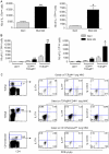
Figure 6. Early IL-17A and F are produced mainly by γδ T cells.
Lymphocytes from the lung of Rorc(γt)-Gfp TG mice were prepared 24 h after BLM or saline administration, and stimulated in vitro with PMA/ionomycin for 4 h. Specific staining of cell markers (CD1d-tetramer-APC, anti-NK1.1-PerCP-Cy5.5, anti-CD4-APCalexa750, anti-CD8-PB, anti-TCRγδ-APC, anti-TCRαβ-APC), or isotype control and intracellular staining for IL-17 (anti-IL-17A-PE, anti-IL-17F-PE, anti-GFP alexa488 or isotype controls) were performed. FACS analysis showed that BLM administration enhanced the total number of IL-17AposRORγtpos and IL-17FposRORγtpos lung mononuclear cells (A). IL-17-producing cells positive for RORγt were essentially γδ T cells and CD4pos αβ T cells but few iNKT cells (B). Among γδ T cells after bleomycin administration, at least 30% produce IL-17A and 5% produce IL-17F (C). Results are mean values ± SD from 4 independent experiments, with pools of 5 mice/group for each experiment, * p<0.05; **, p<0.01).
Figure 7
Figure 7. Remodeling and fibrosis are dependent on IL-23p19.
Analysis of late pulmonary remodeling and fibrosis was performed 14 days after BLM administration (5 mg/kg). MMP-2 activity (A) and TIMP-1 production (B) were reduced in IL-23p19 deficient mice. Active MMP-2 was expressed as relative intensity (RI). Latent TGF-β1 detected in BALF from wild-type mice was reduced in BALF from IL-23p19 deficient mice (C). Total collagen content in lung was significantly reduced in IL-23p19 deficient mice in comparision to wild-type mice (D). Collagen was measured using the Sircol collagen dye binding assay. Lung microscopic sections showed extensive fibrotic areas with collagen deposition in wild-type mice treated with BLM which was attenuated in IL-23p19−/− mice (e). Sirius red (SR) staining, scale bars 1 mm and 200 µm, n = 5 mice. Data represent mean values ± SD from 2 independent experiments (n = 5 mice per group; *, p<0.05; **, p<0.01; ***, p<0.001).
Figure 8
Figure 8. Attenuation of bleomycin-induced lung fibrosis in IL-17RA−/− mice.
Pulmonary remodeling and fibrotic response of wild-type mice or IL-17RA deficient mice 14 days after BLM or saline i.n. instillation. MMP-2 (72 Kd) activity in BALF was analyzed by zymography 14 days after administration of BLM (5 mg/kg i.n.). Active MMP-2 was upregulated at day 14 after BLM in the BALF of wild-type mice but only partially in IL-17RA−/− mice (A). TIMP-1 as indicator of a fibrotic process was upregulated in the lungs of B6, but to a lesser extent in IL-17RA−/− mice on day 14 (B). The latent form of TGF-β1 was present in BALF from B6 mice, 14 days after BLM, but was reduced in BALF from IL-17RA deficient mice as determined by ELISA assay (C). Active MMP-2 was expressed as relative intensity (RI). TIMP-1 and TGF-β1 levels were assessed by ELISA. Total collagen content enhanced 14 days after bleomycin instillation was significantly reduced in lung of IL-17RA deficient mice (D). Collagen was measured using the Sircol collagen dye binding assay. Data represent mean values ± SD from 2 independent experiments (n = 4 mice per group; *, p<0.05; **, p<0.01; ***p<0.001). Lung microscopic sections showed extensive fibrotic areas at day 14 with collagen deposition in wild-type mice treated with BLM (5 mg/kg i.n.) which was attenuated in IL-17RA−/− mice (E). Sirius red (SR) staining, scale bars 1 mm and 200 µm, n = 5 mice).
Figure 9
Figure 9. Attenuation of bleomycin-induced lung fibrosis upon anti-IL-17A treatment.
Pulmonary remodeling and fibrotic responses of wild-type mice treated with neutralizing antibody against IL-17A (150 µg/mouse, i.p. just after BLM and 3 times per week) 14 days after BLM (5 mg/kg i.n.) or saline. MMP-2 (72 Kd) activity in BALF was analyzed by zymography 14 days after administration of BLM. Active MMP-2 upregulated upon BLM in the BALF of wild-type mice was greatly reduced after 6 treatments with neutralizing antibody against IL-17A (A). TIMP-1 in lung was also significantly decreased after treatment with neutralizing antibody against IL-17A in comparison to untreated BLM mice (B). Latent TGF-β1 detected in BALF from wild-type mice was reduced in BALF from wild-type mice treated with anti-IL-17A antibodies (C). Active MMP-2 was expressed as relative intensity (RI). TIMP-1 and TGF-β1 levels were assessed by ELISA. In order to quantify lung collagen deposition, all the slides were numerised by Qimaging and analyzed by imageJ - NIH software. % of SR by lung were shown. Collagen content enhanced after BLM was significantly reduced when the mice were treated with anti-IL-17A antibody (D). Data represent mean values ± SD from 2 independent experiments (n = 5 mice per group; *, p<0.05; ***, p<0.001). Lung microscopic sections showed extensive fibrotic areas at day 14 with collagen deposition in wild-type mice administrated with BLM which was attenuated in mice treated with neutralizing antibodies against IL-17A (E). Sirius red (SR) staining, scale bars 1 mm and 200 µm, n = 5 mice.

Similar articles

Cited by

References

    1. Kondoh Y, Taniguchi H, Kawabata Y, Yokoi T, Suzuki K, et al. Acute exacerbation in idiopathic pulmonary fibrosis. Analysis of clinical and pathologic findings in three cases. Chest. 1993;103:1808–1812. - PubMed
    1. Gasse P, Mary C, Guenon I, Noulin N, Charron S, et al. IL-1R1/MyD88 signaling and the inflammasome are essential in pulmonary inflammation and fibrosis in mice. J Clin Invest. 2007;117:3786–3799. - PMC - PubMed
    1. Gasse P, Riteau N, Charron S, Girre S, Fick L, et al. Uric acid is a danger signal activating NALP3 inflammasome in lung injury inflammation and fibrosis. Am J Respir Crit Care Med. 2009;179:903–913. - PubMed
    1. Ferretti S, Bonneau O, Dubois GR, Jones CE, Trifilieff A. IL-17, produced by lymphocytes and neutrophils, is necessary for lipopolysaccharide-induced airway neutrophilia: IL-15 as a possible trigger. J Immunol. 2003;170:2106–2112. - PubMed
    1. Miyamoto M, Prause O, Sjostrand M, Laan M, Lotvall J, et al. Endogenous IL-17 as a mediator of neutrophil recruitment caused by endotoxin exposure in mouse airways. J Immunol. 2003;170:4665–4672. - PubMed

MeSH terms