. 2011 Nov;25(11):4048-62.
doi: 10.1096/fj.11-191627. Epub 2011 Aug 11.

Small-molecule activators of TMEM16A, a calcium-activated chloride channel, stimulate epithelial chloride secretion and intestinal contraction

Affiliations

Small-molecule activators of TMEM16A, a calcium-activated chloride channel, stimulate epithelial chloride secretion and intestinal contraction

Wan Namkung et al. FASEB J. 2011 Nov.

Abstract

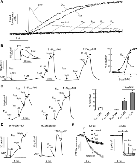
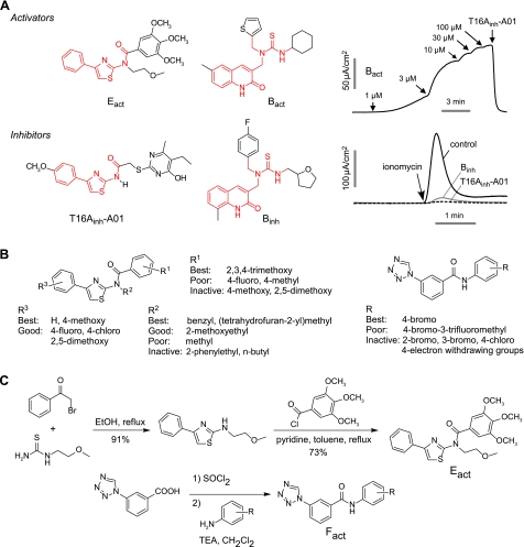
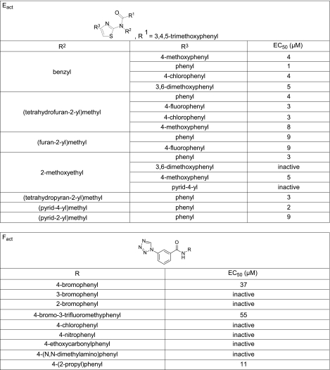
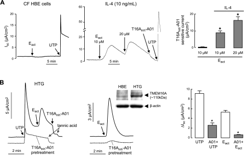
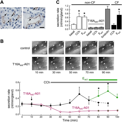
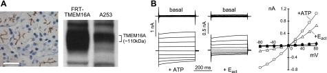
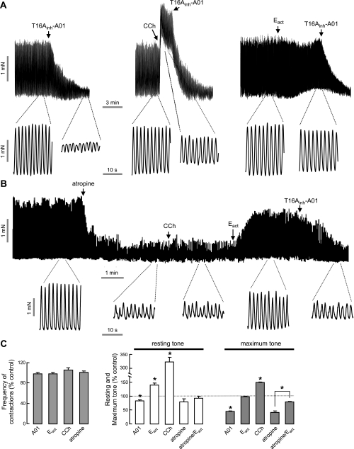
TMEM16A (ANO1) is a calcium-activated chloride channel (CaCC) expressed in secretory epithelia, smooth muscle, and other tissues. Cell-based functional screening of ∼110,000 compounds revealed compounds that activated TMEM16A CaCC conductance without increasing cytoplasmic Ca(2+). By patch-clamp, N-aroylaminothiazole "activators" (E(act)) strongly increased Cl(-) current at 0 Ca(2+), whereas tetrazolylbenzamide "potentiators" (F(act)) were not active at 0 Ca(2+) but reduced the EC(50) for Ca(2+)-dependent TMEM16A activation. Of 682 analogs tested, the most potent activator (E(act)) and potentiator (F(act)) produced large and more sustained CaCC Cl(-) currents than general agonists of Ca(2+) signaling, with EC(50) 3-6 μM and Cl(-) conductance comparable to that induced transiently by Ca(2+)-elevating purinergic agonists. Analogs of activators were identified that fully inhibited TMEM16A Cl(-) conductance, providing further evidence for direct TMEM16A binding. The TMEM16A activators increased CaCC conductance in human salivary and airway submucosal gland epithelial cells, and IL-4 treated bronchial cells, and stimulated submucosal gland secretion in human bronchi and smooth muscle contraction in mouse intestine. Small-molecule, TMEM16A-targeted activators may be useful for drug therapy of cystic fibrosis, dry mouth, and gastrointestinal hypomotility disorders, and for pharmacological dissection of TMEM16A function.


Figures

Figure 1.
Figure 1.
Identification of small-molecule TMEM16A activators by high-throughput screening. A) Screening protocol. FRT cells stably expressing TMEM16A and the halide-sensitive cytoplasmic fluorescent sensor YFP-H148Q/I152L/F46L were incubated for 10 min with test compound. Fluorescence was monitored in response to addition of iodide. B) Fluorescence measured in single wells of 96-well plates, showing vehicle and positive (ionomycin) controls and examples of inactive and active compounds. C) Structures of TMEM16A activators of six different chemical classes.
Figure 2.
Figure 2.
Characterization of TMEM16A activators. A) Cytoplasmic calcium measured by Fluo-4 fluorescence. Arrow indicates addition of 100 μM ATP (gray line) or 10 μM of indicated TMEM16A activators. B) Apical membrane current measured in TMEM16A-expressing FRT cells in the presence of a transepithelial chloride gradient and after basolateral membrane permeabilization. Left and center panels: representative current traces showing ATP (100 μM), Eact- or Fact-stimulated TMEM16A Cl current. T16Ainh-A01 (10 μM) was added where indicated. Inset shows long-time Eact effect. Right panel: concentration-activation data summary (mean±se, n=4–6). C) Synergistic effect of Eact and Fact. Left and center panels: representative current traces showing synergy. Right panel: data summary of low doses of TMEM16A activation (mean±se, n=5). *P < 0.05. D) Apical membrane current measured in FRT cells transfected with mouse TMEM16A or TMEM16B. E) Effect of Eact and Fact on CFTR and ENaC. Left panel: FRT cells expressing wild-type CFTR and YFP indicator were pretreated for 5 min with 10 μM Eact and Fact. Forskolin (10 μM) was added as indicated. Right panel: HBE cells were pretreated for 5 min with 10 μM Eact and Fact, with amiloride (10 μM) added as indicated.
Figure 3.
Figure 3.
Structure-activity analysis and synthesis of TMEM16A activators. A) Structural similarities between TMEM16A inhibitors and activators (common scaffolds shown in red). Apical membrane current measurements show activation of TMEM16A by Bact (top panels), inhibition of ionomycin (1 μM)-induced TMEM16A currents by pretreatment B and E class analogs (each 10 μM; bottom panels). B) Summary of structural determinants for TMEM16A activation. Left panel: Eact class. Right panel: Fact class. C) Synthesis of Eact and Fact analogs (see Materials and Methods).
Figure 4.
Figure 4.
Structure-activity analysis of Eact and Fact analogs. EC50 values were determined from fluorescence plate reader assay.
Figure 5.
Figure 5.
Patch-clamp analysis of Ca2+ requirements for TMEM16A activation by Eact and Fact. A) Apical membrane current measured in TMEM16A-expressing FRT cells. ER calcium stores were depleted by CPA (50 μM, 30 min) and 0 CaCl2 in bath. ATP (100 μM), Fact (10 μM), Eact (10 μM), and T16Ainh-A01 (10 μM) were added as indicated. B, C) Whole-cell TMEM16A currents were recorded at a holding potential at 0 mV, and pulsing to voltages between ± 80 mV (in steps of 20 mV) in the absence and presence of 3 μM Eact (B) or 10 μM Fact (C). Left panels: free calcium concentration of pipette solutions were clamped at 0 μM (black), 0.07 μM (green), 0.15 μM (red), and 1 μM (blue). Eact (3 μM) or Fact (10 μM) was added as indicated. Right panels: current-voltage (I/V) plots of mean currents at the middle of each voltage pulse. D) TMEM16A inhibited by 10 μM T16Ainh-A01 after stimulation by Eact or Fact.
Figure 6.
Figure 6.
Airway epithelial chloride secretion. A) Short-circuit current in CF HBE cells. Left and center panels: Eact and UTP (100 μM) were added in control (left) and IL-4 (10 ng/ml, 24 h, center) treated CF HBE cells. Right panels: summary of Eact-induced, T16Ainh-A01-sensitive peak current (mean±se, n=6–8). *P < 0.05. ENaC was inhibited by 10 μM amiloride. B) Eact (10 μM) and UTP (100 μM) induced CaCC Cl current measured in primary cultures of non-CF human tracheal gland (HTG) serous cells. Left and center panels: TMEM16A, CFTR, and ENaC were inhibited by pretreatment with T16Ainh-A01, CFTRinh-172, and amiloride, respectively. Inset: TMEM16A immunoblot in whole cell homogenates of CF HBE and HTG cells. Right panel: summary of UTP and Eact-induced peak current in the presence and absence of T16Ainh-A01 (mean±se, n=3). *P < 0.05.
Figure 7.
Figure 7.
Airway submucosal gland fluid secretion in human bronchi. A) TMEM16A immunohistochemistry in CF (left panel) and non-CF (right panel) human bronchi showing apical membrane expression in serous gland epithelial cells (arrows). Scale bar = 20 μm. B) Mucus (fluid) secretion in human bronchi. Top panels: images of mucus bubbles formed under oil in response to basolateral application of 300 nM carbachol (CCh) and 20 μM Eact. TMEM16A was inhibited by 30 μM T16Ainh-A01. Individual fluid bubbles marked with arrowheads. Scale bar = 0.5 mm. Bottom panels: CCh and Eact-induced secretion rates. Where indicated, tissues were pretreated with T16Ainh-A01 (30 μM). Each point is the average of measurements made from 20 glands (mean±se). C) Summary of human gland fluid secretion rates measured at 20 min after addition of 20 μM Eact, and 30 min after application of 300 nM carbachol (CCh) and 10 μM forskolin (20–66 glands from 3 tracheas and 4 bronchi). In CF-bronchi, 6 glands from one donor were stimulated by Eact. *P < 0.05.
Figure 8.
Figure 8.
Salivary gland epithelial cell Cl secretion. A) Expression of TMEM16A in human salivary gland. Left panel: TMEM16A immunostaining in human parotid gland. Scale bar = 20 μm. Right panel: immunoblot of TMEM16A in FRT-TMEM16A and A253 cells. B) Whole-cell patch-clamp recordings in A253 cells. Left panel: CaCC and TMEM16A chloride current induced by 100 μM ATP and 10 μM Eact, respectively. Right panel: current/voltage (I/V) plot of mean currents at the middle of each voltage pulse (voltages between −80 and +80 mV in steps of 20 mV).
Figure 9.
Figure 9.
Intestinal smooth muscle contraction. A) Representative traces from mouse ileal segments showing effects of T16Ainh-A01 (10 μM), carbachol (CCh, 1 μM) and Eact (10 μM). B) Effect of CCh (1 μM) and Eact (10 μM) following atropine (1 μM). C) Summary of contraction frequency (left panel) and resting and maximum tone (right panel; mean±se, n=4–7). *P < 0.05.

Similar articles

Cited by

  • The molecular basis of the genesis of basal tone in internal anal sphincter.
    Zhang CH, Wang P, Liu DH, Chen CP, Zhao W, Chen X, Chen C, He WQ, Qiao YN, Tao T, Sun J, Peng YJ, Lu P, Zheng K, Craige SM, Lifshitz LM, Keaney JF Jr, Fogarty KE, ZhuGe R, Zhu MS. Zhang CH, et al. Nat Commun. 2016 Apr 22;7:11358. doi: 10.1038/ncomms11358. Nat Commun. 2016. PMID: 27101932 Free PMC article.
  • Anion efflux mediates transduction in the hair cells of the zebrafish lateral line.
    Lunsford ET, Bobkov YV, Ray BC, Liao JC, Strother JA. Lunsford ET, et al. Proc Natl Acad Sci U S A. 2023 Dec 26;120(52):e2315515120. doi: 10.1073/pnas.2315515120. Epub 2023 Dec 20. Proc Natl Acad Sci U S A. 2023. PMID: 38117855 Free PMC article.
  • Long-term expanding human airway organoids for disease modeling.
    Sachs N, Papaspyropoulos A, Zomer-van Ommen DD, Heo I, Böttinger L, Klay D, Weeber F, Huelsz-Prince G, Iakobachvili N, Amatngalim GD, de Ligt J, van Hoeck A, Proost N, Viveen MC, Lyubimova A, Teeven L, Derakhshan S, Korving J, Begthel H, Dekkers JF, Kumawat K, Ramos E, van Oosterhout MF, Offerhaus GJ, Wiener DJ, Olimpio EP, Dijkstra KK, Smit EF, van der Linden M, Jaksani S, van de Ven M, Jonkers J, Rios AC, Voest EE, van Moorsel CH, van der Ent CK, Cuppen E, van Oudenaarden A, Coenjaerts FE, Meyaard L, Bont LJ, Peters PJ, Tans SJ, van Zon JS, Boj SF, Vries RG, Beekman JM, Clevers H. Sachs N, et al. EMBO J. 2019 Feb 15;38(4):e100300. doi: 10.15252/embj.2018100300. Epub 2019 Jan 14. EMBO J. 2019. PMID: 30643021 Free PMC article.
  • Normal Calcium-Activated Anion Secretion in a Mouse Selectively Lacking TMEM16A in Intestinal Epithelium.
    Vega G, Guequén A, Johansson MEV, Arike L, Martínez-Abad B, Nyström EEL, Scudieri P, Pedemonte N, Millar-Büchner P, Philp AR, Galietta LJ, Hansson GC, Flores CA. Vega G, et al. Front Physiol. 2019 Jun 13;10:694. doi: 10.3389/fphys.2019.00694. eCollection 2019. Front Physiol. 2019. PMID: 31263421 Free PMC article.
  • Variomics screen identifies the re-entrant loop of the calcium-activated chloride channel ANO1 that facilitates channel activation.
    Bill A, Popa MO, van Diepen MT, Gutierrez A, Lilley S, Velkova M, Acheson K, Choudhury H, Renaud NA, Auld DS, Gosling M, Groot-Kormelink PJ, Gaither LA. Bill A, et al. J Biol Chem. 2015 Jan 9;290(2):889-903. doi: 10.1074/jbc.M114.618140. Epub 2014 Nov 25. J Biol Chem. 2015. PMID: 25425649 Free PMC article.

References

    1. Hartzell C., Putzier I., Arreola J. (2005) Calcium-activated chloride channels. Annu. Rev. Physiol. 67, 719–758 - PubMed
    1. Verkman A. S., Galietta L. J. (2009) Chloride channels as drug targets. Nat. Rev. Drug. Discov. 8, 153–171 - PMC - PubMed
    1. Eggermont J. (2004) Calcium-activated chloride channels: (un) known, (un)loved? Proc. Am. Thorac. Soc. 1, 22–27 - PubMed
    1. Yang Y. D., Cho H., Koo J. Y., Tak M. H., Cho Y., Shim W. S., Park S. P., Lee J., Lee B., Kim B. M., Raouf R., Shin Y. K., Oh U. (2008) TMEM16A confers receptor-activated calcium-dependent chloride conductance. Nature 455, 1210–1215 - PubMed
    1. Caputo A., Caci E., Ferrera L., Pedemonte N., Barsanti C., Sondo E., Pfeffer U., Ravazzolo R., Zegarra-Moran O., Galietta L. J. (2008) TMEM16A, a membrane protein associated with calcium-dependent chloride channel activity. Science 322, 590–594 - PubMed

Publication types

LinkOut - more resources