Ectopic myelinating oligodendrocytes in the dorsal spinal cord as a consequence of altered semaphorin 6D signaling inhibit synapse formation
- PMID: 21831918
- PMCID: PMC3160102
- DOI: 10.1242/dev.066076
Ectopic myelinating oligodendrocytes in the dorsal spinal cord as a consequence of altered semaphorin 6D signaling inhibit synapse formation
Abstract
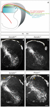
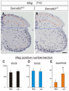
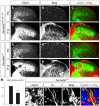
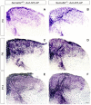
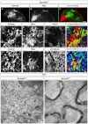
Different types of sensory neurons in the dorsal root ganglia project axons to the spinal cord to convey peripheral information to the central nervous system. Whereas most proprioceptive axons enter the spinal cord medially, cutaneous axons typically do so laterally. Because heavily myelinated proprioceptive axons project to the ventral spinal cord, proprioceptive axons and their associated oligodendrocytes avoid the superficial dorsal horn. However, it remains unclear whether their exclusion from the superficial dorsal horn is an important aspect of neural circuitry. Here we show that a mouse null mutation of Sema6d results in ectopic placement of the shafts of proprioceptive axons and their associated oligodendrocytes in the superficial dorsal horn, disrupting its synaptic organization. Anatomical and electrophysiological analyses show that proper axon positioning does not seem to be required for sensory afferent connectivity with motor neurons. Furthermore, ablation of oligodendrocytes from Sema6d mutants reveals that ectopic oligodendrocytes, but not proprioceptive axons, inhibit synapse formation in Sema6d mutants. Our findings provide new insights into the relationship between oligodendrocytes and synapse formation in vivo, which might be an important element in controlling the development of neural wiring in the central nervous system.
Figures




Similar articles
-
PlexinA1 signaling directs the segregation of proprioceptive sensory axons in the developing spinal cord.Neuron. 2006 Dec 7;52(5):775-88. doi: 10.1016/j.neuron.2006.10.032. Neuron. 2006. PMID: 17145500 Free PMC article.
-
Semaphorin 5B is a repellent cue for sensory afferents projecting into the developing spinal cord.Development. 2014 May;141(9):1940-9. doi: 10.1242/dev.103630. Epub 2014 Apr 9. Development. 2014. PMID: 24718987
-
Axon guidance in the spinal cord: choosin' by exclusion.Neuron. 2006 Dec 7;52(5):745-6. doi: 10.1016/j.neuron.2006.11.013. Neuron. 2006. PMID: 17145495
-
Molecular mechanisms underlying monosynaptic sensory-motor circuit development in the spinal cord.Dev Dyn. 2018 Apr;247(4):581-587. doi: 10.1002/dvdy.24611. Epub 2018 Jan 17. Dev Dyn. 2018. PMID: 29226492 Free PMC article. Review.
-
Molecular Analysis of Sensory Axon Branching Unraveled a cGMP-Dependent Signaling Cascade.Int J Mol Sci. 2018 Apr 24;19(5):1266. doi: 10.3390/ijms19051266. Int J Mol Sci. 2018. PMID: 29695045 Free PMC article. Review.
Cited by
-
RhoA is dispensable for axon guidance of sensory neurons in the mouse dorsal root ganglia.Front Mol Neurosci. 2012 May 22;5:67. doi: 10.3389/fnmol.2012.00067. eCollection 2012. Front Mol Neurosci. 2012. PMID: 22661927 Free PMC article.
-
Transmembrane semaphorins: Multimodal signaling cues in development and cancer.Cell Adh Migr. 2016 Nov;10(6):675-691. doi: 10.1080/19336918.2016.1197479. Epub 2016 Jun 13. Cell Adh Migr. 2016. PMID: 27295627 Free PMC article. Review.
-
Making Connections: Guidance Cues and Receptors at Nonneural Cell-Cell Junctions.Cold Spring Harb Perspect Biol. 2018 Nov 1;10(11):a029165. doi: 10.1101/cshperspect.a029165. Cold Spring Harb Perspect Biol. 2018. PMID: 28847900 Free PMC article. Review.
-
Muscle proprioceptors in adult rat: mechanosensory signaling and synapse distribution in spinal cord.J Neurophysiol. 2017 Nov 1;118(5):2687-2701. doi: 10.1152/jn.00497.2017. Epub 2017 Aug 16. J Neurophysiol. 2017. PMID: 28814636 Free PMC article.
-
Contribution of semaphorins to the formation of the peripheral nervous system in higher vertebrates.Cell Adh Migr. 2016 Nov;10(6):593-603. doi: 10.1080/19336918.2016.1243644. Epub 2016 Oct 7. Cell Adh Migr. 2016. PMID: 27715392 Free PMC article. Review.
References
-
- Alvarez F. J., Villalba R. M., Zerda R., Schneider S. P. (2004). Vesicular glutamate transporters in the spinal cord, with special reference to sensory primary afferent synapses. J. Comp. Neurol. 472, 257-280 - PubMed
-
- Arber S., Ladle D. R., Lin J. H., Frank E., Jessell T. M. (2000). ETS gene Er81 controls the formation of functional connections between group Ia sensory afferents and motor neurons. Cell 101, 485-498 - PubMed
-
- Brown A. G. (1981). Organization in the Spinal Cord. New York: Springer;
-
- Christopherson K. S., Ullian E. M., Stokes C. C., Mullowney C. E., Hell J. W., Agah A., Lawler J., Mosher D. F., Bornstein P., Barres B. A. (2005). Thrombospondins are astrocyte-secreted proteins that promote CNS synaptogenesis. Cell 120, 421-433 - PubMed
Publication types
MeSH terms
Substances
Grants and funding
LinkOut - more resources
Full Text Sources
Medical
Molecular Biology Databases

