. 2011;6(7):e22896.
doi: 10.1371/journal.pone.0022896. Epub 2011 Jul 28.

Redox-induced Src kinase and caveolin-1 signaling in TGF-β1-initiated SMAD2/3 activation and PAI-1 expression

Affiliations

Redox-induced Src kinase and caveolin-1 signaling in TGF-β1-initiated SMAD2/3 activation and PAI-1 expression

Rohan Samarakoon et al. PLoS One. 2011.

Abstract

Background: Plasminogen activator inhibitor-1 (PAI-1), a major regulator of the plasmin-based pericellular proteolytic cascade, is significantly increased in human arterial plaques contributing to vessel fibrosis, arteriosclerosis and thrombosis, particularly in the context of elevated tissue TGF-β1. Identification of molecular events underlying to PAI-1 induction in response to TGF-β1 may yield novel targets for the therapy of cardiovascular disease.

Principal findings: Reactive oxygen species are generated within 5 minutes after addition of TGF-β1 to quiescent vascular smooth muscle cells (VSMCs) resulting in pp60(c-src) activation and PAI-1 expression. TGF-β1-stimulated Src kinase signaling sustained the duration (but not the initiation) of SMAD3 phosphorylation in VSMC by reducing the levels of PPM1A, a recently identified C-terminal SMAD2/3 phosphatase, thereby maintaining SMAD2/3 in an active state with retention of PAI-1 transcription. The markedly increased PPM1A levels in triple Src kinase (c-Src, Yes, Fyn)-null fibroblasts are consistent with reductions in both SMAD3 phosphorylation and PAI-1 expression in response to TGF-β1 compared to wild-type cells. Activation of the Rho-ROCK pathway was mediated by Src kinases and required for PAI-1 induction in TGF-β1-stimulated VSMCs. Inhibition of Rho-ROCK signaling blocked the TGF-β1-mediated decrease in nuclear PPM1A content and effectively attenuated PAI-1 expression. TGF-β1-induced PAI-1 expression was undetectable in caveolin-1-null cells, correlating with the reduced Rho-GTP loading and SMAD2/3 phosphorylation evident in TGF-β1-treated caveolin-1-deficient cells relative to their wild-type counterparts. Src kinases, moreover, were critical upstream effectors of caveolin-1(Y14) phosphoryation and initiation of downstream signaling.

Conclusions: TGF-β1-initiated Src-dependent caveolin-1(Y14) phosphorylation is a critical event in Rho-ROCK-mediated suppression of nuclear PPM1A levels maintaining, thereby, SMAD2/3-dependent transcription of the PAI-1 gene.


Conflict of interest statement

Competing Interests: The authors have declared that no competing interests exist.

Figures

Figure 1
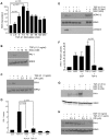
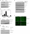
Figure 1. PAI-1 induction in response to TGF-β1 involves reactive oxygen species (ROS).
DCF fluorescence measurements (as described in Methods) were used to determine ROS generation (per equivalent number of cells) and expressed relative to unstimulated cultures (set as a.u. = 1). ROS levels increase within 5 minutes after addition of TGF-β1 (1 ng/ml) to serum-deprived quiescent VSMCs (A). ROS generation appears to be important in TGF-β1-stimulated PAI-1 expression since PAI-1 induction is effectively suppressed by even low concentrations of the established inhibitors of free radical generation NAC (B) and DPI (C). NAC pretreatment also attenuates (at 2 mM) and completely eliminates (at concentrations ≥5 mM) TGF-β1-dependent ERK1/2 and SMAD2/3 phosphorylation but has no effect of EGF-stimulated ERK1/2 activation (E). Both NAC (B,F,G) and DPI (C,H) pretreatment (30 mins) served to assess the role of ROS in TGF-β1- and EGF-mediated PAI-1 induction. ERK2 provided a loading control. Data plots (A,D,F) represent the mean ± S.D. of three independent experiments; statistical significance among the indicated groups was calculated by t-test.
Figure 2
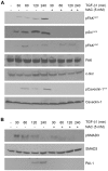
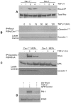
Figure 2. Inhibition of ROS generation attenuates TGF-β1 signaling in VSMC.
Quiescent VSMCs were stimulated with TGF-β1 (1 ng/ml) for the times indicated with or without NAC (5 mM) pretreatment for 1 hour. Increases in pSrc Y416, pFAKY577 and pCaveolinY14 (targets of c-Src kinases) in response to TGF-β1 is completely inhibited by NAC, suggesting an upstream role for ROS generation in activation of Src/FAK/caveolin-1 signaling pathways (A). FAKY397 phosphorylation by TGF-β1 (at least within the time frame of 2 hours) is relatively unaffected by NAC blockade of ROS generation. Total levels of c-Src, FAK and caveolin-1 are largely unchanged over the time course of TGF-β1 exposure serving as loading controls (A). To assess the role of ROS generation in SMAD3 activation, TGF-β1-stimulated SMAD3 phosphorylation over time was compared to an identical window with NAC pretreatment. Blots were probed with antibodies to determine both pSMAD3 and total SMAD3 levels (B).
Figure 3
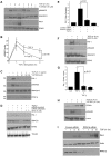
Figure 3. Downstream signaling events initiated by TGF-β1-activated Src kinase.
SYF+/+/+ and SYF−/−/− fibroblasts were serum-deprived for 1 day prior to stimulation with TGF-β1 (0.1 ng/ml) for the times indicated and lysates subject to western analysis. Src activation (assessed using phospho- Src Y416 antibodies) and increased EGFR phosphorylation at the Src kinase target Y845 site, are both evident in TGF-β1-stimulated wild-type (SYF+/+/+) MEFs but not Src, Fyn, Yes triple-null (SYF−/−/−) cells (A). The level (at 15 and 30 minutes) and maintenance (at 4 hrs) of SMAD3 phosphorylation is significantly reduced in SYF−/−/− fibroblasts compared to their wild-type counterparts (B). In contrast to the typical time course-dependency of PAI-1 induction in response to TGF-β1 in SYF+/+/+ cells, PAI-1 was not detectable in Src-deficient MEFs regardless of the duration of TGF-β1 exposure. The absence of PAI-1 expression and attenuated SMAD3 phosphorylation reflected increased PPM1A levels in SYF−/−/− as compared to SYF+/+/+ fibroblasts (B). Pretreatment of VSMCs with the Src kinase inhibitor SU6656 (2 µM) blocked the long-term maintenance (but not the initiation) of SMAD2/3 phosphorylation in response to TGF-β1 while total SMAD levels remain unchanged (C). Src Y416 phosphorylation by TGF-β1 was completely eliminated by SU6656 confirming the effectiveness of this inhibitor (C). Transient transfection of VSMCs with a dominant-negative pp60c-src (DN-Src) expression construct (or a GFP control vector) 72 hours prior to incubation with TGF-β1 for 6 hours was followed by western analysis for PAI-1. TGF-β1-stimulated PAI-1 induction was effectively suppressed by the DN-Src but not the GFP construct (D). SYF−/−/− cells genetically-engineered to express wild-type pp60c-src (SYF−/−/−+WT Src) rescued PAI-1 inducibility in response to TGF-β1 (E). ERK2 (A,D,E) and SMAD3 (C) serve as a loading controls.
Figure 4
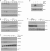
Figure 4. FAK is a downstream target of Src kinases and is required for PAI-1 induction by TGF-β1.
MEFs were serum-deprived for 1 day prior to addition of TGF-β1 (0.1 ng/ml). TGF-β1 stimulates FAK phosphorylation at the Y577 and Y861 sites in SYF+/+/+ but not SYF−/−/−cells consistent with an upstream role of Src kinases in FAK activation. TGF-β1-induced FAKY397 autophosphorylation, in contrast, is unaffected by genetic ablation of src family kinases (A). To assess the role of FAK in TGF-β1-induced PAI-1 and CTGF expression, serum-deprived FAK+/+ and FAK−/− MEFs were stimulated with TGF-β1 and blots probed with antibodies to PAI-1 and CTGF (B). TGF-β1 stimulates FAK phosphorylation at Y397, Y561 and Y861 only in wild-type but not, as anticipated, in FAK-null fibroblasts (C) providing antibody specificity controls for panels A–C. TGF-β1-stimulated c-Src and EGFR activation is significantly attenuated in FAK−/− cells relative to FAK+/+ MEFs (C). SMAD3 C-terminal phosphorylation in response to TGF-β1 is reduced in FAK−/− as compared to FAK+/+ cells; total SMAD2/3 levels were unchanged regardless of FAK genetic status (D). Western analysis was used to evaluate the effect of FAK genetic status (FAK−/− vs. FAK+/+) on TGF-β1-induced caveolin-1Y14 phosphorylation (D). Consistent with previous observations , total caveolin-1 is lower in FAK−/− MEFs compared to wild-type cultures (D). Assessment of total FAK (A,B), ERK2 (B,C) and SMAD3 (D) provided loading controls.
Figure 5
Figure 5. Caveolin-1 is required for TGF-β1-induced PAI-1 expression.
Serum-deprived (1 day) caveolin-1+/+ and caveolin-1−/− MEFs were stimulated with TGF-β1 (0.1 ng/ml) for 2 or 4 hours and blots probed with antibodies to PAI-1, pSMAD2 and pERK1/2. PAI-1 induction is apparent in wild-type but not in caveolin-1-deficient cells (A,B). TGF-β1-induced SMAD2 phosphorylation is decreased while ERK1/2 activation is increased in caveolin-1-null compared to wild-type fibroblasts at comparable time points (A,C). Exposure to TGF-β1 (T) was for 4 hours in (B) and for 2 or 4 hours in (C). Introduction of a wild-type caveolin-1 construct (+WT Cav-1) in caveolin-1-null cells rescues TGF-β1 inducibility of PAI-1 unlike caveolin-1−/− MEFs expressing GFP (+GFP) (D). VSMCs were transfected with control or caveolin-1 siRNA constructs and, after a brief period of serum deprivation, stimulated with TGF-β1 for 4 hours. Cellular lysates were separated by electrophoresis and blots probed with antibodies to PAI-1, caveolin-1 and actin (as a loading control) (E). Histograms (B,C) depict the mean ± S.D. of three independent experiments.
Figure 6
Figure 6. c-Src is an upstream regulator of caveolin-1Y14 phosphorylation.
MEFs were serum-deprived for 1 day prior to incubation with TGF-β1 (0.1 ng/ml) for the times indicated. Western analysis indicated that TGF-β1 stimulated caveolin-1 phosphorylation at the Y14 c-Src kinase target site in caveolin-1+/+ fibroblasts but not, as expected, in caveolin-1−/− cells (A,B). CaveolinY14 phosphorylation is similarly evident extracts of SYF+/+/+ but not in SYF−/−/− MEFs (C). Stable expression of a pp60c-src construct (+WT Src) in SYF−/−/− fibroblasts is sufficient to rescue caveolinY14 phosphorylation in response to TGF-β1 (but not in empty vector expressing SYF−/−/− cells) despite comparable caveolin-1 expression in both cell types (D). Pretreatment of serum-deprived VSMC with the Src kinase inhibitor SU6656 (2 µM) prior to addition of TGF-β1 (1 ng/ml) eliminated TGF-β1-induced Src Y416 activation, caveolinY14 phosphorylation and PAI-1 expression (E). Total ERK2 (A,B), caveolin-1 (C,D,E) and c-Src (E) were approximately constant under all culture conditions providing internal loading controls. Data plotted in (B) represent the mean ± S.D. of three independent experiments. To assess potential growth factor-associated changes in caveolin-1 localization, subconfluent serum-deprived MEFs were stimulated with TGF-β1 (0.1 ng/ml) for 2 hrs and the distribution of phospho-caveolin-1Y14 and total caveolin-1 assessed by immunocytochemistry; control cells remained untreated (F).
Figure 7
Figure 7. RhoA both interacts with caveolin-1Y14 in response to TGF-β1 and required for PAI-1 induction.
A Rho-GTPase assay (as described in Methods) was used to assess relative RhoA activation by TGF-β1 in fibroblasts. RhoA-GTP loading increased within 2–4 hours of TGF-β1 addition (0.1 ng/ml) to 1-day serum-deprived wild-type MEFs. In contrast, the level and duration of RhoA activation during this 4 hour window is markedly reduced in caveolin-1-null fibroblasts compared to caveolin-1+/+ cells (A). Immunoprecipitation (IP) of RhoA followed by phospho-caveolin-1Y14 western analysis disclosed a time-dependent association between phospho-caveolin-1Y14 and endogenous RhoA in response to TGF-β1 while total levels of caveolin-1 remain unchanged (B). IP of caveolin-1 followed by western blotting for RhoA similarly confirmed increased interaction between both proteins in wild-type (WT) MEFs upon a 2 to 4 hr stimulation with TGF-β1 but not in caveolin-null cells (C). Transfection of a dominant-negative RhoA construct prior to addition of TGF-β1 effectively inhibited PAI-1 expression while introduction of a GFP control vector was without effect (D) indicating that RhoA is required for TGF-β1-induced PAI-1 expression.
Figure 8
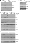
Figure 8. Rho-ROCK pathway regulates nuclear levels of PPM1A and maintains SMAD3 activation.
VSMCs maintained under serum-deprived conditions for 1 day were TGF-β1-stimulated (1 ng/ml) with or without the ROCK inhibitor, Y-27632 (10 µm) and cellular lysates probed for pSMAD2, total SMAD2/3, PAI-1 and ERK2 (A). Late-stage (4 hour) pSMAD2 levels were markedly attenuated and PAI-1 expression completely inhibited by ROCK blockade (A). TGF-β1-induced SMAD phosphorylation at the late time points (4 hours) is significantly reduced by inhibition of ROCK signaling. Serum-deprived VSMCs were pretreated for 30 minutes with Y-27632 (at indicated concentrations) prior to exposure to TGF-β1 for 4 hours. Cell lysates were probed for PAI-1, SMAD2/3, pSMAD2/3 and PPM1A (C). PAI-1 expression in response to TGF-β1 was completely blocked by Y-27632 pre-exposure (10 µM final concentration) despite the initial increase in SMAD2 phosphorylation in Y-27632-treated cells. Concentrations of Y-27632 that effectively inhibit PAI-1 induction and suppress SMAD2 phosphorylation also increase PPMIA levels (C). Transient knock-down of SMAD3 with siRNA constructs (as detailed in Methods) (D,E) or pre-incubation with the small molecule inhibitor of SMAD3 phosphorylation SIS3 (5 µM) (D,F,G) eliminates TGF-β1-induced PAI-1 expression in VSMCs (D,E) and MEFs (F,G). Cell fractionation studies confirmed that nuclear accumulation of pSMAD3 in response to TGF-β1 is blocked while nuclear PPM1A content increased upon pre-incubation with Y-27632 (H). TGF-β1 stimulation for 4 hours actually reduced nuclear PPM1A levels, which was restored by Y-27632 pretreatment (H). siRNA-mediated PPM1A knockdown in VSMCs resulted in a significantly increased TGF-β1-induced PAI-1 response compared to cells transfected with control siRNA constructs (I). ERK2 (A,B,D–G), SMAD2/3 (A,C,F), tubulin (D), lamin (H) and actin (I) provide loading controls. Data plotted in (B,E,G) is the mean ± S.D. of three independent experiments.
Figure 9
Figure 9. A model for TGF-β1 stimulated maintenance of SMAD3 phosphorylation and PAI-1 induction via Src/FAK/Caveolin-1 signaling.
TGF-β1 stimulates caveolin-1Y14 phosphorylation in a reactive oxygen species-FAK/c-Src dependent manner removing repressive influences on EGFR signaling (in red) leading to EGFR transactivation (also by c-Src), thereby, initiating signaling events leading to the MEK-ERK pathway activation necessary for PAI-1 induction. Src kinase phosphorylation of caveolin-1Y14 also stimulates Rho-GTP loading and ROCK (an established downstream target of Rho) activation is necessary for PAI-1 induction. pCaveolin-1Y14-Rho-ROCK mediated signaling leads to inhibition of PTEN-PPM1A interactions resulting in a reduction of nuclear PPM1A phosphatase (black pathway), thereby, maintaining the pSMAD2/3 levels (highlighted in blue) required for PAI-1 induction by TGF-β1 (see text). PAI-1 is elevated in atherosclerotic plaques frequently colocalizing with α-smooth muscle actin-expressing cells, presumably VSMCs (insert).

Similar articles

Cited by

References

    1. Vaughan DE. PAI-1 and atherothrombosis. J Thromb Haemost. 2005;3:1879–1883. - PubMed
    1. Weisberg AD, Albornoz F, Griffin JP, Crandall DL, Elokdah H, et al. Pharmacological inhibition and genetic deficiency of PAI-1 attenuates angiotensin II/salt-induced aortic remodeling. Arterioscler Thromb Vasc Biol. 2005;25:365–371. - PubMed
    1. Smith LH, Dixon JD, Stringham JR, Eren M, Elokdah H, et al. Pivotal role of PAI-1 in a murine model of hepatic vein thrombosis. Blood. 2006;107:132–134. - PMC - PubMed
    1. Samarakoon R, Goppelt-Struebe M, Higgins PJ. Linking cell structure to gene regulation: signaling events and expression controls on the model genes PAI-1 and CTGF. Cell Signal. 2010;22:1413–1419. - PMC - PubMed
    1. Wang TJ, Gona P, Larson MG, Tofler GH, Levy D, et al. Multiple biomarkers for the prediction of first major cardiovascular events and death. N Engl J Med. 2006;355:2631–2639. - PubMed

Publication types

MeSH terms