. 2011 Sep;138(17):3625-37.
doi: 10.1242/dev.064162.

Satellite cells, connective tissue fibroblasts and their interactions are crucial for muscle regeneration

Affiliations

Satellite cells, connective tissue fibroblasts and their interactions are crucial for muscle regeneration

Malea M Murphy et al. Development. 2011 Sep.

Abstract

Muscle regeneration requires the coordinated interaction of multiple cell types. Satellite cells have been implicated as the primary stem cell responsible for regenerating muscle, yet the necessity of these cells for regeneration has not been tested. Connective tissue fibroblasts also are likely to play a role in regeneration, as connective tissue fibrosis is a hallmark of regenerating muscle. However, the lack of molecular markers for these fibroblasts has precluded an investigation of their role. Using Tcf4, a newly identified fibroblast marker, and Pax7, a satellite cell marker, we found that after injury satellite cells and fibroblasts rapidly proliferate in close proximity to one another. To test the role of satellite cells and fibroblasts in muscle regeneration in vivo, we created Pax7(CreERT2) and Tcf4(CreERT2) mice and crossed these to R26R(DTA) mice to genetically ablate satellite cells and fibroblasts. Ablation of satellite cells resulted in a complete loss of regenerated muscle, as well as misregulation of fibroblasts and a dramatic increase in connective tissue. Ablation of fibroblasts altered the dynamics of satellite cells, leading to premature satellite cell differentiation, depletion of the early pool of satellite cells, and smaller regenerated myofibers. Thus, we provide direct, genetic evidence that satellite cells are required for muscle regeneration and also identify resident fibroblasts as a novel and vital component of the niche regulating satellite cell expansion during regeneration. Furthermore, we demonstrate that reciprocal interactions between fibroblasts and satellite cells contribute significantly to efficient, effective muscle regeneration.


Figures
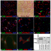
Fig. 1.
Fig. 1.
Tcf4 is highly expressed in muscle connective tissue (MCT) fibroblasts during muscle regeneration. (A-C) Tcf4+ cells at 5 dpi (BaCl2) are interstitial to regenerating laminin+ myofibers within Sirius Red+ MCT. A and B show adjacent sections. (D-F) In muscle at 5 dpi, some Tcf4+ cells are PDGFRα+ (D, arrows), and all are Pax7 and MyoD (E) and F4/80 (F). (G,H) MCT fibroblasts isolated from TAs at 5 dpi are Tcf4+, PDGFRα+ (G) and αSMA+ (H). (I) Semi-quantitative PCR shows that neonatal MCT fibroblasts, but not myoblasts, express Tcf4 and Col6a3. Scale bars: in B, 50 μm for A,B; in C, 12.5 μm; in F, 25 μm for D-F; in H, 10 μm for G,H.
Fig. 2.
Fig. 2.
After BaCl2 injury, Pax7+ satellite cells and Tcf4+ fibroblasts expand rapidly in close proximity to one another and regenerating myofibers. (A-G,CC) MyHCemb+ regenerating myofibers. (H-N,DD,GG) Pax7+ satellite cells. (O-U,EE,HH) Tcf4+ muscle connective tissue (MCT) fibroblasts. (V-BB,FF) Sirius Red+ MCT. Arrows in I, P and Q label a few of the PHH3+ Pax7+ or PHH3+Tcf4+ cells. (II) Tibialis anterior (TA) cross-sectional area measured on Sirius Red-stained sections 30-40 μm from TA origin. At each time point, adjacent sections are shown. Scale bar: 100 μm for A-BB. For all graphs, mean ± s.e.m. are plotted.
Fig. 3.
Fig. 3.
Pax7CreERT2 mice efficiently label satellite cells. (A) Pax7CreERT2 targeting strategy. (B-D) In myofibers isolated from uninjured tibialis anterior muscles (TAs) from Pax7CreERT2/+;R26RYFP/+ mice one day after five daily tamoxifen doses, 95% of Pax7+ satellite cells are YFP+ (B) and YFP+ cells are Syndecan4+ (C) and CD34+ (D). (E) In fibers isolated from uninjured TAs from Pax7CreERT2/CreERT2;R26RYFP/+ mice one day after five tamoxifen doses, normal numbers of CD34+ satellite cells are present. (F-O) In cryosections of uninjured TAs from Pax7CreERT2/+;R26RmTmG/+ mice one day after three tamoxifen doses, nearly all Pax7+ cells are GFP+ (F-I), lie within laminin+ myofiber basement membrane (K-N), and are Tcf4 (J). Tomato in Cre-myofibers was quenched by antigen retrieval. Arrows indicate Pax7+ satellite cells. (O) At 14 dpi, all regenerating myofibers are β-galactosidase+ in Pax7CreERT2/+;Polr2anlacZ/+. Scale bars: in B, 20 μm; in E, 10 μm for C-E; in M, 25 μm for F-H,K-M; in J, 12.5 μm; in N, 6.25 μm for I,N; in O, 50 μm.
Fig. 4.
Fig. 4.
Ablation of Pax7+ satellite cells leads to complete loss of muscle regeneration. (A-I) At 5 dpi, 91% of Pax7+ cells are ablated (A-C), resulting in fewer MyHCemb+ regenerating myofibers (D-F) and Tcf4+ fibroblasts (G-I) in Pax7CreERT2/+;R26RDTA/+ mice. (J-R) At 28 dpi, tibialis anterior (TA) cross-sectional area (L,O,R) and MyHCemb+ regenerated myofibers (J-L) are reduced, whereas the proportion of Sirius Red+ MCT (M-O), and the density of Tcf4+ fibroblasts (P-R) is increased in Pax7CreERT2/+;R26RDTA/+ mice. Insets in J and K show residual, incompletely injured myofibers with peripheral nuclei in Pax7CreERT2/+;R26RDTA/+ mice compared with regenerated myofibers with centralized nuclei in Pax7+/+;R26RDTA/+. (S-Z) At 28 dpi, {BaCl2 or cardiotoxin (CTX)], injured TAs are completely fibrotic or edemic in Pax7CreERT2/+;R26RDTA/+ mice, in whole mount (S,T,W,X) and Sirius Red-stained cross-sections (U,V,Y,Z). (AA-DD) Ablation of satellite cells prior to CTX injury leads to loss of regenerated muscle. In all tamoxifen/injury strategy schema, gray bars represent one day and black bars one week, tamoxifen (TMX) administration is indicated by blue arrowheads and BaCl2 or CTX application is indicated by red arrows. Whole mount images have been flipped so the injured limb (R) is on the right. TA weights include attached extensor digitorum longus (EDL). Scale bars: in Q, 100 μm for A,B,D,E,G,H,J,K,M,N,P,Q; in DD, 500 μm for U,V,Y,Z,CC,DD. For all graphs, mean ± s.e.m. are plotted.
Fig. 5.
Fig. 5.
Tcf4CreERT2 mice label muscle connective tissue (MCT) fibroblasts. (A) Tcf4CreERT2 targeting strategy. (B) At 14 dpi, several myofibers in uninjured tibialis anterior muscles (TAs) and all myofibers in injured TAs of Pax7CreERT2/+;R26RlacZ/+ mice are β-galactosidase+, but in TAs of Tcf4CreERT2/+;R26RlacZ/+ mice no myofibers are β-galactosidase+. (C-K) In cryosections of TAs at 5 dpi (BaCl2) from Tcf4CreERT2/+;R26RmTmG/+ mice (five tamoxifen doses), Tcf4+ fibroblasts are GFP+ (F-K), lie in between laminin+ regenerating myofibers (F-H) and are not F4/80+ (I-K). (L-N) MCT fibroblasts isolated from TAs at 5 dpi (BaCl2) from Tcf4CreERT2/+;R26RmTmG/+ mice (five tamoxifen doses) are GFP+ (L-N), Tcf4+ (L), αSMA+ (M) and PDGFRα+ (N). (O) In preparations of all mononuclear cells from the same Tcf4CreERT2/+;R26RmTmG/+ mice as shown in L-N, GFP+ cells are Pax7 and MyoD. Scale bars: in K, 25 μm for C-K; in O, 10 μm for L-O.
Fig. 6.
Fig. 6.
During muscle regeneration, ablation of Tcf4+ fibroblasts leads to premature satellite cell differentiation and smaller regenerated myofibers. (A-L) At 3 dpi, Tcf4+ cells are reduced (J-L), with no change in Pax7+ cells (A-C), but with an increase in MyoD+ progenitors/myoblasts (D-F) and MyHCemb+ regenerating myofibers (G-I) in Tcf4CreERT2/+;R26RDTA/+ mice. (M-X) At 5 dpi, 42% of Tcf4+ cells were calculated to be ablated (V-X), resulting in fewer Pax7+ cells (M-O), MyoD+ progenitors/myoblasts (P-R) and MyHCemb+ regenerating myofibers (S-U) in Tcf4CreERT2/+;R26RDTA/+ mice. (Y-KK) At 28 dpi, despite Tcf4+ fibroblast ablation (HH-JJ), Pax7+ cells recover (Y-AA), muscle largely regenerates (BB-GG), but diameter of myofibers is reduced (BB,CC,EE,FF,KK) in Tcf4CreERT2/+;R26RDTA/+ versus Tcf4+/+;R26RDTA/+ mice. (LL-PP) By 56 dpi, regenerated muscle is recovered in Tcf4CreERT2/+;R26RDTA/+ mice. Scale bars: in OO,100 μm for all photomicrographs. For all graphs, mean ± s.e.m. are plotted.
Fig. 7.
Fig. 7.
Model of the role of Pax7+ satellite cells and Tcf4+ muscle connective tissue (MCT) fibroblasts and their interactions during muscle regeneration. (A-C) Summary of cells and their interactions after injury during normal regeneration (A), with ablation of satellite cells (B) and with partial ablation of MCT fibroblasts (C). (A) During normal regeneration, satellite cells and fibroblasts rapidly proliferate. Satellite cells are absolutely required for normal regeneration of myofibers. Reciprocal interactions between satellite cells and fibroblasts are also important for regeneration. (B) With ablation of satellite cells, no regeneration of myofibers occurs and muscle is largely replaced by MCT and fibroblasts. (C) Partial ablation of fibroblasts leads to premature differentiation of satellite cells and results in smaller muscles with smaller myofibers at 28 dpi, when muscle regeneration is normally complete.

Similar articles

Cited by

References

    1. Alexakis C., Partridge T., Bou-Gharios G. (2007). Implication of the satellite cell in dystrophic muscle fibrosis: a self-perpetuating mechanism of collagen overproduction. Am. J. Physiol. Cell Physiol. 293, C661-C669 - PubMed
    1. Bailey A. J., Shellswell G. B., Duance V. C. (1979). Identification and change of collagen types in differentiating myoblasts and developing chick muscle. Nature 278, 67-69 - PubMed
    1. Beauchamp J. R., Heslop L., Yu D. S., Tajbakhsh S., Kelly R. G., Wernig A., Buckingham M. E., Partridge T. A., Zammit P. S. (2000). Expression of CD34 and Myf5 defines the majority of quiescent adult skeletal muscle satellite cells. J. Cell Biol. 151, 1221-1234 - PMC - PubMed
    1. Caldwell C. J., Mattey D. L., Weller R. O. (1990). Role of the basement membrane in the regeneration of skeletal muscle. Neuropathol. Appl. Neurobiol. 16, 225-238 - PubMed
    1. Cerletti M., Jurga S., Witczak C. A., Hirshman M. F., Shadrach J. L., Goodyear L. J., Wagers A. J. (2008). Highly efficient, functional engraftment of skeletal muscle stem cells in dystrophic muscles. Cell 134, 37-47 - PMC - PubMed

Publication types

MeSH terms

Substances