. 2011 Sep 1;187(5):2783-93.
doi: 10.4049/jimmunol.1100804. Epub 2011 Aug 3.

Identification of a novel proinflammatory human skin-homing Vγ9Vδ2 T cell subset with a potential role in psoriasis

Affiliations

Identification of a novel proinflammatory human skin-homing Vγ9Vδ2 T cell subset with a potential role in psoriasis

Ute Laggner et al. J Immunol. .

Abstract

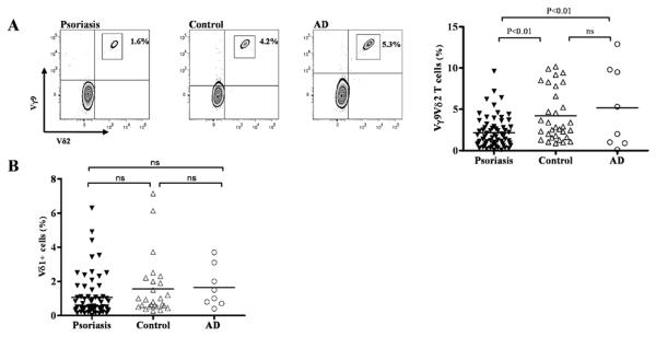
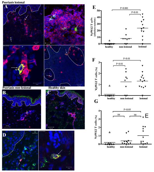
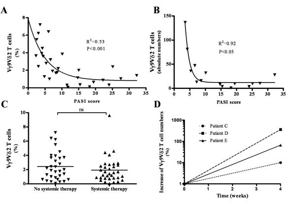
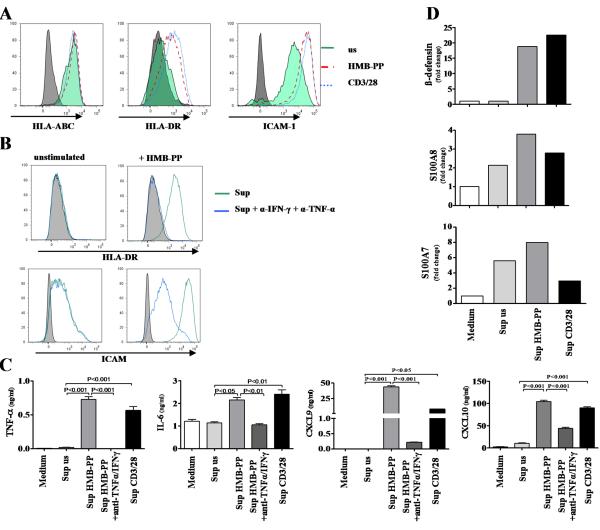
γδ T cells mediate rapid tissue responses in murine skin and participate in cutaneous immune regulation including protection against cancer. The role of human γδ cells in cutaneous homeostasis and pathology is characterized poorly. In this study, we show in vivo evidence that human blood contains a distinct subset of proinflammatory cutaneous lymphocyte Ag and CCR6-positive Vγ9Vδ2 T cells, which is rapidly recruited into perturbed human skin. Vγ9Vδ2 T cells produced an array of proinflammatory mediators including IL-17A and activated keratinocytes in a TNF-α- and IFN-γ-dependent manner. Examination of the common inflammatory skin disease psoriasis revealed a striking reduction of circulating Vγ9Vδ2 T cells in psoriasis patients compared with healthy controls and atopic dermatitis patients. Decreased numbers of circulating Vγ9Vδ2 T cells normalized after successful treatment with psoriasis-targeted therapy. Taken together with the increased presence of Vγ9Vδ2 T cells in psoriatic skin, these data indicate redistribution of Vγ9Vδ2 T cells from the blood to the skin compartment in psoriasis. In summary, we report a novel human proinflammatory γδ T cell involved in skin immune surveillance with immediate response characteristics and with potential clinical relevance in inflammatory skin disease.


Figures

Figure 1
Figure 1. Vγ9Vδ2 T cells are decreased in peripheral blood of psoriasis patients
Vγ9Vδ2 T cells in peripheral blood of age-matched psoriasis patients suffering from plaque type psoriasis (n=66), healthy controls (n=32) and atopic dermatitis (AD) patients (n=8) were stained for CD3, Vγ9 and Vδ2 and examined by flow cytometry. One representative staining for each group is shown in A. Vγ9Vδ2 T cells within the CD3 subset were significantly decreased in psoriasis patients (2.16% (+0.23%) compared to healthy controls (4.21% (±0.55%), p<0.01) and AD patients (5.18% (±1.75%), p<0.01) (A). Vδ1 T cells were not different between psoriasis patients, controls and AD patients (1.06% (±0.15) vs. 1.55% (±0.33%) vs. 1.65% (±0.42%) (B).
Figure 2
Figure 2. Identification of a novel subset of skin homing Vγ9Vδ2 T cells expressing CLA and skin homing chemokine receptors
Psoriasis patients (n=32) and healthy controls (n=19) were analyzed for CLA expression on peripheral Vγ9Vδ2 T cells by flow cytometry. The isotype for CLA and chemokine receptors was set with specific isotype controls for each marker, gated on CD3 and Vδ2. A subset of Vγ9Vδ2 T cells in patients and controls both expressed CLA, a marker for skin homing T cells (A). Percentage of CLA+Vγ9Vδ2 T cells gated on CD3+ cells (blue border and symbols) were significantly lower in psoriasis patients compared to healthy controls (0.94% (±0.11%) vs. 2.22% (±0.38%); p=0.001) whereas the CLA- subset was not significantly changed in patients (1.74% (±0.29%) vs. 2.67% (±0.58%) indicating a selective decrease of CLA+Vγ9Vδ2 T cells in psoriasis patients (B). The skin associated chemokine receptors CCR4, CCR6 and CCR10 gated on peripheral blood Vγ9Vδ2 T cells were analyzed by flow cytometry in patients and controls (C). Vγ9Vδ2 T cells expressed high levels of CCR6 (purple border and symbols) (patients: 16.5% (+3.5%-73%) vs. controls: 30.61% (7.3%-59%); p<0.05) while expressing variably levels of CCR4 (green border and symbols) (patients: 2.75% (0%-14.9%) vs. controls: (2.8% (1%-17%), ns) and little CCR10 (turquoise border and symbols) (patients: 1.6% (0%-15%) vs. controls: 5% (1%-10%), p<0.05) (D). Staining of one representative patient (Patient F) and one representative control (Control A) is shown for the expression of CLA, CCR4, CCR6 and CCR10.
Figure 3
Figure 3. CLA+ Vγ9Vδ2 T cells are recruited to human skin
Suction blisters were performed on normal skin (“non-perturbed”) and skin that was injected 24 hours before with sterile saline (“perturbed”) (n=6, matched) of healthy volunteers (A). Blister fluid was harvested 16-20 hours after blister induction and assessed for absolute and relative Vγ9Vδ2 T cells and non-Vγ9Vδ2 CD3+ cells. Perturbed skin contained significantly higher percentages of Vγ9Vδ2 T cells than non-perturbed skin (p<0.05) (B). Absolute numbers of Vγ9Vδ2 T cells and non-Vγ9Vδ2 CD3+ cells were both significantly higher in perturbed compared to non-perturbed skin (both p<0.05) (C+D) but Vγ9Vδ2 T cells increased to a significantly greater extent than non-Vγ9Vδ2 CD3+ cells (p<0.05) (E). Vγ9Vδ2 T cells in perturbed skin were significantly enriched for CLA+Vγ9Vδ2 T cells as compared to circulating Vγ9Vδ2 T cells of the same individuals (p<0.01) (F+G).
Figure 4
Figure 4. Vγ9Vδ2 T cells are increased in skin of psoriasis patients
Frozen skin sections were stained with anti-CD3 mAb (red) and anti-Vδ2 mAb (green), identifying Vδ2 T cells as yellow. Nuclei were stained with To-Pro-3 (blue) and dermo-epidermal barrier is indicated with a white line. Vδ2 T cells were present in lesional psoriatic dermis as well as scattered in the epidermis (A). Vδ2 T cells were found in non-lesional psoriatic skin (B) while in healthy skin, Vδ2 T cells were rare (C). Co-staining of anti-CLA (green) and anti-Vδ2 (red) revealed CLA expression on skin Vγ9Vδ2 (D). Psoriatic skin harbored significantly higher absolute numbers of Vδ2 T cells than non-lesional (p<0.01) or healthy skin (p<0.001) as calculated in 1 cm of skin section (E). Relative quantification of Vγ9Vδ2 T cells in skin sections revealed that a higher percentage of T cells in non-lesional (p<0.01) as well as lesional (p<0.01) psoriatic skin express Vδ2 than in normal skin (F). We established T cell lines from lesional (n=12), edge (n=11) and healthy (n=8) skin by culturing skin pieces in the presence of IL-2 (60 IU/ml) and IL-15 (12 ng/ml) for 2-3 weeks and harvesting cells that migrated out of the tissue. Skin T cell lines were analyzed for γδ T cell subsets after gating on CD3+ cells. In lesional skin there were more Vγ9Vδ2 T cells (1.03% (±0.31%)) than in edge (0.39% (±0.16%), ns) or healthy skin (0.21% (±0.18 %), p<0.05) (G).
Figure 5
Figure 5. Clinical correlation between psoriasis severity and circulating Vγ9Vδ2 T cells
Lower percentages of circulating Vγ9Vδ2 T cells significantly correlated with severe clinical disease as reflected in a higher PASI score (p<0.001, R2=0.53, n=30) (A). Absolute Vγ9Vδ2 T cell numbers were obtained in a subpopulation of patients (n=16) and also correlated with disease severity (p<0.05, R2=0.92) (B). Comparing percentage of peripheral Vγ9Vδ2 T cells of patients without systemic treatment to treated patients with the same PASI score we did not observe any significant difference (2.44% (+0.35%) vs. 1.93 (+0.31%), ns) (C). We analysed 3 patients before and during the course of their treatment at week 0 and week 4. In all of these patients PASI had dropped by at least 40% after 4 weeks of systemic treatment (Patient C: 4 weeks - 40%; Patient D: 4 weeks - 48%, Patient E: 45%) In all three patients, the absolute numbers of peripheral Vγ9Vδ2 T cell increased with successful treatment (Patient C: 4 weeks – 10%; Patient D: 4 weeks - 360%; Patient E: 4 weeks - 67%) (D).
Figure 6
Figure 6. CLA+Vγ9Vδ2 T cells produce psoriasis-relevant inflammatory mediators
CLA+Vγ9Vδ2 T cells from healthy controls were sorted and stimulated with HMB-PP, CD3/28 beads or PMA/Ionomycin before analyzing supernatant by multiplex bead assay. Activated CLA+Vγ9Vδ2 T cells up-regulated their IFN-γ and TNF-α production (representative experiment, n=8) (A). IL-17A production was assessed in circulating Vγ9Vδ2 T cells by flow cytometry after gating on the Vγ9 subset. A small percentage of Vγ9+ T cells produced IL-17A which was enriched in the CLA+Vγ9+ subset (B). CLA+Vγ9Vδ2 T cells produced the chemokine IL-8 as assessed by multiplex bead assay (C). The CC-chemokines CCL3, CCL4 and CCL5 were produced already constitutively. The production of CCL3 and CCL4 was increased upon activation, whereas CCL5 production could not be further up-regulated (all one representative experiment, n=11) (C) IGF-1 production was assessed by flow cytometry. Unstimulated cells cultured with medium alone showed a subtle constitutive production of IGF-1 (light grey). PMA/Ionomycin (red) and HMB-PP (dark grey) led to up-regulation of IGF-1 production in CLA+Vγ9Vδ2 T cells (D). Relative IGF-1 expression as assessed by quantitative PCR showed an up-regulation of IGF-1 expression upon activation also with IFN-α (n=4) (D).
Figure 7
Figure 7. CLA+Vγ9Vδ2 T cells activate keratinocytes
Keratinocytes were cultured for 48 hours with the supernatant of stimulated and resting CLA+Vγ9Vδ2 T cells. Supernatant of Vγ9Vδ2 T cells activated with HMB-PP (red) or CD3/28 beads (blue) strongly up-regulated the activation markers HLA-DR and ICAM-1 on keratinocytes, while HLA-ABC levels were comparable to the supernatant of unstimulated cells (green) (A). Up-regulation of HLA-DR and ICAM was inhibited when adding anti-TNF-α and α-IFN-γ antibodies to the culture (green: supernatant of HMB-PP activated CLA+Vγ9Vδ2 T cells only, blue: supernatant of HMB-PP activated CLA+Vγ9Vδ2 T cells + α-IFN-γ and α-TNF-α antibodies) (B). Keratinocytes exposed to supernatant of HMB-PP activated CLA+Vγ9Vδ2 T cells produced high amounts of the CXC chemokine CXCL9 and CXCL10 in a largely TNF-α and IFN-γ dependent fashion (C). Activated CLA+Vγ9Vδ2 T cell supernatant induced production of β-defensin, S100A7 and S100A8 but not LL37 in keratinocytes at RNA level (representative experiment, n=6) (D).

Similar articles

Cited by

References

    1. Hayday AC. [gamma][delta] cells: a right time and a right place for a conserved third way of protection. Annu Rev Immunol. 2000;18:975–1026. - PubMed
    1. Hayday A, Tigelaar R. Immunoregulation in the tissues by gammadelta T cells. Nat Rev Immunol. 2003;3:233–242. - PubMed
    1. Girardi M, Lewis J, Glusac E, Filler RB, Geng L, Hayday AC, Tigelaar RE. Resident skin-specific gammadelta T cells provide local, nonredundant regulation of cutaneous inflammation. J Exp Med. 2002;195:855–867. - PMC - PubMed
    1. Strid J, Roberts SJ, Filler RB, Lewis JM, Kwong BY, Schpero W, Kaplan DH, Hayday AC, Girardi M. Acute upregulation of an NKG2D ligand promotes rapid reorganization of a local immune compartment with pleiotropic effects on carcinogenesis. Nat Immunol. 2008;9:146–154. - PubMed
    1. Girardi M, Oppenheim DE, Steele CR, Lewis JM, Glusac E, Filler R, Hobby P, Sutton B, Tigelaar RE, Hayday AC. Regulation of cutaneous malignancy by gammadelta T cells. Science. 2001;294:605–609. - PubMed

Publication types

MeSH terms

Substances