. 2011 Oct;31(19):4007-21.
doi: 10.1128/MCB.05810-11. Epub 2011 Aug 1.

Ribosomal protein L11 recruits miR-24/miRISC to repress c-Myc expression in response to ribosomal stress

Affiliations

Ribosomal protein L11 recruits miR-24/miRISC to repress c-Myc expression in response to ribosomal stress

Kishore B Challagundla et al. Mol Cell Biol. 2011 Oct.

Abstract

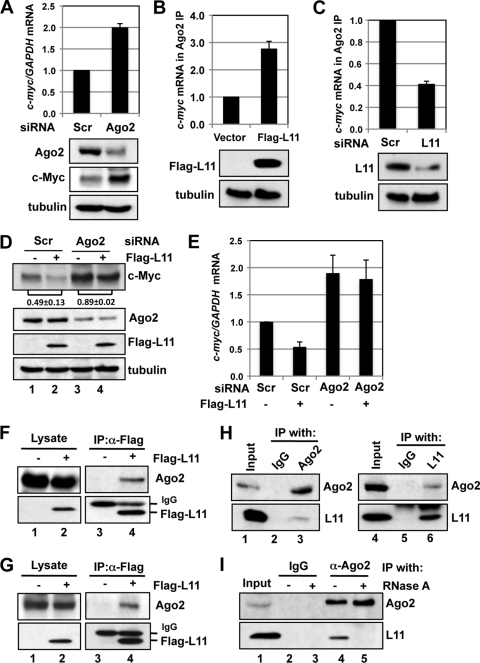
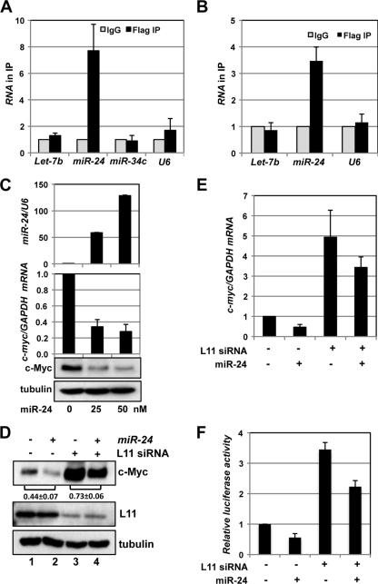
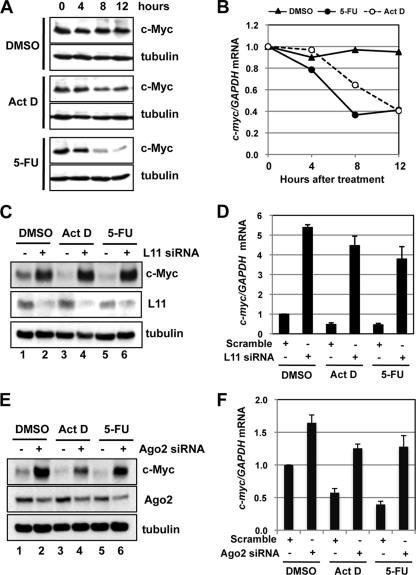
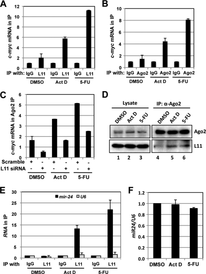
c-Myc promotes cell growth by enhancing ribosomal biogenesis and translation. Deregulated expression of c-Myc and aberrant ribosomal biogenesis and translation contribute to tumorigenesis. Thus, a fine coordination between c-Myc and ribosomal biogenesis is vital for normal cell homeostasis. Here, we show that ribosomal protein L11 regulates c-myc mRNA turnover. L11 binds to c-myc mRNA at its 3' untranslated region (3'-UTR), the core component of microRNA-induced silencing complex (miRISC) argonaute 2 (Ago2), as well as miR-24, leading to c-myc mRNA reduction. Knockdown of L11 drastically increases the levels and stability of c-myc mRNA. Ablation of Ago2 abrogated the L11-mediated reduction of c-myc mRNA, whereas knockdown of L11 rescued miR-24-mediated c-myc mRNA decay. Interestingly, treatment of cells with the ribosomal stress-inducing agent actinomycin D or 5-fluorouracil significantly decreased the c-myc mRNA levels in an L11- and Ago2-dependent manner. Both treatments enhanced the association of L11 with Ago2, miR-24, and c-myc mRNA. We further show that ribosome-free L11 binds to c-myc mRNA in the cytoplasm and that this binding is enhanced by actinomycin D treatment. Together, our results identify a novel regulatory paradigm wherein L11 plays a critical role in controlling c-myc mRNA turnover via recruiting miRISC in response to ribosomal stress.


Figures

Fig. 1.
Fig. 1.
L11 regulates c-myc mRNA levels and stability. (A) Knockdown of L11 increases the levels of c-myc mRNA and c-Myc protein. U2OS cells were transfected with scrambled (Scr) siRNA or one of two L11 siRNAs (L11si-1 or L11si-2). The cells were assayed for expression of c-myc mRNA by the use of RT-qPCR and of c-Myc protein by the use of IB assays. (B) Knockdown of L11 stabilizes c-myc mRNA. U2OS cells transfected with scrambled or L11 siRNA were treated with 2 μM Act D. The cells were harvested at the indicated time points and assayed for relative levels of c-myc mRNA normalized to the expression of GAPDH mRNA by the use of RT-qPCR assays. The average c-myc mRNA half-life value is shown. *, P < 0.01 compared to t1/2 of c-myc mRNA in scrambled-RNA transfected cells. (C) Knockdown of L11 does not affect c-myc mRNA translation. U2OS cells transfected with scrambled or L11 siRNA were pulse-labeled with [35S]methionine. Equal amounts of cell lysates were immunoprecipitated with anti-c-Myc (C33) antibody-conjugated beads followed by autography (top right panel) and IB with anti-c-Myc antibody (Y69) (middle right panel). The total lysates were also loaded on an SDS-PAGE gel followed by autography (left panel). The relative translation efficiency of c-myc mRNA was calculated based on the ratio of radiolabeled to total immunoprecipitated c-Myc protein and is plotted in the bottom right panel. (D) Overexpression of L11 reduces the levels of c-myc mRNA and c-Myc protein. U2OS cells transfected with control or Flag-L11 vector were assayed for expression of c-myc mRNA by the use of RT-qPCR and of protein by the use of IB assays. (E and F) Knockdown of L11 increases the levels of c-myc mRNA and c-Myc protein in 293 and WI38 cells. 293 (E) and WI38 (F) cells were transfected with scrambled or L11 siRNA. The cells were assayed for expression of c-myc mRNA by the use of RT-qPCR and of c-Myc protein by the use of IB assays.
Fig. 2.
Fig. 2.
L11 regulates c-myc by targeting the 3′-UTR of c-myc mRNA. (A) L11 binds to c-myc mRNA. Lysates from 293 (left panel) and U2OS (right panel) cells transfected with Flag-L11 were immunoprecipitated with control mouse IgG or anti-Flag antibody. RNA extracted from the immunoprecipitates was retrotranscribed and assayed for expression of c-myc mRNA by the use of RT-qPCR assays. (B) L11 binds to the c-myc 3′-UTR. U2OS cells transfected with Flag-L11 together with control pGL3 or pGL3-myc3′UTR vector were immunoprecipitated with anti-Flag or mouse IgG. The immunoprecipitates were assayed for expression of the luciferase mRNA by RT-qPCR assays. (C) L11 reduction of luciferase mRNA levels and suppression of luciferase activity are dependent on the c-myc 3′-UTR. 293 cells transfected with the indicated plasmids together with β-gal plasmid were assayed for relative luciferase activity levels normalized to β-gal expression (top panel) and relative luciferase mRNA levels normalized to GAPDH mRNA by the use of RT-qPCR assays (middle panel). The protein expression results are shown in the bottom panels. (D) Knockdown of L11 increases the levels of luciferase mRNA and luciferase activity in a manner dependent on the c-myc 3′-UTR. U2OS cells transfected with β-gal and pGL3 or pGL3-myc3′UTR plasmids and siRNAs were assayed for relative luciferase activity levels normalized to β-gal expression (top panel) and relative luciferase mRNA levels normalized to GAPDH mRNA by the use of RT-qPCR assays (middle panel). The protein expression results are shown in the bottom panels. (E) Diagram of the control pGL3, pGL3-myc3′UTR, and pGL3-myc3′UTR fragment (F1, F2, F3, and F4) vectors. The relative positions of the full-length (FL) c-myc 3′UTR and its fragments (F1 through F4) are indicated, with the first nucleotide after the stop codon labeled “1.” The position of the PCR product used to examine expression of the luciferase mRNA is indicated in the coding region of the luciferase gene. pA indicates a poly(A) tail. (F) L11 binds to the 3′ end of the c-myc 3′-UTR. 293 cells transfected with Flag-L11 together with control pGL3 or pGL3-myc3′UTR or its fragments were immunoprecipitated with anti-Flag or mouse IgG. The immunoprecipitates were assayed for expression of luciferase mRNA by RT-qPCR assays. (G) L11 suppression of luciferase activity is dependent on the 3′ end of the c-myc 3′-UTR. 293 cells transfected with the indicated plasmids together with a β-gal plasmid were assayed for relative luciferase activity levels normalized to β-gal expression. The protein expression results are shown in the bottom panels.
Fig. 3.
Fig. 3.
Mutual dependency of L11 and ago2 in regulating c-myc mRNA. (A) Ago2 regulates c-myc mRNA levels. U2OS cells transfected with scrambled or Ago2 siRNA were assayed for expression of c-Myc protein (bottom panels) and mRNA (top panel). (B) Overexpression of L11 increases association of Ago2 with c-myc mRNA. U2OS cells were transfected with control or Flag-L11 plasmid. The cell lysates were immunoprecipitated with anti-Ago2 antibodies followed by an RT-qPCR assay to determine the levels of c-myc mRNA. The protein expression results are shown in the bottom panels. (C) Knockdown of L11 reduces the association of Ago2 with c-myc mRNA. Lysates from U2OS cells transfected with scrambled or L11 siRNA were immunoprecipitated with anti-Ago2 antibodies followed by an RT-qPCR assay to determine the levels of c-myc mRNA. The protein expression results are shown in the bottom panels. (D and E) L11 suppression of c-myc mRNA requires Ago2. U2OS cells transfected with control or Flag-L11 plasmid together with scrambled or Ago2 siRNA were assayed for c-Myc protein expression by immunoblotting (IB) (D) and for mRNA expression by RT-qPCR assays (E). The c-Myc bands were quantified and normalized to tubulin. The ratios of lane 2 to lane 1 and of lane 4 to lane 3 for the results from three independent experiments are indicated in panel D. (F and G) Ectopically expressed L11 interacts with Ago2. 293 (F) or U2OS (G) cells transfected with control or Flag-L11 plasmid were subjected to IP with anti-Flag antibody followed by IB using anti-Ago2 antibodies. (H) Endogenous L11 interacts with endogenous Ago2. 293 cell lysates were immunoprecipitated with control mouse IgG or anti-Ago2 (left panels) or rabbit IgG or anti-L11 (right panels) antibody followed by IB detection performed using anti-L11 or anti-Ago2 antibodies. (I) Interaction of L11 with Ago2 requires RNA. U2OS cell lysates were immunoprecipitated with control mouse IgG or anti-Ago2 antibodies in the absence (lanes 2 and 4) or presence (lanes 3 and 5) of RNase followed by IB detection performed using anti-L11 or anti-Ago2 antibodies.
Fig. 4.
Fig. 4.
L11 recruits miR-24 to target c-myc mRNA. (A and B) L11 binds to miR-24 in cells. Lysates from 293 (A) or U2OS (B) cells transfected with Flag-L11 plasmid were immunoprecipitated with anti-Flag antibody or control IgG followed by detection of the indicated miRNAs by the use of RT-qPCR assays. (C) Overexpression of miR-24 decreases c-Myc levels. U2OS cells transfected with a control or with different doses of miR-24 mimics were assayed for the relative levels of expression of miR-24 normalized with U6 snRNA (top panel) and of c-myc mRNA normalized with GAPDH mRNA (middle panel) by the use of RT-qPCR assays as well as c-Myc protein (bottom panel) by the use of IB. (D and E) Knockdown of L11 attenuates c-myc downregulation by miR-24. U2OS cells transfected with scrambled or L11 siRNA together with control or miR-24 mimics were assayed for c-Myc protein levels by IB (D) and c-myc mRNA levels by the use of RT-qPCR (E). The c-Myc bands were quantified and normalized to tubulin. The ratios of lane 2 to lane 1 and of lane 4 to lane 3 for the results from three independent experiments are indicated in panel D. (F) Knockdown of L11 attenuates the miR-24-mediated suppression of luciferase activity. U2OS cells were transfected with pGL3-myc3′UTR and β-gal expression plasmids together with siRNA and/or miRNAs as indicated. The cells were assayed for relative luciferase activity levels normalized to β-gal expression.
Fig. 5.
Fig. 5.
L11 downregulates c-myc mRNA in response to ribosomal stress. (A and B) c-Myc is downregulated by treatment with Act D or 5-FU. U2OS cells were treated with DMSO, Act D (5 nM), or 5-FU (10 μg/ml) for the indicated hours. The cells were assayed for c-Myc protein expression by IB (A) and c-myc mRNA expression by RT-qPCR (B) assays. (C and D) Knockdown of L11 rescues the downregulation of c-Myc by treatment with Act D or 5-FU. U2OS cells transfected with scrambled or L11 siRNA were treated with DMSO, Act D (5 nM), or 5-FU (10 μg/ml) for 12 h. The cells were assayed for c-Myc protein expression by IB (C) and c-myc mRNA expression by RT-qPCR (D) assays. (E and F) Knockdown of Ago2 rescues the downregulation of c-Myc by treatment with Act D or 5-FU. U2OS cells transfected with scrambled or Ago2 siRNA were treated with DMSO, Act D (5 nM), or 5-FU (10 μg/ml) for 12 h. The cells were assayed for c-Myc protein expression by IB (E) and c-myc mRNA expression by RT-qPCR (F) assays.
Fig. 6.
Fig. 6.
L11 recruits miRISC to c-myc mRNA in response to ribosomal stress. (A) Treatment with Act D or 5-FU increases the binding of L11 to c-myc mRNA. U2OS cells were treated with DMSO, Act D (5 nM), or 5-FU (10 μg/ml) for 12 h. The cell lysates were immunoprecipitated with control IgG or anti-L11 antibodies followed by RT-qPCR detection of c-myc mRNA. (B) Treatment with Act D or 5-FU increases the binding of Ago2 to c-myc mRNA. U2OS cells were treated with DMSO, Act D (5 nM), or 5-FU (10 μg/ml) for 12 h. The cell lysates were immunoprecipitated with anti-Ago2 antibodies or control IgG followed by RT-qPCR detection of c-myc mRNA. (C) Knockdown of L11 abolishes Ago2 binding to c-myc mRNA in response to ribosomal stress. U2OS cells transfected with scrambled or L11 siRNA were treated with DMSO, Act D (5 nM), or 5-FU (10 μg/ml) for 12 h. The cell lysates were immunoprecipitated with anti-Ago2 antibodies or control IgG followed by RT-qPCR detection of c-myc mRNA. The data were normalized to the c-myc mRNA in IP with control IgG. (D) Treatment with Act D or 5-FU increases the binding of L11 to Ago2. U2OS cells were treated with DMSO, Act D (5 nM), or 5-FU (10 μg/ml) for 12 h. The cell lysates were immunoprecipitated with anti-Ago2 antibodies followed by IB using anti-L11 antibodies. (E) Treatment with Act D or 5-FU increases the association of L11 with miR-24. U2OS cells were treated with DMSO, Act D (5 nM), or 5-FU (10 μg/ml) for 12 h and subjected to IP with anti-L11 or control IgG, followed by RT-qPCR detection of miR-24 and U6 snRNA. (F) Treatment with Act D or 5-FU does not change the total levels of miR-24 in cells. U2OS cells treated with DMSO, Act D (5 nM), or 5-FU (10 μg/ml) for 12 h were examined for expression of miR-24 normalized to that of U6 snRNA by the use of RT-qPCR assays.
Fig. 7.
Fig. 7.
Ribosome-free L11 associates with c-myc mRNA in the cytoplasm. (A) L11 binds to the c-myc 3′-UTR in the cytoplasm. 293 cells transfected with pGL3-myc3′UTR together with control Flag or Flag-L11 vector were fractionated into the cytoplasm (Cyto) and the nucleus (Nuc) fractions, followed by IP with anti-Flag antibody or mouse IgG. The immunoprecipitates were assayed for expression of luciferase mRNA (left panel) and c-myc mRNA (middle panel) by the use of RT-qPCR assays. Cellular fractionation was verified by detecting the nuclear SP1 and cytoplasmic tubulin by the use of IB (right panels). Actin was used as a loading control. (B) Act D treatment enhances the interaction of endogenous L11 with the c-myc mRNA in the cytoplasm. The cytoplasmic and the nuclear fractions were isolated from U2OS cells treated with DMSO or Act D (5 nM) for 12 h, followed by IP with anti-L11 antibodies or control rabbit IgG. The immunoprecipitates were assayed for expression of c-myc mRNA by the use of RT-qPCR assays (left panel). Cellular fractionation was verified by IB detection of SP1 and tubulin proteins (right panels). (C) Treatment with Act D releases L11 from the nucleolus into the nucleoplasm and the cytoplasm. U2OS cells treated with DMSO or Act D (5 nM) for 12 h were subjected to isolation of the cytoplasm (Cyto), nucleoplasm (Np), and the nucleolus (No) fractions, followed by IB detection of the indicated proteins. Nucleophosmin (B23) and nucleolin (C23) were used as nucleolar markers. (D) Ribosome-free L11 binds to c-myc mRNA. Nonribosome supernatants containing free RPs were isolated from 293 cells transfected using control or Flag-L11 plasmid and sucrose centrifugation as described in Materials and Methods. The supernatants were immunoprecipitated with anti-Flag antibody or mouse IgG followed by RT-qPCR detection of c-myc mRNA in the immunoprecipitates. (E) Act D treatment enhances the interaction of free endogenous L11 with c-myc mRNA. Nonribosome supernatants containing free RPs were isolated from U2OS cells treated with DMSO or Act D (5 nM) for 12 h. The supernatants were immunoprecipitated with anti-L11 antibodies or control IgG followed by RT-qPCR detection of c-myc mRNA (left panel). The L11 protein was detected using IB (upper right panels). S.E, short exposure; L.E, longer exposure. The rRNAs from polysome pellets as shown in the lower right panel were revealed by ethidium bromide staining. (F) L11 interacts with c-myc mRNA in the presence of EDTA. U2OS cells transfected with Flag-L11 were subjected to IP in the absence or presence of 20 mM EDTA. RNA IPs were conducted using anti-Flag antibody or control IgG followed by RT-qPCR detection of c-myc mRNA. (G) Sequential co-IP of the L11–c-Myc protein complex. 293 cells were transfected with V5–c-Myc in the presence or absence of Flag-L11. The cell lysates were first immunoprecipitated with anti-Flag antibody. The Flag-L11-associated protein complexes were eluted with Flag peptide. Half of the elution was assayed for V5–c-Myc and Flag-L11 proteins by the use of IB. The other half was subjected to a second IP using anti-V5 antibody followed by IB detection of V5–c-Myc and Flag-L11 proteins. (H) Sequential co-IP of the L11–c-myc mRNA complex. 293 cells were transfected with V5–c-Myc in the presence or absence of Flag-L11. The cell lysates were first immunoprecipitated with anti-Flag antibody or control IgG followed by elution with Flag peptide. Half of the elution was assayed for c-myc mRNA by the use of RT-qPCR assays. The other half was subjected to a second IP using anti-V5 antibody or control IgG, followed by RT-qPCR detection of the c-myc mRNA.
Fig. 8.
Fig. 8.
L11 regulation of c-myc mRNA turnover is specific. (A) Knockdown of L23, L26, or L29 does not enhance c-Myc levels. U2OS cells transfected with scrambled, L23, L26, or L29 siRNA were subjected to detection of c-myc mRNA by the use of RT-qPCR and of c-Myc protein by the use of IB assays. The results of knockdown of the individual RPs shown in the bottom panels were determined by IB (L11 and L23) or RT-PCR (L26 and L29). (B) Overexpression of L23, L26, or L29 does not reduce c-Myc levels. U2OS cells transfected with control (Ctl), Flag-L23, Flag-L26, or Flag-L29 plasmid were subjected to detection of c-myc mRNA by the use of RT-qPCR (top panel) and of c-Myc protein by the use of IB assays (bottom panels). (C) L23 and L29 do not bind to c-myc mRNA in cells. 293 cells were transfected with control pGL3 or pGL3-myc3′UTR together with Flag-L11, Flag-L23, or Flag-L29. The cell lysates were immunoprecipitated with anti-Flag antibody or control IgG, followed by RT-qPCR detection of luciferase mRNA. (D) Knockdown of L11 does not evoke upregulation of a set of other mRNAs. U2OS cells transfected with scrambled or L11 siRNA were subjected to detection of the indicated mRNAs by the use of RT-qPCR assays.
Fig. 9.
Fig. 9.
Schematic model of L11 regulation of c-myc mRNA decay via miRISC in response to ribosomal stress. Under normal conditions (top panel), the 40S and 60S mature ribosome subunits are assembled in the nucleolus and transported into the cytoplasm to mediate translation of c-myc mRNA. Upon perturbation of ribosomal biogenesis (ribosomal stress; bottom panel), individual small (RPS) and large (RPL) ribosomal proteins, including L11, are released from the nucleolus into the nucleoplasm, where L11 binds to c-Myc protein and suppresses its transactivation activity (not shown), and into the cytoplasm, where L11 recruits miR-24-loaded miRISC to the c-myc 3′-UTR, leading to c-myc mRNA decay.

Similar articles

Cited by

References

    1. Adhikary S., Eilers M. 2005. Transcriptional regulation and transformation by Myc proteins. Nat. Rev. Mol. Cell Biol. 6:635–645 - PubMed
    1. Andersen J. S., et al. 2002. Directed proteomic analysis of the human nucleolus. Curr. Biol. 12:1–11 - PubMed
    1. Arabi A., et al. 2005. c-Myc associates with ribosomal DNA and activates RNA polymerase I transcription. Nat. Cell Biol. 7:303–310 - PubMed
    1. Bhat K. P., Itahana K., Jin A., Zhang Y. 2004. Essential role of ribosomal protein L11 in mediating growth inhibition-induced p53 activation. EMBO J. 23:2402–2412 - PMC - PubMed
    1. Bhattacharyya S. N., Habermacher R., Martine U., Closs E. I., Filipowicz W. 2006. Relief of microRNA-mediated translational repression in human cells subjected to stress. Cell 125:1111–1124 - PubMed

Publication types

MeSH terms

LinkOut - more resources