Monoacylglycerol lipase exerts dual control over endocannabinoid and fatty acid pathways to support prostate cancer
- PMID: 21802006
- PMCID: PMC3149849
- DOI: 10.1016/j.chembiol.2011.05.009
Monoacylglycerol lipase exerts dual control over endocannabinoid and fatty acid pathways to support prostate cancer
Abstract
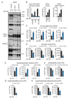
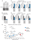
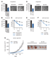
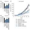
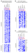
Cancer cells couple heightened lipogenesis with lipolysis to produce fatty acid networks that support malignancy. Monoacylglycerol lipase (MAGL) plays a principal role in this process by converting monoglycerides, including the endocannabinoid 2-arachidonoylglycerol (2-AG), to free fatty acids. Here, we show that MAGL is elevated in androgen-independent versus androgen-dependent human prostate cancer cell lines, and that pharmacological or RNA-interference disruption of this enzyme impairs prostate cancer aggressiveness. These effects were partially reversed by treatment with fatty acids or a cannabinoid receptor-1 (CB1) antagonist, and fully reversed by cotreatment with both agents. We further show that MAGL is part of a gene signature correlated with epithelial-to-mesenchymal transition and the stem-like properties of cancer cells, supporting a role for this enzyme in protumorigenic metabolism that, for prostate cancer, involves the dual control of endocannabinoid and fatty acid pathways.
Copyright © 2011 Elsevier Ltd. All rights reserved.
Figures
Similar articles
-
Dual blockade of FAAH and MAGL identifies behavioral processes regulated by endocannabinoid crosstalk in vivo.Proc Natl Acad Sci U S A. 2009 Dec 1;106(48):20270-5. doi: 10.1073/pnas.0909411106. Epub 2009 Nov 16. Proc Natl Acad Sci U S A. 2009. PMID: 19918051 Free PMC article.
-
Characterization of monoacylglycerol lipase inhibition reveals differences in central and peripheral endocannabinoid metabolism.Chem Biol. 2009 Jul 31;16(7):744-53. doi: 10.1016/j.chembiol.2009.05.009. Chem Biol. 2009. PMID: 19635411 Free PMC article.
-
Inhibition of the endocannabinoid-regulating enzyme monoacylglycerol lipase elicits a CB1 receptor-mediated discriminative stimulus in mice.Neuropharmacology. 2017 Oct;125:80-86. doi: 10.1016/j.neuropharm.2017.06.032. Epub 2017 Jun 30. Neuropharmacology. 2017. PMID: 28673548 Free PMC article.
-
A review on the monoacylglycerol lipase: at the interface between fat and endocannabinoid signalling.Curr Med Chem. 2010;17(24):2588-607. doi: 10.2174/092986710791859414. Curr Med Chem. 2010. PMID: 20491633 Review.
-
The serine hydrolases MAGL, ABHD6 and ABHD12 as guardians of 2-arachidonoylglycerol signalling through cannabinoid receptors.Acta Physiol (Oxf). 2012 Feb;204(2):267-76. doi: 10.1111/j.1748-1716.2011.02280.x. Epub 2011 Apr 22. Acta Physiol (Oxf). 2012. PMID: 21418147 Free PMC article. Review.
Cited by
-
Monoglyceride lipase: Structure and inhibitors.Chem Phys Lipids. 2016 May;197:13-24. doi: 10.1016/j.chemphyslip.2015.07.011. Epub 2015 Jul 26. Chem Phys Lipids. 2016. PMID: 26216043 Free PMC article. Review.
-
Association between cannabinoid CB₁ receptor expression and Akt signalling in prostate cancer.PLoS One. 2013 Jun 5;8(6):e65798. doi: 10.1371/journal.pone.0065798. Print 2013. PLoS One. 2013. PMID: 23755281 Free PMC article.
-
The fatty acid amide hydrolase inhibitor URB597 exerts anti-inflammatory effects in hippocampus of aged rats and restores an age-related deficit in long-term potentiation.J Neuroinflammation. 2012 Apr 26;9:79. doi: 10.1186/1742-2094-9-79. J Neuroinflammation. 2012. PMID: 22537429 Free PMC article.
-
The Interactions between Insulin and Androgens in Progression to Castrate-Resistant Prostate Cancer.Adv Urol. 2012;2012:248607. doi: 10.1155/2012/248607. Epub 2012 Apr 3. Adv Urol. 2012. PMID: 22548055 Free PMC article.
-
The pharmacological landscape and therapeutic potential of serine hydrolases.Nat Rev Drug Discov. 2012 Jan 3;11(1):52-68. doi: 10.1038/nrd3620. Nat Rev Drug Discov. 2012. PMID: 22212679 Free PMC article. Review.
References
-
- Alexander A, Smith PF, Rosengren RJ. Cannabinoids in the treatment of cancer. Cancer Lett. 2009;285:6–12. - PubMed
-
- Bankert RB, Hess SD, Egilmez NK. SCID mouse models to study human cancer pathogenesis and approaches to therapy: potential, limitations, and future directions. Front Biosci. 2002;2:c44–c62. - PubMed
-
- Bifulco M, Malfitano AM, Pisanti S, Laezza C. Endocannabinoids in endocrine and related tumours. Endocr Relat Cancer. 2008;15:391–408. - PubMed
Publication types
MeSH terms
Substances
Grants and funding
LinkOut - more resources
Full Text Sources
Other Literature Sources
Medical

