S100A4-induced cell motility and metastasis is restricted by the Wnt/β-catenin pathway inhibitor calcimycin in colon cancer cells
- PMID: 21795396
- PMCID: PMC3172260
- DOI: 10.1091/mbc.E10-09-0739
S100A4-induced cell motility and metastasis is restricted by the Wnt/β-catenin pathway inhibitor calcimycin in colon cancer cells
Abstract
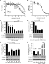
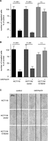
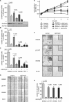
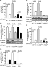
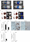
The calcium-binding protein S100A4 is a central mediator of metastasis formation in colon cancer. S100A4 is a target gene of the Wnt/β-catenin pathway, which is constitutively active in the majority of colon cancers. In this study a high-throughput screen was performed to identify small-molecule compounds targeting the S100A4-promoter activity. In this screen calcimycin was identified as a transcriptional inhibitor of S100A4. In colon cancer cells calcimycin treatment reduced S100A4 mRNA and protein expression in a dose- and time-dependent manner. S100A4-induced cellular processes associated with metastasis formation, such as cell migration and invasion, were inhibited by calcimycin in an S100A4-specific manner. Calcimycin reduced β-catenin mRNA and protein levels despite the expression of Δ45-mutated β-catenin. Consequently, calcimycin inhibited Wnt/β-catenin pathway activity and the expression of prominent β-catenin target genes such as S100A4, cyclin D1, c-myc, and dickkopf-1. Finally, calcimycin treatment of human colon cancer cells inhibited metastasis formation in xenografted immunodeficient mice. Our results demonstrate that targeting the expression of S100A4 with calcimycin provides a functional strategy to restrict cell motility in colon cancer cells. Therefore calcimycin may be useful for studying S100A4 biology, and these studies may serve as a lead for the development of treatments for colon cancer metastasis.
Figures

Similar articles
-
Novel effect of antihelminthic Niclosamide on S100A4-mediated metastatic progression in colon cancer.J Natl Cancer Inst. 2011 Jul 6;103(13):1018-36. doi: 10.1093/jnci/djr190. Epub 2011 Jun 17. J Natl Cancer Inst. 2011. PMID: 21685359
-
Intervening in β-catenin signaling by sulindac inhibits S100A4-dependent colon cancer metastasis.Neoplasia. 2011 Feb;13(2):131-44. doi: 10.1593/neo.101172. Neoplasia. 2011. PMID: 21403839 Free PMC article.
-
The metastasis-associated gene S100A4 is a novel target of beta-catenin/T-cell factor signaling in colon cancer.Gastroenterology. 2006 Nov;131(5):1486-500. doi: 10.1053/j.gastro.2006.08.041. Epub 2006 Aug 22. Gastroenterology. 2006. PMID: 17101323
-
Wnt up your mind - intervention strategies for S100A4-induced metastasis in colon cancer.Gen Physiol Biophys. 2009;28 Spec No Focus:F55-64. Gen Physiol Biophys. 2009. PMID: 20093727 Review.
-
S100A4 in Cancer Metastasis: Wnt Signaling-Driven Interventions for Metastasis Restriction.Cancers (Basel). 2016 Jun 20;8(6):59. doi: 10.3390/cancers8060059. Cancers (Basel). 2016. PMID: 27331819 Free PMC article. Review.
Cited by
-
Differential effect of plakoglobin in restoring the tumor suppressor activities of p53-R273H vs. p53-R175H mutants.PLoS One. 2024 Oct 3;19(10):e0306705. doi: 10.1371/journal.pone.0306705. eCollection 2024. PLoS One. 2024. PMID: 39361615 Free PMC article.
-
Systemic shRNA mediated knock down of S100A4 in colorectal cancer xenografted mice reduces metastasis formation.Oncotarget. 2012 Aug;3(8):783-97. doi: 10.18632/oncotarget.572. Oncotarget. 2012. PMID: 22878175 Free PMC article.
-
microRNAs and Corresponding Targets Involved in Metastasis of Colorectal Cancer in Preclinical In Vivo Models.Cancer Genomics Proteomics. 2020 Sep-Oct;17(5):453-468. doi: 10.21873/cgp.20204. Cancer Genomics Proteomics. 2020. PMID: 32859626 Free PMC article. Review.
-
Friend or Foe: S100 Proteins in Cancer.Cancers (Basel). 2020 Jul 24;12(8):2037. doi: 10.3390/cancers12082037. Cancers (Basel). 2020. PMID: 32722137 Free PMC article. Review.
-
Insulin/IGF Axis and the Receptor for Advanced Glycation End Products: Role in Meta-inflammation and Potential in Cancer Therapy.Endocr Rev. 2023 Jul 11;44(4):693-723. doi: 10.1210/endrev/bnad005. Endocr Rev. 2023. PMID: 36869790 Free PMC article. Review.
References
-
- Ambartsumian N, et al. The metastasis-associated Mts1(S100A4) protein could act as an angiogenic factor. Oncogene. 2001;20:4685–4695. - PubMed
-
- Ambartsumian NS, Grigorian MS, Larsen IF, Karlstrom O, Sidenius N, Rygaard J, Georgiev G, Lukanidin E. Metastasis of mammary carcinomas in GRS/A hybrid mice transgenic for the mts1 gene. Oncogene. 1996;13:1621–1630. - PubMed
-
- Barker N, Clevers H. Mining the Wnt pathway for cancer therapeutics. Nat Rev Drug Discov. 2006;5:997–1014. - PubMed
Publication types
MeSH terms
Substances
LinkOut - more resources
Full Text Sources
Other Literature Sources
Molecular Biology Databases
Research Materials

