. 2011 Oct;85(19):10319-31.
doi: 10.1128/JVI.00787-11. Epub 2011 Jul 27.

Enterovirus 71 and coxsackievirus A16 3C proteases: binding to rupintrivir and their substrates and anti-hand, foot, and mouth disease virus drug design

Affiliations

Enterovirus 71 and coxsackievirus A16 3C proteases: binding to rupintrivir and their substrates and anti-hand, foot, and mouth disease virus drug design

Guangwen Lu et al. J Virol. 2011 Oct.

Abstract

Enterovirus 71 (EV71) and coxsackievirus A16 (CVA16) are the major causative agents of hand, foot, and mouth disease (HFMD), which is prevalent in Asia. Thus far, there are no prophylactic or therapeutic measures against HFMD. The 3C proteases from EV71 and CVA16 play important roles in viral replication and are therefore ideal drug targets. By using biochemical, mutational, and structural approaches, we broadly characterized both proteases. A series of high-resolution structures of the free or substrate-bound enzymes were solved. These structures, together with our cleavage specificity assay, well explain the marked substrate preferences of both proteases for particular P4, P1, and P1' residue types, as well as the relative malleability of the P2 amino acid. More importantly, the complex structures of EV71 and CVA16 3Cs with rupintrivir, a specific human rhinovirus (HRV) 3C protease inhibitor, were solved. These structures reveal a half-closed S2 subsite and a size-reduced S1' subsite that limit the access of the P1' group of rupintrivir to both enzymes, explaining the reported low inhibition activity of the compound toward EV71 and CVA16. In conclusion, the detailed characterization of both proteases in this study could direct us to a proposal for rational design of EV71/CVA16 3C inhibitors.


Figures

Fig. 1.
Fig. 1.
Overview of the domain organization within the viral polyproteins of EV71 and CVA16. The single ORF of EV71 or CVA16 could be further divided into three regions (P1, P2, and P3). The four structural subunits encoded by the P1 region and the seven nonstructural proteins encoded by the P2 and P3 regions are colored cyan, light pink, and gray, respectively. The eight cleavage junction sites that are to be processed by the 3C protease are indicated, with each joining sequence spanning from the P10 to P6′ residues listed above or below the schematic boxes representing viral protein subunits Vp4 to 3D. The scissor bonds are highlighted in red.
Fig. 2.
Fig. 2.
Overall structures of the 3C proteases from CVA16 and EV71. (A) Cartoon representation of the structure of CVA16 3C. Domain I (strands aI to gI) and domain II (strands aII to iII) are colored green and magenta, respectively. The α-helices are marked A to D according to their occurrence along the primary structure. The long loop over the “rear” surface connecting domains I and II is highlighted in blue. The catalytic triad, which is composed of H40, E71, and C147, is shown as cyan sticks. The N and C termini are indicated. (B) Structure of the C147A mutant enzyme from EV71 (EV71-C147A 3C). The structural elements involved are colored and labeled as in the CVA16 3C protease. The extra strand that is located next to strand fI of the protease molecule is indicated. (C) In the solved structure of EV71-C147A 3C, residues KPVLRTA (orange) of the extra strand could form 6 hydrogen bonds with E61, V63, and R134 of the parental (green) protease molecule and could form 5 H-bonds with D99, S111, M112, and F113 of the neighboring (magenta) enzyme molecule. The dashed lines indicate H bonds, and the residues involved are labeled. (D) Superimposition of the 3Cs from CVA16 (cyan) and EV71 (orange). Alignment of the two proteases yields an RMSD of 0.365 Å for all equivalent Cα pairs, demonstrating strong similarities between the enzymes. The well-aligned catalytic triads are highlighted. (E) Superimposition of the free-enzyme structures (cyan for CVA16 3C, orange for EV71 3C) in this study on a recently reported 3-Å unliganded structure of EV71 3C (green). The substrate-recognizing β-ribbon, which exhibits major differences, and the well-aligned cII and fII strands are highlighted.
Fig. 3.
Fig. 3.
Noncovalent complex structure of CVA16-C147A 3C with peptide SP-1 (“P” portion). (A) Surface representation to highlight the substrate binding groove of the protease. The peptide, shown in stick form is contoured for an electron density at 1.0 σ above the mean using the 2|Fo|−|Fc| map. (B) Detailed interaction between the peptide and the protease. The residues involved are labeled. Hydrogen bonds are shown as dashed lines.
Fig. 4.
Fig. 4.
Chemical structure of rupintrivir. Overall, rupintrivir could be described as a peptide-mimic inhibitor containing a lactam at P1, a fluorophenylalanine at P2, a Val at P3, a 5-methyl-3-isoxazole at P4, and an α,β-unsaturated ester at P1′. The five groups (P4 to P1′) of the inhibitor are indicated.
Fig. 5.
Fig. 5.
Rupintrivir exhibits the same mode of binding to the EV71 and CVA16 3C proteases. (A and B) Overall structure of the enzyme-inhibitor complex. The structures of rupintrivir bound to the 3C proteases from CVA16 and EV71 are shown in panels A and B, respectively. Rupintrivir is in orange, and Cys147 is cyan. The covalent bond formed between the inhibitor and the protease is highlighted with an arrow. The inhibitor is shown in stick representation with a 1.0-σ contoured map. There are 8 molecules per asymmetric unit in the structure of rupintrivir bound to EV71 3C. These molecules have essentially the same structure, and molecule A was selected for depiction or comparison. (C) Superimposition of the two complex structures reveals the same binding mode of rupintrivir to the EV71 (green) and CVA16 (cyan) 3C proteases. The inhibitor moieties in the EV71 and CVA16 enzymes are shown as orange and magenta sticks, respectively.
Fig. 6.
Fig. 6.
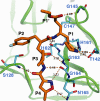
Detailed interactions between rupintrivir and CVA16 3C. H-bond interactions are shown as dashed lines. The five groups of the inhibitor are labeled P1′ to P4 and colored orange. The residues in the enzyme that interact with rupintrivir are shown as thin cyan sticks and are labeled.
Fig. 7.
Fig. 7.
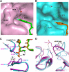
Rupintrivir is not as well accommodated in CVA16 3C as in HRV 3C. (A) Comparison of rupintrivir in CVA16 (magenta) and HRV (cyan) 3C proteases. The 1.7-Å uplift of the P2 group and the tilted P1′ ester chain of the inhibitor in CVA16 3C relative to those of the compound in HRV 3C are indicated. (B) Contributions of the P1′ group to the enzyme-inhibitor interaction. Two H bonds are formed between the ester group and the protein when the compound binds to HRV 3C (magenta), whereas only one is observed in the case of CVA16 3C (cyan). The inhibitor moiety in the CVA16 and HRV enzymes is shown as green and orange sticks, respectively.
Fig. 8.
Fig. 8.
The half-closed S2 subsite and an S1′ pocket with reduced size in CVA16 3C. (A and B) Surface representation of the S2 subsites in the CVA16 (A) and HRV (B) 3C proteases. The residues referred to in the text are presented as thin sticks and are labeled. The P2 groups of rupintrivir in the CVA16 and HRV enzymes are shown as green and orange sticks, respectively. The dashed lines indicate H-bond or polar interactions. The red balls represent water molecules. (C) Superimposition of the 3C proteases from CVA16 (magenta) and HRV (cyan) at the S1′ subsite. The residues constituting the specificity pocket in the respective proteases are indicated. The 0.9-Å shift observed for the main-chain carbonyl of His24 in CVA16 3C relative to that of the corresponding residue (Lys24) in the rhinoviral protease is highlighted. The P1′ group of rupintrivir, which exhibits great conformational differences in the respective proteases, is shown in stick representation (green for rupintrivir in CVA16-3C and orange for the compound in HRV-3C). (D) Structural features leading to the reduced size of the CVA16 3C S1′ subsite. The residues involved and the aII-bII loop (the loop connecting the aII and bII strands) are indicated.
Fig. 9.
Fig. 9.
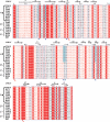
Multiple-sequence alignment of the 3C proteases from human enteroviruses A to D compared to that of HRV 3C. The spiral lines indicate α-helices, and the horizontal arrows represent β-strands. Residue numbers for CVA16 3C are given above the sequence. The catalytic triad is indicated by red arrows. The residues comprising the half-closed S2 subsite and the smaller S1′ pocket in CVA16 3C are marked with blue triangles and are colored cyan to highlight their conservation among the human enterovirus group A viral proteases. The four species (human enteroviruses A to D) to which the enteroviruses belong are labeled on the left as A, B, C, and D, respectively. CVA, human coxsackievirus A; CVB, human coxsackievirus B; EV, human enterovirus; HRV, human rhinovirus 2. Strain selections were as follows: CVA16, Beijing0907; EV71, Anhui1-09-China; CVA4, CA4; CVA5, Swartz; CVA8, Donovan; CVA10, Kowalik; EV90, F950027; EV91, BAN00-10406; EV92, RJG7; CVB2, OH; CVB3, 28; CVB5, 2000/CSF/KOR; CVA13, Flores; CVA15, G-9; CVA17, BAN01-10577; EV68, Fermon; and EV94, 19/04.

Similar articles

Cited by

References

    1. Adams P. D., et al. 2010. PHENIX: a comprehensive Python-based system for macromolecular structure solution. Acta Crystallogr. D Biol. Crystallogr. 66:213–221 - PMC - PubMed
    1. Alexander J. P., Jr., Baden L., Pallansch M. A., Anderson L. J. 1994. Enterovirus 71 infections and neurologic disease—United States, 1977–1991. J. Infect. Dis. 169:905–908 - PubMed
    1. Anand K., et al. 2002. Structure of coronavirus main proteinase reveals combination of a chymotrypsin fold with an extra alpha-helical domain. EMBO J. 21:3213–3224 - PMC - PubMed
    1. Berger A., Schechter I. 1970. Mapping the active site of papain with the aid of peptide substrates and inhibitors. Phil. Trans. R. Soc. Lond. B Biol. Sci. 257:249–264 - PubMed
    1. Bible J. M., et al. 2008. Molecular epidemiology of human enterovirus 71 in the United Kingdom from 1998 to 2006. J. Clin. Microbiol. 46:3192–3200 - PMC - PubMed

Publication types

MeSH terms

LinkOut - more resources