

Activation of G protein-coupled estrogen receptor induces endothelium-independent relaxation of coronary artery smooth muscle
- PMID: 21791623
- PMCID: PMC3213995
- DOI: 10.1152/ajpendo.00037.2011
Activation of G protein-coupled estrogen receptor induces endothelium-independent relaxation of coronary artery smooth muscle
Erratum in
- Am J Physiol Endocrinol Metab. 2012 Dec 15;303(12):E1502-3
Abstract
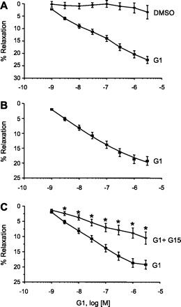
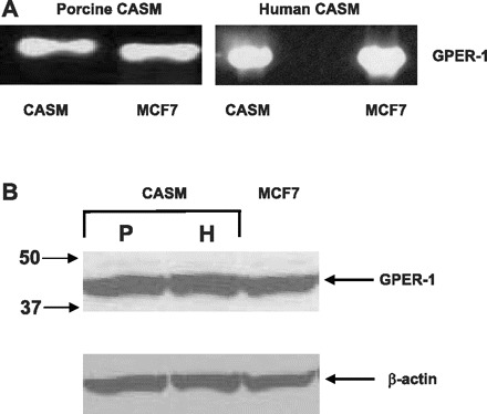
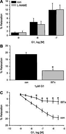
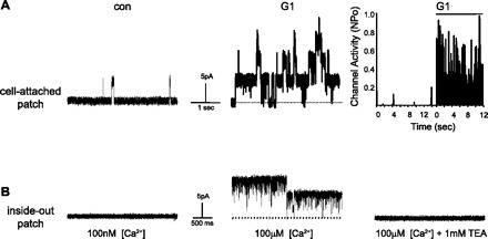
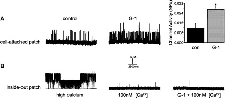
Estrogens can either relax or contract arteries via rapid, nongenomic mechanisms involving classic estrogen receptors (ER). In addition to ERα and ERβ, estrogen may also stimulate G protein-coupled estrogen receptor 1 (GPER) in nonvascular tissue; however, a potential role for GPER in coronary arteries is unclear. The purpose of this study was to determine how GPER activity influenced coronary artery reactivity. In vitro isometric force recordings were performed on endothelium-denuded porcine arteries. These studies were augmented by RT-PCR and single-cell patch-clamp experiments. RT-PCR and immunoblot studies confirmed expression of GPER mRNA and protein, respectively, in smooth muscle from either porcine or human coronary arteries. G-1, a selective GPER agonist, produced a concentration-dependent relaxation of endothelium-denuded porcine coronary arteries in vitro. This response was attenuated by G15, a GPER-selective antagonist, or by inhibiting large-conductance calcium-activated potassium (BK(Ca)) channels with iberiotoxin, but not by inhibiting NO signaling. Last, single-channel patch-clamp studies demonstrated that G-1 stimulates BK(Ca) channel activity in intact smooth muscle cells from either porcine or human coronary arteries but had no effect on channels isolated in excised membrane patches. In summary, GPER activation relaxes coronary artery smooth muscle by increasing potassium efflux via BK(Ca) channels and requires an intact cellular signaling mechanism. This novel action of estrogen-like compounds may help clarify some of the controversy surrounding the vascular effects of estrogens.
Figures
Similar articles
-
Essential role of the 90-kilodalton heat shock protein in mediating nongenomic estrogen signaling in coronary artery smooth muscle.J Pharmacol Exp Ther. 2009 Jun;329(3):850-5. doi: 10.1124/jpet.108.149112. Epub 2009 Mar 17. J Pharmacol Exp Ther. 2009. PMID: 19293389 Free PMC article.
-
Adaptive increases in expression and vasodilator activity of estrogen receptor subtypes in a blood vessel-specific pattern during pregnancy.Am J Physiol Heart Circ Physiol. 2015 Nov 15;309(10):H1679-96. doi: 10.1152/ajpheart.00532.2015. Epub 2015 Sep 25. Am J Physiol Heart Circ Physiol. 2015. PMID: 26408543 Free PMC article.
-
Endothelium-derived nitric oxide inhibits the relaxation of the porcine coronary artery to natriuretic peptides by desensitizing big conductance calcium-activated potassium channels of vascular smooth muscle.J Pharmacol Exp Ther. 2010 Jul;334(1):223-31. doi: 10.1124/jpet.110.166652. Epub 2010 Mar 23. J Pharmacol Exp Ther. 2010. PMID: 20332186
-
G-protein-coupled estrogen receptor as a new therapeutic target for treating coronary artery disease.World J Cardiol. 2014 Jun 26;6(6):367-75. doi: 10.4330/wjc.v6.i6.367. World J Cardiol. 2014. PMID: 24976908 Free PMC article. Review.
-
The G protein-coupled estrogen receptor GPER/GPR30 as a regulator of cardiovascular function.Vascul Pharmacol. 2011 Jul-Sep;55(1-3):17-25. doi: 10.1016/j.vph.2011.06.003. Epub 2011 Jul 5. Vascul Pharmacol. 2011. PMID: 21742056 Free PMC article. Review.
Cited by
-
Activation of GPER Induces Differentiation and Inhibition of Coronary Artery Smooth Muscle Cell Proliferation.PLoS One. 2013 Jun 19;8(6):e64771. doi: 10.1371/journal.pone.0064771. Print 2013. PLoS One. 2013. PMID: 23840305 Free PMC article.
-
Bradycardic effects mediated by activation of G protein-coupled estrogen receptor in rat nucleus ambiguus.Exp Physiol. 2013 Mar;98(3):679-91. doi: 10.1113/expphysiol.2012.069377. Epub 2012 Oct 26. Exp Physiol. 2013. PMID: 23104934 Free PMC article.
-
Diabetes impairs the vascular effects of aldosterone mediated by G protein-coupled estrogen receptor activation.Front Pharmacol. 2015 Mar 2;6:34. doi: 10.3389/fphar.2015.00034. eCollection 2015. Front Pharmacol. 2015. PMID: 25784875 Free PMC article.
-
Cycling matters: Sex hormone regulation of vascular potassium channels.Channels (Austin). 2023 Dec;17(1):2217637. doi: 10.1080/19336950.2023.2217637. Channels (Austin). 2023. PMID: 37243715 Free PMC article. Review.
-
G protein-coupled estrogen receptor agonist improves cerebral microvascular function after hypoxia/reoxygenation injury in male and female rats.Stroke. 2013 Mar;44(3):779-85. doi: 10.1161/STROKEAHA.112.678177. Epub 2013 Jan 29. Stroke. 2013. PMID: 23362079 Free PMC article.
References
-
- Canonaco M, Giusi G, Madeo A, Facciolo RM, Lappano R, Canonaco A, Maggiolini M. A sexually dimorphic distribution pattern of the novel estrogen receptor G-protein-coupled receptor 30 in some brain areas of the hamster. J Endocrinol 196: 131–138, 2008 - PubMed
-
- Chester AH, Jiang C, Borland JA, Yacoub MH, Collins P. Oestrogen relaxes human epicardial coronary arteries through non-endothelium-dependent mechanisms. Coron Artery Dis 6: 417–422, 1995 - PubMed
-
- Darkow DJ, Lu L, White RE. Estrogen relaxation of coronary artery smooth muscle is mediated by nitric oxide and cGMP. Am J Physiol Heart Circ Physiol 272: H2765–H2773, 1997 - PubMed
Publication types
MeSH terms
Substances
Grants and funding
LinkOut - more resources
Full Text Sources