

Amyloid-related imaging abnormalities in amyloid-modifying therapeutic trials: recommendations from the Alzheimer's Association Research Roundtable Workgroup
- PMID: 21784348
- PMCID: PMC3693547
- DOI: 10.1016/j.jalz.2011.05.2351
Amyloid-related imaging abnormalities in amyloid-modifying therapeutic trials: recommendations from the Alzheimer's Association Research Roundtable Workgroup
Abstract
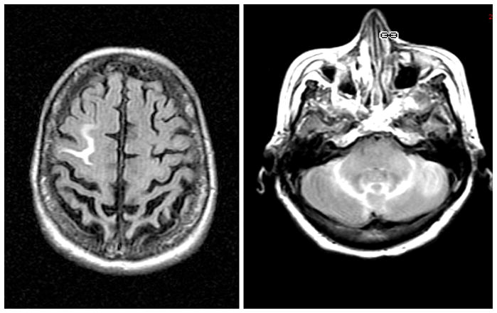
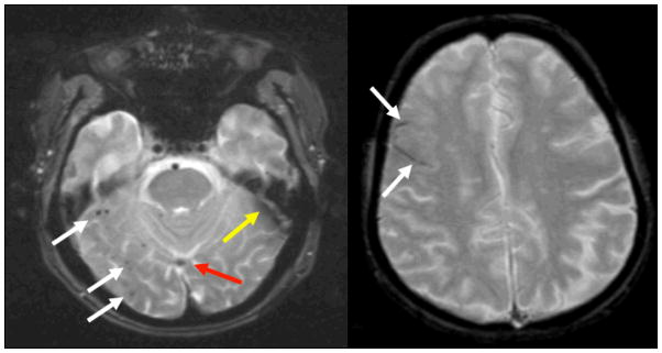
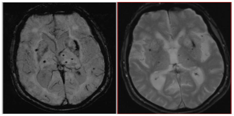
Amyloid imaging related abnormalities (ARIA) have now been reported in clinical trials with multiple therapeutic avenues to lower amyloid-β burden in Alzheimer's disease (AD). In response to concerns raised by the Food and Drug Administration, the Alzheimer's Association Research Roundtable convened a working group to review the publicly available trial data, attempts at developing animal models, and the literature on the natural history and pathology of related conditions. The spectrum of ARIA includes signal hyperintensities on fluid attenuation inversion recoverysequences thought to represent "vasogenic edema" and/or sulcal effusion (ARIA-E), as well as signal hypointensities on GRE/T2* thought to represent hemosiderin deposits (ARIA-H), including microhemorrhage and superficial siderosis. The etiology of ARIA remains unclear but the prevailing data support vascular amyloid as a common pathophysiological mechanism leading to increased vascular permeability. The workgroup proposes recommendations for the detection and monitoring of ARIA in ongoing AD clinical trials, as well as directions for future research.
Copyright © 2011 The Alzheimer's Association. Published by Elsevier Inc. All rights reserved.
Figures


Similar articles
-
MR imaging features of amyloid-related imaging abnormalities.AJNR Am J Neuroradiol. 2013 Oct;34(10):1958-65. doi: 10.3174/ajnr.A3500. Epub 2013 Apr 11. AJNR Am J Neuroradiol. 2013. PMID: 23578674 Free PMC article.
-
Detection and Management of Amyloid-Related Imaging Abnormalities in Patients with Alzheimer's Disease Treated with Anti-Amyloid Beta Therapy.J Prev Alzheimers Dis. 2022;9(2):211-220. doi: 10.14283/jpad.2022.21. J Prev Alzheimers Dis. 2022. PMID: 35542992 Review.
-
The value of subtraction MRI in detection of amyloid-related imaging abnormalities with oedema or effusion in Alzheimer's patients: An interobserver study.Eur Radiol. 2018 Mar;28(3):1215-1226. doi: 10.1007/s00330-017-5022-6. Epub 2017 Sep 27. Eur Radiol. 2018. PMID: 28956123 Free PMC article. Clinical Trial.
-
Amyloid-Related Imaging Abnormalities in the Era of Anti-Amyloid Beta Monoclonal Antibodies for Alzheimer's Disease: Recent Updates on Clinical and Imaging Features and MRI Monitoring.Korean J Radiol. 2024 Aug;25(8):726-741. doi: 10.3348/kjr.2024.0105. Korean J Radiol. 2024. PMID: 39109501 Free PMC article. Review.
-
Chronic Verubecestat Treatment Suppresses Amyloid Accumulation in Advanced Aged Tg2576-AβPPswe Mice Without Inducing Microhemorrhage.J Alzheimers Dis. 2017;59(4):1393-1413. doi: 10.3233/JAD-170056. J Alzheimers Dis. 2017. PMID: 28800329 Free PMC article.
Cited by
-
Amyloid-related imaging abnormalities from trials of solanezumab for Alzheimer's disease.Alzheimers Dement (Amst). 2016 Mar 2;2:75-85. doi: 10.1016/j.dadm.2016.02.004. eCollection 2016. Alzheimers Dement (Amst). 2016. PMID: 27239538 Free PMC article.
-
Molecular biomarkers for vascular cognitive impairment and dementia.Nat Rev Neurol. 2023 Dec;19(12):737-753. doi: 10.1038/s41582-023-00884-1. Epub 2023 Nov 13. Nat Rev Neurol. 2023. PMID: 37957261 Review.
-
Subcortical signal alteration of corticospinal tracts. A radiologic manifestation of ARIA: A case report.Radiol Case Rep. 2022 Nov 6;18(1):275-279. doi: 10.1016/j.radcr.2022.10.023. eCollection 2023 Jan. Radiol Case Rep. 2022. PMID: 36388611 Free PMC article.
-
Prevalence of cerebral amyloid angiopathy: A systematic review and meta-analysis.Alzheimers Dement. 2022 Jan;18(1):10-28. doi: 10.1002/alz.12366. Epub 2021 May 31. Alzheimers Dement. 2022. PMID: 34057813 Free PMC article.
-
Bapineuzumab alters aβ composition: implications for the amyloid cascade hypothesis and anti-amyloid immunotherapy.PLoS One. 2013;8(3):e59735. doi: 10.1371/journal.pone.0059735. Epub 2013 Mar 21. PLoS One. 2013. PMID: 23555764 Free PMC article.
References
-
- Sperling R, Salloway S, Fox N, Barackos J, Morris K, Francis G, et al., editors. Risk Factors and Clinical Course Associated with Vasogenic Edema in a Phase II Trial of Bapineuzumab. American Academy of Neurology; Seattle, Washington: 2009.
-
-
#XX1 RA.
-
-
- Pfeifer M, Boncristiano S, Bondolfi L, Stalder A, Deller T, Staufenbiel M, et al. Cerebral hemorrhage after passive anti-Abeta immunotherapy. Science. 2002 Nov 15;298(5597):1379. - PubMed
Publication types
MeSH terms
Substances
Grants and funding
LinkOut - more resources
Full Text Sources
Other Literature Sources
Medical
