. 2011 Jul 22;43(2):217-28.
doi: 10.1016/j.molcel.2011.06.009.

The ClpS adaptor mediates staged delivery of N-end rule substrates to the AAA+ ClpAP protease

Affiliations

The ClpS adaptor mediates staged delivery of N-end rule substrates to the AAA+ ClpAP protease

Giselle Román-Hernández et al. Mol Cell. .

Abstract

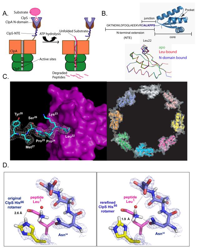
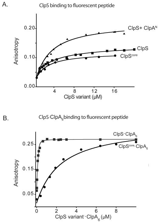
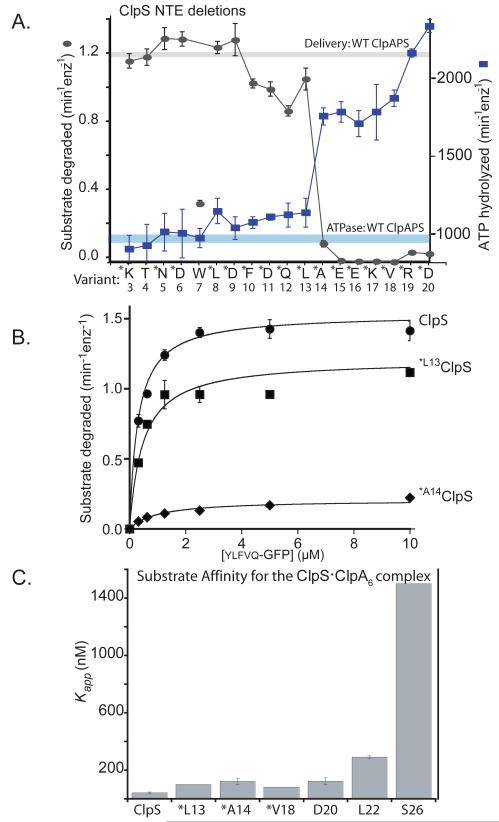
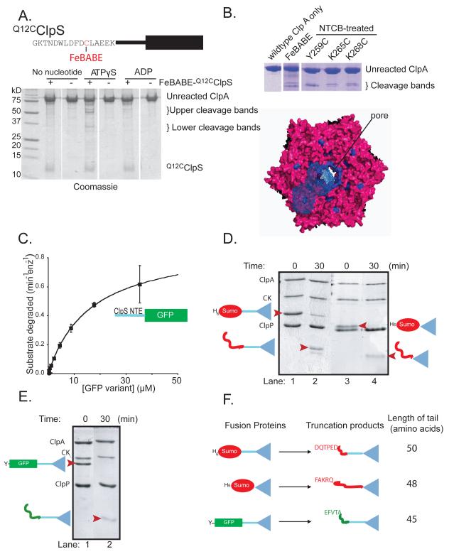
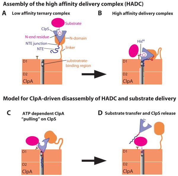
The ClpS adaptor delivers N-end rule substrates to ClpAP, an energy-dependent AAA+ protease, for degradation. How ClpS binds specific N-end residues is known in atomic detail and clarified here, but the delivery mechanism is poorly understood. We show that substrate binding is enhanced when ClpS binds hexameric ClpA. Reciprocally, N-end rule substrates increase ClpS affinity for ClpA(6). Enhanced binding requires the N-end residue and a peptide bond of the substrate, as well as multiple aspects of ClpS, including a side chain that contacts the substrate α-amino group and the flexible N-terminal extension (NTE). Finally, enhancement also needs the N domain and AAA+ rings of ClpA, connected by a long linker. The NTE can be engaged by the ClpA translocation pore, but ClpS resists unfolding/degradation. We propose a staged-delivery model that illustrates how intimate contacts between the substrate, adaptor, and protease reprogram specificity and coordinate handoff from the adaptor to the protease.


Figures

Figure 1
Figure 1. N-end-rule substrate recognition
A) In bacteria, the ClpS adaptor (light blue) recognizes and binds N-end-rule substrates (pink) and delivers them for degradation by the ClpAP protease. B) Top: ClpS has a flexible NTE required for N-end-rule substrate delivery and a folded ClpScore domain, which binds N-end-rule substrates. The ALKPPS sequence at the NTE-core junction is important for adaptor function. Bottom: Backbone Cα superposition (r.m.s.d. < 0.5 Å) of ClpScore (3O1F, green), a peptide-bound ClpS structure (2W9R, red), and ClpS from a complex with the ClpA N domain (1R6O, blue). C) Left panel: In the rerefined 2WA9 structure, the side chain of Leu22 from an adjacent ClpS subunit was bound in the N-end-rule binding pocket, and density (1σ) for Leu22, Lys23, Pro24, and Pro25 was continuous with that for Ser26, Met27, Tyr28, and Lys29, which are part of ClpScore. Right panel: The rerefined map for the 2WA9 structure contained density (1.5 σ) for eight ClpS subunits in the asymmetric unit, arranged head to tail in a ring. The original 2WA9 structure (Schuenemann et al., 2009) had seven ClpS subunits, each with a bound N-end-rule peptide. D) In the rerefined 2W9R structure (right panel), the correct rotamer of the His66 side chain make hydrogen bonds (dashed lines) with the α-NH3 group of the bound N-end peptide and fits nicely into the electron density. In the original 2W9R structure (Schuenemann et al., 2009; left panel), His66 rotamer chosen makes a poor hydrogen bond with the carbonyl oxygen of the first peptide residue and does not fit optimally into the electron density. In both panels, the electron density (1.25σ) is from the our rerefined map.
Figure 2
Figure 2. N-end-rule degrons bind more tightly to the ClpS-ClpA6 complex
A) A fluorescent N-end-rule peptide (LLYVQRDSKEC-fl; 200 nM) was bound with similar affinities (KD ~3 μM) by ClpS, by ClpScore, and by ClpS in complex with the ClpA N domain, as assayed by changes in anisotropy. The molecular weights and maximum anisotropies of each complex differ. B) Increasing concentrations of 1:1 molar mixtures of ClpA6 and ClpS or ClpScore were titrated against the LLYVQRDSKEC-fl peptide (100 nM). The ClpS-ClpA6 complex bound more tightly (Kapp = 42 ± 6 nM) than the ClpScore-ClpA6 complex (Kapp = 1.5 ± 0.25 μM), demonstrating that the ClpS NTE is required for affinity enhancement. Assays contained 4 mM ATPγS to promote ClpA hexamer formation.
Figure 3
Figure 3. ClpS binds ClpA6 more tightly in the presence of N-end-rule peptides
A) As assayed by anisotropy, ClpA6 bound 200 nM fluorescent ClpS*F tightly in the presence 20 μM Trp-conh2, LLYVQRDSKEC, or FV N-end-rule peptides (Kapp < 20 nM) and more weakly in the absence of peptide or with 20 μM MLYVQRDSKEC peptide (Kapp ~ 180 nM). B) The H66 residue of ClpS is one of the side chains involved in the formation of one of the three hydrogen bonds that the adaptor forms with the α-amino group of the N-end degron. Overlay of the apo (green, PDB 3O1F) and the peptide-bound (blue, 2W9R) crystal structures of ClpS reveal no major global changes occur upon peptide binding. The most substantial change is the rotation of the H66 side chain, which appear to need to move away from the pocket in the apo form to accommodate the N-degron in the peptide binding site. C) ClpA6 bound ClpS*F (KD = 200 ± 6 nM) and H66AClpS*F (KD = 345 ± 3 nM) with similar affinities. Addition of 20 μM LLYVQRDSKEC N-end-rule peptide enhanced ClpA6 affinity for ClpS*F substantially (Kapp = 20 ± 10 nM) but increased affinity for H66AClpS*F only modestly (Kapp = 178 ± 4 nM). D) H66AClpS-ClpA6 complex binds more weakly to an N-end-rule fluorescent peptide (LLYVQRDSKEC-fl) when compared to ClpS-ClpA6 (Kapp = 560 nM vs Kapp = 42 ± 6 nM for wild type). E) An N-end-rule peptide (LLYVQRDSKEC; 20 μM) enhanced binding of ClpS*F to ClpA6 but not to ΔLClpA6, which has shorter linker between the N and D1 domains (Cranz-Mileva et al., 2008). F) Michaelis-Menten plots showed that substituting ΔLClpA6 (4-residue linker) for ClpA6 (26-residue linker) decreased KM and Vmax for ClpAPS degradation (100 nM ClpA6 or ΔLClpA6; 200 nm ClpP14; 600 nm ClpS) of the N-end-rule substrate ylfvqela-GFP.
Figure 4
Figure 4. A minimal NTE length is required for ClpS function
A) ClpS variants (1 μM) with N-terminal truncations were assayed for delivery of ylfvqela-GFP (1 μM) for ClpAP degradation (gray curve) and for effects on ClpAP ATP hydrolysis (blue curve) using 100 nM ClpA6 and 270 nM ClpP14 for both assays. Data points represent averages (n=3) ± 1 SD. Each ClpS variant is named by the first wild-type residue in the construct. Those marked with an asterisk contain an additional N-terminal methionine and are therefore one residue longer than the labels indicate; these mutants were expressed as SUMO-fusion proteins and cleaved in vitro (see Experimental Procedures) or were expressed as standard non-fusion proteins but retained the initiator Met (verified by mass spectrometry). The T4 and W7 variants were also expressed as standard non-fusion proteins but mass spectrometry and/or N-terminal sequencing showed that the initiator Met was removed from both of these proteins. Note the sharp activity transitions between *L13 ClpS (starting Met12Leu13) and *A14 ClpS (starting Met13Ala14). The processing of the W7 variant is inconsistent with canonical methionine aminopeptidase activity and generates a good N-end-rule residue, which may be responsible for the poor activity of this ClpS variant in delivering other N-end-rule substrates. B) Michaelis-Menten plots of ylfvqela-GFP degradation by ClpAP and ClpS or variants (100 nM ClpA6; 200 nm ClpP14; 600 nm ClpS or variants). Wild-type ClpS and *L13 ClpS (beginning Met12Leu13Ala14) supported roughly similar steady-state degradation kinetics, but delivery by *A14 ClpS (beginning Met13Ala14Glu15) resulted in a substantial decrease in Vmax. Thus, the NTE must have a critical minimal length to support efficient substrate delivery. The solid lines are a global fit to a model in which the ClpS-substrate complex binds ClpA in an initial bimolecular step (K1 = 1.1 μM) and then is engaged for degradation in a second unimolecular step (K2), which depends on NTE length. In this model, apparent Vmax = Etotalkdeg/(1+K2) and apparent KM = K1K2/(1+K2). For the fits shown, the kdeg value was 2.1 min−1 and the K2 values were 0.37 (wild-type ClpS), 0.74 (*L13 ClpS), and 9.2 (*A14 ClpS). C) Binding to an N-end-rule peptide (LLYVQRDSKEC-fl; 150 nM) by complexes of ClpA6 with ClpS variants (1 ClpS per ClpA6) showed that ClpS junction residues are important for formation of the high-affinity delivery complex. Variants marked * have an additional N-terminal methionine. Apparent affinity constants were 43 nM (wild-type ClpS), 100 nM (*L13), 130 nM (*A14), 83 nM (*V18), 130 nM (D20), 290 nM (L22), and 1500 nM (S26).
Figure 5
Figure 5. The ClpS NTE contacts ClpA near the axial pore
A) Top: FeBABE was attached to residue 12 of Q12CClpS variant for cleavage studies. Bottom: As assayed by SDS-PAGE, cleavage of ClpA required FeBABE-modified Q12CClpS and ATPγS. B) ClpA residues 259-268 are highlighted in blue in a top view of a model of the hexameric D1 ring (Guo et al., 2002). In one ClpA subunit, blue-wire shading shows regions within 12 Å of residues 259-268, which represents the approximate reach of the tethered FeBABE. C) A substrate consisting of residues 2-26 of ClpS fused to GFP was efficiently degraded by ClpAP, as shown by Michaelis-Menten analysis (KM = 16.4 μM; Vmax = 0.62 min−1 enz−1). D) Assays monitored by SDS-PAGE showed that ClpAP only partially degraded the H6-Sumo-ClpS and H6-Sumo-ClpScore fusion proteins, resulting in truncated products of a lower molecular weight (marked by red arrowheads in lanes 2 & 4). E) ClpAP partially degraded the ylfvqela-GFP-ClpS fusion protein, resulting in a lower molecular weight truncation product (marked by a red arrowhead in lane 2). F) Depiction of the ClpS fusion proteins used to test degradation by ClpAP (left) and the corresponding truncation products produced by degradation (right). N-terminal sequencing of the truncation products revealed that the new N-termini corresponded to an internal sequence in the protein fused to ClpS (either Sumo or GFP). The truncation products consisted of the ClpS core and an additional N-terminal tail of 45-50 amino acids.
Figure 6
Figure 6. Model for staged delivery of N-end-rule substrates
A) Independent binding of ClpS to the ClpA N domain and of the substrate N-end residue in the ClpS pocket results in a low-affinity ternary complex. B) A high-affinity delivery complex is stabilized by additional interactions between the D1 ring of ClpA and NTE-junction residues and between the D1 ring, the His66 side chain of ClpS, and the N-end residue of the substrate. C) Translocation-mediated ClpA tugging on the NTE distorts the ClpScore structure, weakens ClpS interactions with the N-end residue, and facilitates transfer of the N-degron of the substrate to a site in the ClpA pore. D) ClpS slips from the grasp of ClpA, clearing the pore and allowing subsequent degradation of the N-end-rule substrate.

Similar articles

Cited by

References

    1. Adams PD, Grosse-Kunstleve RW, Hung LW, Ioerger TR, McCoy AJ, Moriarty NW, Read RJ, Sacchettini JC, Sauter NK, Terwilliger TC. PHENIX: building new software for automated crystallographic structure determination. Acta Crystallogr D Biol Crystallogr. 2002;58:1948–1954. - PubMed
    1. Bachmair A, Finley D, Varshavsky A. In vivo half-life of a protein is a function of its amino-terminal residue. Science. 1986;234:179–186. - PubMed
    1. Baker TA, Sauer RT. ATP-dependent proteases of bacteria: recognition logic and operating principles. Trends Biochem Sci. 2006;31:647–653. - PMC - PubMed
    1. Bolon DN, Grant RA, Baker TA, Sauer RT. Nucleotide-dependent substrate handoff from the SspB adaptor to the AAA+ ClpXP protease. Mol Cell. 2004;16:343–350. - PubMed
    1. Burton RE, Siddiqui SM, Kim YI, Baker TA, Sauer RT. Effects of protein stability and structure on substrate processing by the ClpXP unfolding and degradation machine. EMBO J. 2001;20:3092–100. - PMC - PubMed

Publication types

MeSH terms

LinkOut - more resources