. 2011 Jul 22;43(2):192-202.
doi: 10.1016/j.molcel.2011.06.019.

ATR autophosphorylation as a molecular switch for checkpoint activation

Affiliations

ATR autophosphorylation as a molecular switch for checkpoint activation

Shizhou Liu et al. Mol Cell. .

Abstract

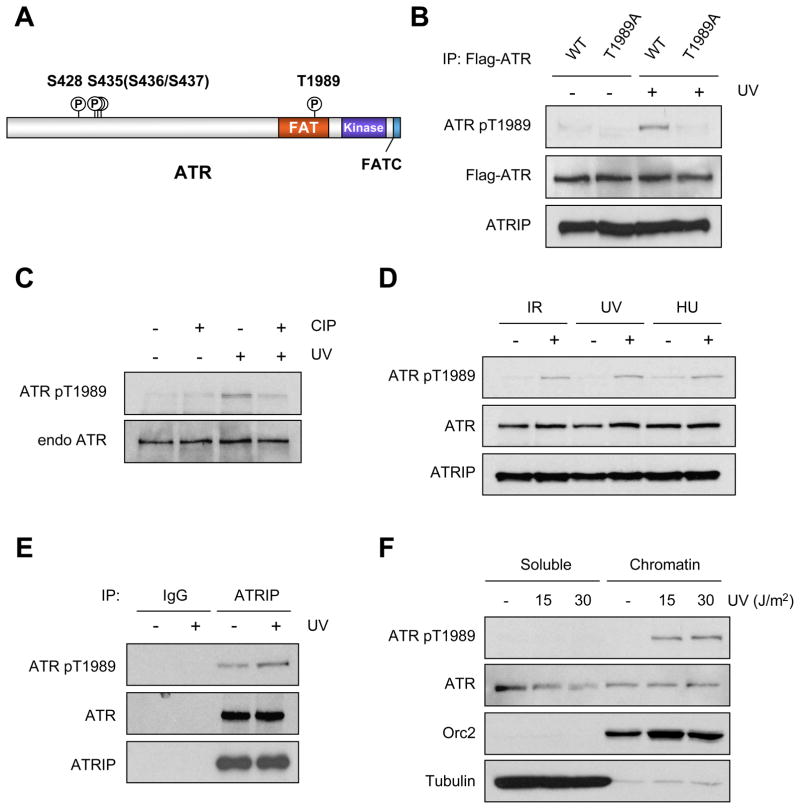
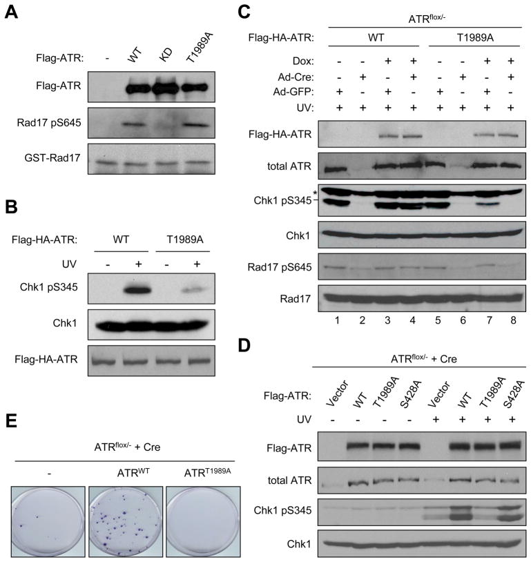
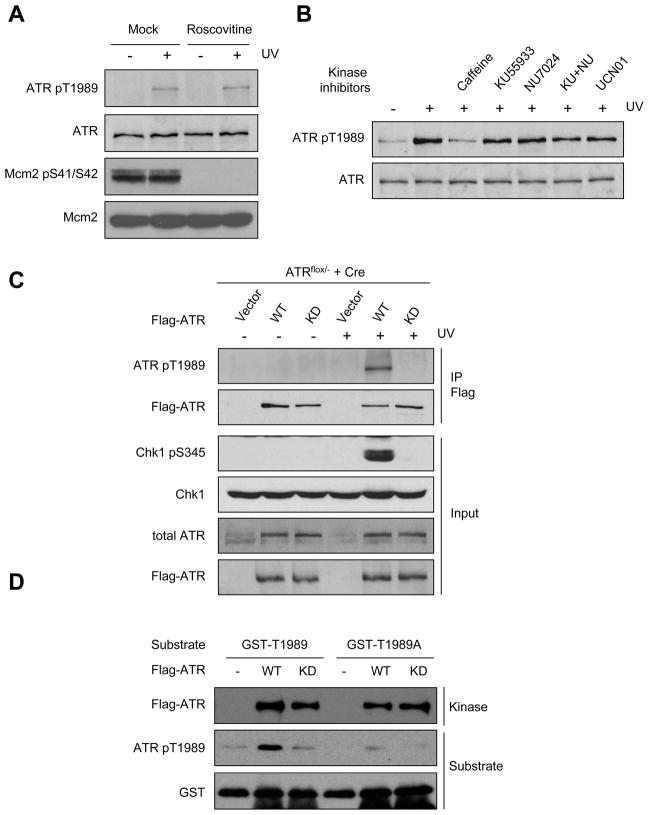
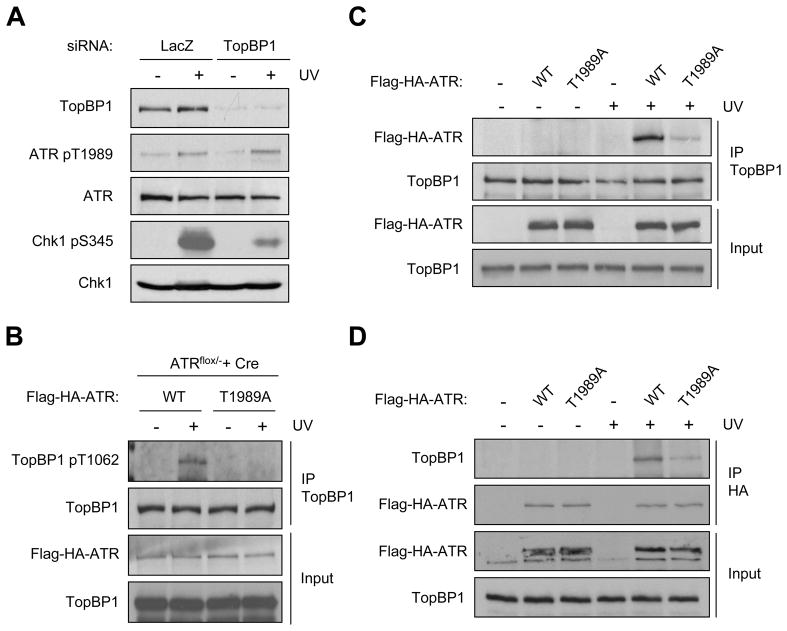
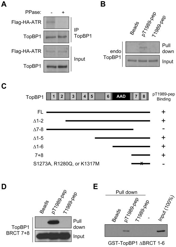
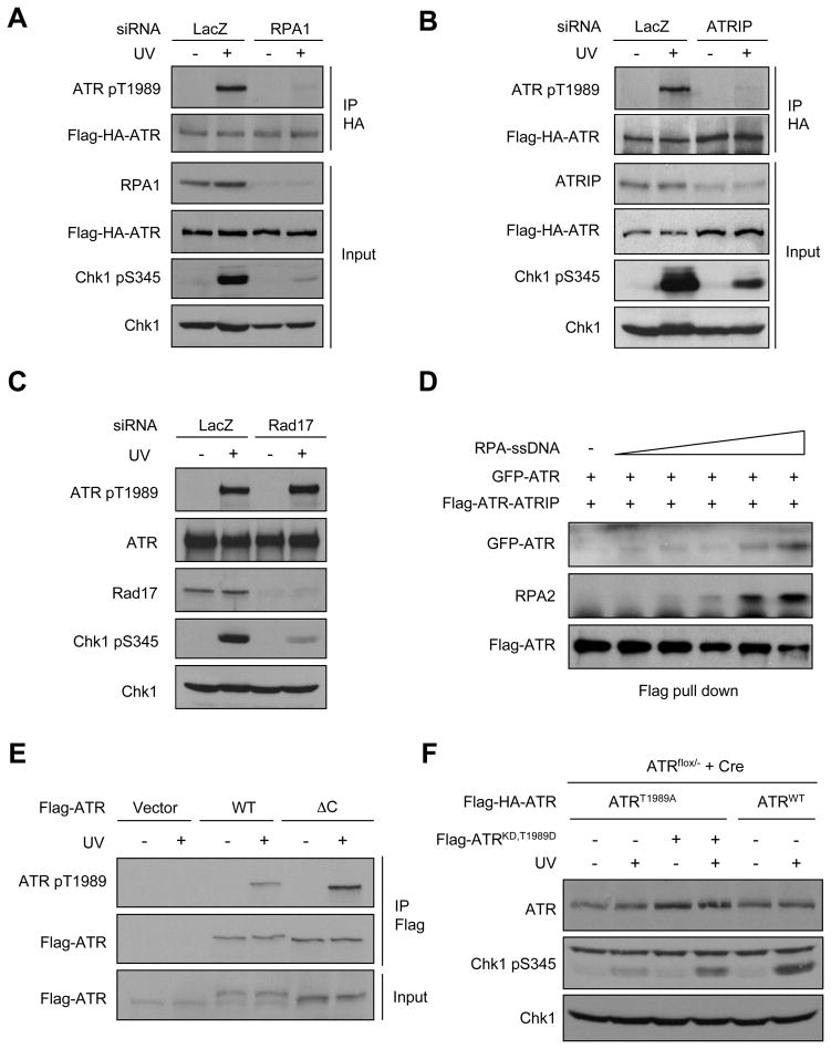
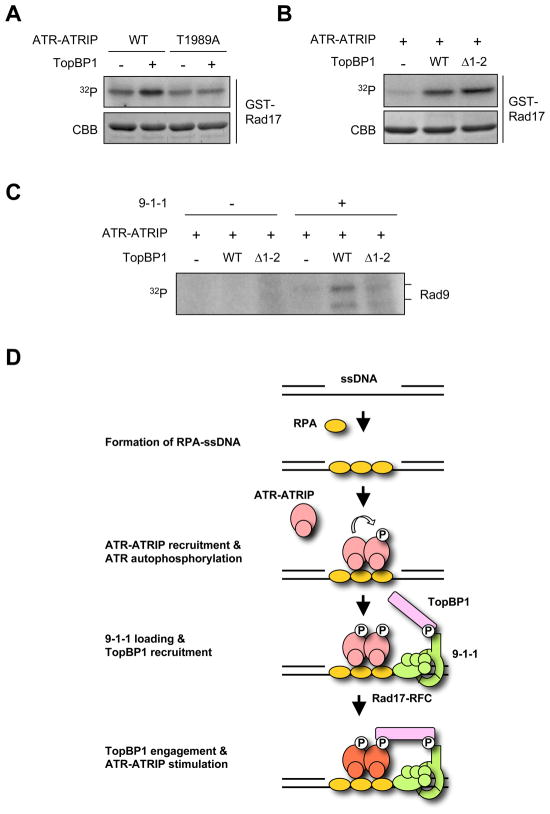
The ataxia telangiectasia-mutated and Rad3-related (ATR) kinase is a master checkpoint regulator safeguarding the genome. Upon DNA damage, the ATR-ATRIP complex is recruited to sites of DNA damage by RPA-coated single-stranded DNA and activated by an elusive process. Here, we show that ATR is transformed into a hyperphosphorylated state after DNA damage, and that a single autophosphorylation event at Thr 1989 is crucial for ATR activation. Phosphorylation of Thr 1989 relies on RPA, ATRIP, and ATR kinase activity, but unexpectedly not on the ATR stimulator TopBP1. Recruitment of ATR-ATRIP to RPA-ssDNA leads to congregation of ATR-ATRIP complexes and promotes Thr 1989 phosphorylation in trans. Phosphorylated Thr 1989 is directly recognized by TopBP1 via the BRCT domains 7 and 8, enabling TopBP1 to engage ATR-ATRIP, to stimulate the ATR kinase, and to facilitate ATR substrate recognition. Thus, ATR autophosphorylation on RPA-ssDNA is a molecular switch to launch robust checkpoint response.


Figures

Figure 1
Figure 1. ATR is phosphorylated at Thr 1989 after DNA damage
A,. Schematics of the phosphorylation sites of ATR. Note that mass spectrometry data only identified Ser 436 and S437 as potential phosphorylation sites. B, 293T cells transiently expressing Flag-ATRWT or Flag-ATRT1989A were treated with 50 J/m2 of UV or left untreated, and subjected to immunoprecipitation with anti-Flag antibody in 2h. The levels of precipitated ATR, ATR pT1989, and ATRIP were analyzed using the indicated antibodies. C, Endogenous ATR was immunoprecipitated from HCT116 cell extracts that were treated with calf intestinal phosphatase (CIP) or mock treated. D, HCT116 cells were treated with 10 Gy of IR, 50 J/m2 of UV, or 2 mM HU, and the phosphorylation of endogenous ATR was analyzed 2 h after the treatment. E, Endogenous ATRIP was immunoprecipitated from HCT116 cell extracts. The levels of precipitated ATRIP, ATR, and ATR pT1989 were analyzed by Western blot. F, HCT116 cells were treated with UV or left untreated, and extracts were separated into soluble and chromatin fractions in 2 h. The levels of ATR and ATR pT1989 in these fractions were analyzed by Western blot. Orc2 and tubulin serve as markers of chromatin and soluble fractions, respectively.
Figure 2
Figure 2. Thr 1989 is critical for ATR function
A, The kinase activities of Flag-ATRWT, Flag-ATRKD, and Flag-ATRT1989A were tested in vitro using GST-Rad17 as a substrate. The phosphorylation of Rad17 S645 was analyzed using phospho-specific antibody. B, Flag-HA-ATRWT and Flag-HA-ATRT1989A were inducibly expressed in HCT116-derived stable cell lines. Cells were treated with 15 J/m2 of UV, and the effects of ATRWT and ATRT1989A on Chk1 phosphorylation were analyzed in 2 h. Flag-HA-ATR was detected with the HA antibody. C, ATRflox/−-derivative lines were infected with Ad-Cre to delete endogenous ATR, or infected with Ad-GFP as controls. Where indicated, cells were treated with Dox to induce Flag-HA-ATRWT or Flag-HA-ATRT1989A. The phosphorylation of Chk1 and Rad17 were analyzed using phospho-specific antibodies. Total ATR includes both endogenous and exogenous ATR. *, a band cross-reacting to the phospho-Chk1 antibody. D, ATRflox/− cells were treated with Ad-Cre and transfected with plasmids expressing Flag-ATRWT, Flag-ATRT1989A, or Flag-ATRS428A. E, ATRflox/−-derivative lines were infected with Ad-Cre and treated with Dox as in C. The ability of ATRWT and ATRT1989A to support colony formation was analyzed.
Figure 3
Figure 3. ATR autophosphorylates Thr 1989
A, HCT116 cells were treated with 50 μM roscovitine for 12 h and then irradiated with 15 J/m2 UV. The phosphorylation of ATR and Mcm2 was analyzed using phospho-specific antibodies 2 h after UV treatment. B, HCT116 cells were treated with the indicated kinase inhibitors (4 mM caffeine, 10 μM KU55933, 10 μM NU7026, 1 μM UCN-01) for 1 h and then irradiated with 15 J/m2 UV. The phosphorylation of ATR was analyzed 2 h after UV treatment. C, ATRflox/− cells were treated with Ad-Cre and transfected with plasmids expressing Flag-ATRWT and Flag-ATRKD. The phosphorylation of Flag-ATR and Chk1 was analyzed using phospho-specific antibodies. D, Purified GST-T1989 or GST-T1989A was incubated with Flag-ATRWT or Flag-ATRKD in the presence of ATP. The levels of ATR and substrates were monitored using anti-Flag and anti-GST antibodies. The phosphorylation of substrates was analyzed using the phospho-T1989 antibody.
Figure 4
Figure 4. Phosphorylated T1989 functions upstream of TopBP1
A, HCT116 cells were transfected with control or TopBP1 siRNA. The phosphorylation of ATR and Chk1 was analyzed using phospho-specific antibodies. B, ATRflox/−-derivative cell lines were infected with Ad-Cre and treated with Dox to induce Flag-HA-ATRWT or Flag-HA-ATRT1989A. The phosphorylation of TopBP1 was analyzed using phospho-specific antibody. C–D, Flag-HA-ATRWT and Flag-HA-ATRT1989A were inducibly expressed in ATRflox/−-derivative stable cell lines. Flag-HA-ATR and endogenous TopBP1 were immunoprecipitated from the chromatin fractions. The TopBP1 that coprecipitated with ATR and the ATR that coprecipitated with TopBP1 were analyzed by Western blot.
Figure 5
Figure 5. TopBP1 directly engages phospho-T1989 via BRCT 7–8
A, ATRflox/−-derivative cells expressing Flag-HA-ATRWT were irradiated with UV. The chromatin fractions of cells were solubilized with benzonase, treated with phosphatase or mock treated, and then subjected to TopBP1 immunoprecipitation. The Flag-HA-ATRWT coprecipitated with TopBP1 was analyzed by Western blot. B, Endogenous TopBP1 in cell extracts specifically bound to the phospho-T1989 peptide but not the unphospho-peptide. C, Flag-tagged TopBP1 fragments lacking various BRCT domains were transiently expressed in 293T cells. The ability of the indicated TopBP1 fragments to bind the phospho-T1989 peptide was summarized. D, Transiently expressed TopBP1 BRCT7+8 specifically bound to the phospho-T1989 peptide in cell extracts. E, TopBP1 BRCT Δ1–6 purified from E. coli. directly bound to the phospho-T1989 peptide.
Figure 6
Figure 6. ATR phosphorylates Thr 1989 in trans and promotes the TopBP1-ATR interaction
A–B, ATRflox/−-derivative cells were treated with control, ATRIP, or RPA1 siRNA, and induced to express Flag-HA-ATRWT. The UV-induced phosphorylation of Chk1 and Flag-HA-ATRWT was analyzed using phospho-specific antibodies. C, HCT116 cells were transfected with control or Rad17 siRNA. The phosphorylation of endogenous Chk1 and ATR was analyzed using phospho-specific antibodies. D, Purified Flag-tagged ATR-ATRIP was incubated with increasing amounts of RPA-ssDNA and constant amounts of extracts containing GFP-ATR. The levels of GFP-ATR and RPA2 pulled down by Flag-ATR were analyzed. E, HCT116 cells expressing Flag-ATRWT or Flag-ATRΔC were treated with UV or left untreated. Flag-ATRWT and Flag-ATRΔC were precipitated, and the phosphorylation of T1989 was analyzed using phospho-specific antibodies. F. ATRflox/−-derivative cells infected with Ad-Cre were treated with Dox to induce Flag-HA-ATRWT or Flag-HA-ATRT1989A. Cells expressing ATRT1989A were transfected with a control plasmid or a plasmid encoding Flag-ATRKD,T1989D. The UV-induced Chk1 phosphorylation was analyzed in the indicated cell populations.
Figure 7
Figure 7. TopBP1 engages ATR-ATRIP to stimulate the kinase and facilitate substrate recognition
A, Purified ATRWT-ATRIP and ATRT1989A-ATRIP complexes were incubated with GST-Rad17 in the presence of 32P-labeled ATP. Where indicated, purified full-length TopBP1 was included in the reactions. B, Purified ATRWT-ATRIP was incubated with GST-Rad17, 32P-labeled ATP, and TopBP1WT or TopBP1ΔBRCT1–2. TopBP1WT and TopBP1ΔBRCT1–2 were normalized to give similar stimulation of ATR-ATRIP. C, The in vitro kinase assays were performed as in C except that the 9-1-1 complex was used as substrate. D, A model for the role of ATR autophosphorylation in checkpoint activation. The change of color of ATR-ATRIP from light red to dark red indicates stimulation of the ATR-ATRIP kinase by TopBP1.

Similar articles

Cited by

References

    1. Bakkenist CJ, Kastan MB. DNA damage activates ATM through intermolecular autophosphorylation and dimer dissociation. Nature. 2003;421:499–506. - PubMed
    1. Ball HL, Myers JS, Cortez D. ATRIP binding to replication protein A-single-stranded DNA promotes ATR-ATRIP localization but is dispensable for Chk1 phosphorylation. Mol Biol Cell. 2005;16:2372–2381. - PMC - PubMed
    1. Berkovich E, Monnat RJ, Jr, Kastan MB. Roles of ATM and NBS1 in chromatin structure modulation and DNA double-strand break repair. Nat Cell Biol. 2007;9:683–690. - PubMed
    1. Brown EJ, Baltimore D. ATR disruption leads to chromosomal fragmentation and early embryonic lethality. Genes Dev. 2000;14:397–402. - PMC - PubMed
    1. Byun TS, Pacek M, Yee MC, Walter JC, Cimprich KA. Functional uncoupling of MCM helicase and DNA polymerase activities activates the ATR-dependent checkpoint. Genes Dev. 2005;19:1040–1052. - PMC - PubMed

Publication types

MeSH terms