Systematic analysis of the amino acid residues of human papillomavirus type 16 E7 conserved region 3 involved in dimerization and transformation
- PMID: 21775462
- PMCID: PMC3196409
- DOI: 10.1128/JVI.00643-11
Systematic analysis of the amino acid residues of human papillomavirus type 16 E7 conserved region 3 involved in dimerization and transformation
Abstract
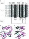
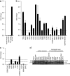
The human papillomavirus (HPV) E7 oncoprotein exists as a dimer and acts by binding to many cellular factors, preventing or retargeting their function and thereby making the infected cell conducive for viral replication. Dimerization of E7 is attributed primarily to the C-terminal domain, referred to as conserved region 3 (CR3). CR3 is highly structured and is necessary for E7's transformation ability. It is also required for binding of numerous E7 cellular targets. To systematically analyze the molecular mechanisms by which HPV16 E7 CR3 contributes to carcinogenesis, we created a comprehensive panel of mutations in residues predicted to be exposed on the surface of CR3. We analyzed our novel collection of mutants, as well as mutants targeting predicted hydrophobic core residues of the dimer, for the ability to dimerize. The same set of mutants was also assessed functionally for transformation capability in a baby rat kidney cell assay in conjugation with activated ras. We show that some mutants of HPV16 E7 CR3 failed to dimerize yet were still able to transform baby rat kidney cells. Our results identify several novel E7 mutants that abrogate transformation and also indicate that E7 does not need to exist as a stable dimer in order to transform cells.
Figures


Similar articles
-
The human papillomavirus E7 proteins associate with p190RhoGAP and alter its function.J Virol. 2014 Apr;88(7):3653-63. doi: 10.1128/JVI.03263-13. Epub 2014 Jan 8. J Virol. 2014. PMID: 24403595 Free PMC article.
-
Conserved region 3 of human papillomavirus 16 E7 contributes to deregulation of the retinoblastoma tumor suppressor.J Virol. 2012 Dec;86(24):13313-23. doi: 10.1128/JVI.01637-12. Epub 2012 Sep 26. J Virol. 2012. PMID: 23015707 Free PMC article.
-
A Conserved Amino Acid in the C Terminus of Human Papillomavirus E7 Mediates Binding to PTPN14 and Repression of Epithelial Differentiation.J Virol. 2020 Aug 17;94(17):e01024-20. doi: 10.1128/JVI.01024-20. Print 2020 Aug 17. J Virol. 2020. PMID: 32581101 Free PMC article.
-
The high-risk HPV16 E7 oncoprotein mediates interaction between the transcriptional coactivator CBP and the retinoblastoma protein pRb.J Mol Biol. 2014 Dec 12;426(24):4030-4048. doi: 10.1016/j.jmb.2014.10.021. Epub 2014 Nov 1. J Mol Biol. 2014. PMID: 25451029 Free PMC article.
-
Molecular mechanisms of transformation by the human papillomaviruses.Princess Takamatsu Symp. 1989;20:199-206. Princess Takamatsu Symp. 1989. PMID: 2562182 Review.
Cited by
-
The Not-So-Good, the Bad and the Ugly: HPV E5, E6 and E7 Oncoproteins in the Orchestration of Carcinogenesis.Viruses. 2021 Sep 22;13(10):1892. doi: 10.3390/v13101892. Viruses. 2021. PMID: 34696321 Free PMC article. Review.
-
Modeling and Molecular Dynamics of the 3D Structure of the HPV16 E7 Protein and Its Variants.Int J Mol Sci. 2021 Jan 30;22(3):1400. doi: 10.3390/ijms22031400. Int J Mol Sci. 2021. PMID: 33573298 Free PMC article.
-
The Human Papillomavirus 16 E7 Oncoprotein Attenuates AKT Signaling To Promote Internal Ribosome Entry Site-Dependent Translation and Expression of c-MYC.J Virol. 2016 May 27;90(12):5611-5621. doi: 10.1128/JVI.00411-16. Print 2016 Jun 15. J Virol. 2016. PMID: 27030265 Free PMC article.
-
The human papillomavirus E7 oncoprotein as a regulator of transcription.Virus Res. 2017 Mar 2;231:56-75. doi: 10.1016/j.virusres.2016.10.017. Epub 2016 Nov 8. Virus Res. 2017. PMID: 27818212 Free PMC article. Review.
-
The human papillomavirus E7 proteins associate with p190RhoGAP and alter its function.J Virol. 2014 Apr;88(7):3653-63. doi: 10.1128/JVI.03263-13. Epub 2014 Jan 8. J Virol. 2014. PMID: 24403595 Free PMC article.
References
-
- Adams A., Gottschling D. E., Kaiser C. A., Stearns T. 1997. Methods in yeast genetics. Cold Spring Harbor Laboratory Press, Cold Spring Harbor, NY
-
- Alonso L. G., et al. 2004. The HPV16 E7 viral oncoprotein self-assembles into defined spherical oligomers. Biochemistry 43:3310–3317 - PubMed
-
- Avvakumov N., Torchia J., Mymryk J. S. 2003. Interaction of the HPV E7 proteins with the pCAF acetyltransferase. Oncogene 22:3833–3841 - PubMed
-
- Baker D., Sali A. 2001. Protein structure prediction and structural genomics. Science 294:93–96 - PubMed
Publication types
MeSH terms
Substances
Grants and funding
LinkOut - more resources
Full Text Sources

