. 2011 Sep 9;286(36):31830-8.
doi: 10.1074/jbc.M111.229518. Epub 2011 Jul 19.

SLC6A14 (ATB0,+) protein, a highly concentrative and broad specific amino acid transporter, is a novel and effective drug target for treatment of estrogen receptor-positive breast cancer

Affiliations

SLC6A14 (ATB0,+) protein, a highly concentrative and broad specific amino acid transporter, is a novel and effective drug target for treatment of estrogen receptor-positive breast cancer

Senthil Karunakaran et al. J Biol Chem. .

Abstract

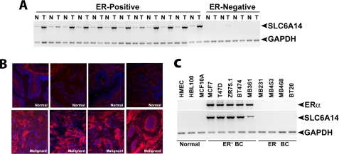
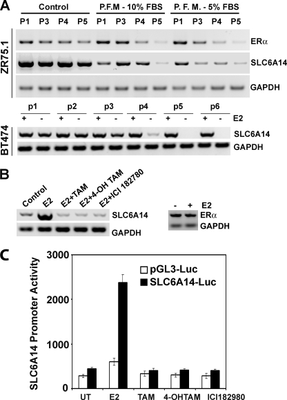
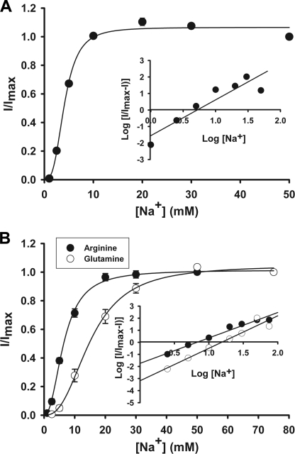
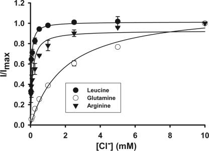
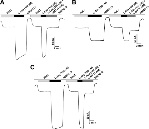
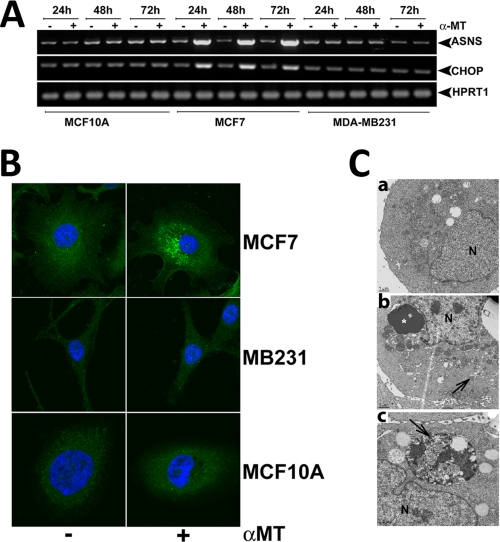
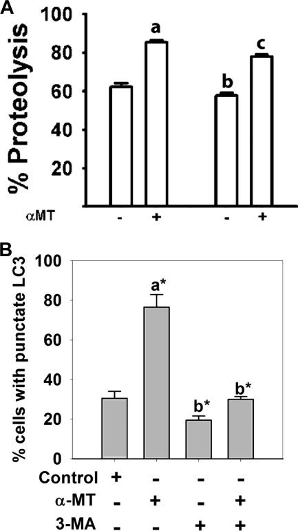
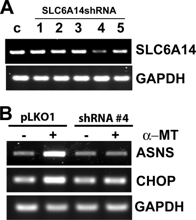
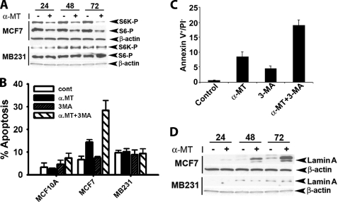
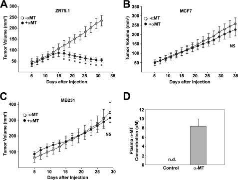
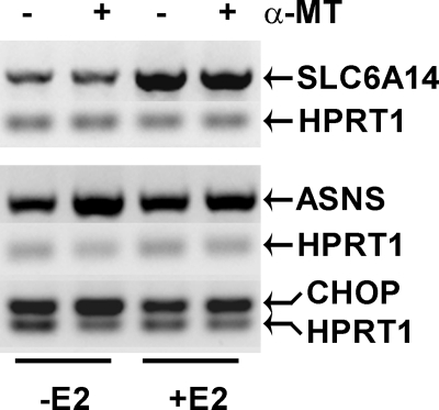
SLC6A14, also known as ATB(0,+), is an amino acid transporter with unique characteristics. It transports 18 of the 20 proteinogenic amino acids. However, this transporter is expressed only at low levels in normal tissues. Here, we show that the transporter is up-regulated specifically in estrogen receptor (ER)-positive breast cancer, demonstrable with primary human breast cancer tissues and human breast cancer cell lines. SLC6A14 is an estrogen/ER target. The transport features of SLC6A14 include concentrative transport of leucine (an activator of mTOR), glutamine (an essential amino acid for nucleotide biosynthesis and substrate for glutaminolysis), and arginine (an essential amino acid for tumor cells), suggesting that ER-positive breast cancer cells up-regulate SLC6A14 to meet their increased demand for these amino acids. Consequently, treatment of ER-positive breast cancer cells in vitro with α-methyl-DL-tryptophan (α-MT), a selective blocker of SLC6A14, induces amino acid deprivation, inhibits mTOR, and activates autophagy. Prolongation of the treatment with α-MT causes apoptosis. Addition of an autophagy inhibitor (3-methyladenine) during α-MT treatment also induces apoptosis. These effects of α-MT are specific to ER-positive breast cancer cells, which express the transporter. The ability of α-MT to cause amino acid deprivation is significantly attenuated in MCF-7 cells, an ER-positive breast cancer cell line, when SLC6A14 is silenced with shRNA. In mouse xenograft studies, α-MT by itself is able to reduce the growth of the ER-positive ZR-75-1 breast cancer cells. These studies identify SLC6A14 as a novel and effective drug target for the treatment of ER-positive breast cancer.


Figures

FIGURE 1.
FIGURE 1.
Up-regulation of SLC6A14 in ER-positive breast cancer. A, RT-PCR analysis of SLC6A14 mRNA in primary human breast cancer tissues and the corresponding adjacent normal tissues. N, normal; T, tumor. B, immunohistochemical analysis of SLC6A14 protein in ER-positive breast cancer tissues and normal breast tissues. C, RT-PCR analysis of SLC6A14 and ERα mRNAs in human breast cancer cell lines. ER+ BC, ER-positive breast cancer cell lines; ER BC, ER-negative breast cancer cell lines.
FIGURE 2.
FIGURE 2.
Regulation of SLC6A14 expression by estrogen/ER. A, ZR-75-1 and BT-474 cells were cultured for five to six passages in control medium (10% fetal bovine serum with phenol red) and phenol red-free medium (P.F.M.; 10 or 5% charcoal-stripped fetal bovine serum), and the levels of ERα and SLC6A14 mRNAs were determined by RT-PCR. P1, P3–P5, and p1–p6 refer to passage numbers. B, BT-474 cells were treated with estradiol (E2; 10 nm) in the presence or absence of anti-estrogens (1 μm) for 24 h and then used for RT-PCR analysis of SLC6A14 and ERα mRNAs. ICI 182780 is also known as Faslodex. TAM, tamoxifen; 4-OH TAM, 4-hydroxytamoxifen. C, BT-474 cells were transfected with either the SLC6A14 promoter (∼3 kb)-luciferase (Luc) construct or empty vector and then treated with estradiol (10 nm) in the presence and absence of anti-estrogens (1 μm). Following the treatment, the reporter activity was measured. Data represent values after normalization with β-galactosidase activity for differences in transfection efficiency. UT, untreated.
FIGURE 3.
FIGURE 3.
Na+ activation kinetics of SLC6A14-mediated transport of leucine (A) and arginine and glutamine (B). Human SLC6A14 was expressed heterologously in X. laevis oocytes by microinjection of cRNA. 4 days following injection, oocytes were used for electrophysiological studies using the two-microelectrode voltage-clamp method. Uninjected oocytes showed negligible currents when perifused with leucine, arginine, or glutamine (1 mm). cRNA-injected oocytes showed marked inward currents when perifused with these three amino acids, and the magnitude of the currents increased with increasing concentrations of Na+. (The concentration of Cl was kept constant at 100 mm.) Because the expression levels varied from oocyte to oocyte, resulting in varying magnitudes of amino acid-induced currents, the currents were normalized by taking the magnitude of the currents induced at 100 mm Na+ as 1 in each oocyte and calculating the magnitude of the currents induced at other concentrations of Na+ as a fraction of this maximal current. The experiments were done with three different oocytes, and the results are given as means ± S.E. Insets, Hill plots.
FIGURE 4.
FIGURE 4.
Cl activation kinetics of SLC6A14-mediated transport of leucine, arginine, and glutamine. The experiments were done as described for the Na+ activation kinetics in the legend to Fig. 3. cRNA-injected oocytes showed marked inward currents when perifused with these three amino acids, and the magnitude of the currents increased with increasing concentrations of Cl. (The concentration of Na+ was kept constant at 100 mm.) The experiments were done with three different oocytes, and the results are given as means ± S.E.
FIGURE 5.
FIGURE 5.
Blockade of SLC6A14-mediated transport of leucine (A), glutamine (B), and arginine (C) by α-MT. Oocytes injected with human SLC6A14 cRNA were perifused with 100 μm leucine, glutamine, or arginine under varying experimental conditions as indicated. Perifusion of the oocytes with the amino acids induced inward currents in the presence of NaCl, and the currents disappeared when NaCl was replaced with N-methyl-d-glucamine (NMDG) chloride. Re-perifusion of the same oocytes with the amino acids again induced inward currents in the presence of NaCl, but these currents disappeared when perifusion with the same amino acids was done in the same buffer but in the presence of 1 mm α-MT.
FIGURE 6.
FIGURE 6.
Effects of SLC6A14 blockade with α-MT in breast cancer cells. A, MCF-10A (a non-malignant mammary epithelial cell line), MCF-7 (an ER-positive breast cancer cell line), and MB-231 (an ER-negative breast cancer cell line) cells were treated with or without 2.5 mm α-MT for 24, 48, or 72 h and then used for analysis of asparagine synthetase (ASNS) and CHOP mRNA levels. B, MCF-7, MB-231, and MCF-10A cells were treated with or without 2.5 mm α-MT for 48 h and then used for immunocytochemical analysis of constitutively expressed LC3. C, MCF-7 cells were treated with (panels b and c) or without (panel a) α-MT (2.5 mm) for 48 h and then used for electron microscopy. Arrows indicate sequestration of the cytoplasm and organelles in pre-autophagosomes and autophagosomes, and the asterisk indicates condensed chromatin. N, nucleus.
FIGURE 7.
FIGURE 7.
Induction of autophagy in MCF-7 cells by α-MT. A, autophagosomal proteolysis of 14C-labeled proteins. The concentrations of α-MT and 3-MA, when present, were 2.5 and 10 mm. The treatment time was 48 h. The extent of proteolysis was determined by the radioactivity present in the protein-free supernatant. a, significantly different from proteolysis in the absence of α-MT and 3-MA (p < 0.001); b, significantly different from proteolysis in the absence of α-MT and 3-MA (p < 0.05); c, significantly different from proteolysis in the presence of α-MT alone (p < 0.01). B, MCF-7 cells were transfected with an GFP-LC3 expression plasmid, and 24 h later, the cells were treated with or without α-MT (2.5 mm) in the presence or absence of 3-MA (10 mm) for 48 h. At the end of the treatment, cells were fixed, and the percent of cells with punctate localization of ectopically expressed GFP-LC3 was quantified. a, compared with the control; b, compared with treatment with α-MT alone; c, compared with treatment with α-MT alone; *, p < 0.001.
FIGURE 8.
FIGURE 8.
Influence of α-MT on asparagine synthetase and CHOP mRNA levels in control MCF-7 cells and in MCF-7 cells with shRNA-induced silencing of SLC6A14. A, five different SLC6A14-specific shRNAs were tested in MCF-7 cells using a lentivirus-based system to monitor their efficacy to silence SLC6A14. SLC6A14 mRNA levels were measured by RT-PCR with GAPDH as an internal control. c, control. B, SLC6A14 was silenced in MCF-7 cells with shRNA-4 using a lentivirus-based system and then treated without or with α-MT (2.5 mm, 48 h). RNA isolated from the cells was used for RT-PCR to monitor the levels of asparagine synthetase (ASNS) and CHOP mRNAs.
FIGURE 9.
FIGURE 9.
A, influence of α-MT on mTOR activity. MCF-7 and MB-231 cells were treated without or with α-MT (2.5 mm) for 24, 48, or 72 h, and the cell lysates were used for Western blotting. S6K-P, phosphorylated S6 kinase. B–D, influence of α-MT with or without 3-MA on cell death. B, MCF-7, MB-231, and MCF-10A cells were treated for 48 h without or with α-MT (2.5 mm), 3-MA (10 mm), or α-MT (2.5 mm) plus 3-MA (10 mm) and analyzed for cell death by propidium iodide (PI) staining and FACS-based cell cycle analysis. C, MCF-7 cells were treated for 48 h without or with α-MT (2.5 mm), 3-MA (10 mm), or α-MT (2.5 mm) plus 3-MA (10 mm) and then analyzed for apoptotic cell death by annexin V labeling. D, MCF-7 and MB-231 cells were treated without or with 2.5 mm α-MT for 24, 48, or 72 h. Cell lysates were then used for Western blot analysis of lamin A degradation product using an antibody that recognizes specifically the proteolytic cleavage product. β-Actin was used as an internal control.
FIGURE 10.
FIGURE 10.
In vivo efficacy of α-MT in the treatment of ER-positive breast cancer. ZR-75-1 (A), MCF-7 (B), and MB-231 (C) cells were injected subcutaneously into BALB/c nude mice (10 × 106 cells/injection site). For MCF-7 cells, slow-releasing estrogen pellets were implanted to support the tumor growth. For each cell line, the control group received water with sucrose, and the treatment group received α-MT (2 mg/ml) in water with sucrose. Tumor size was measured periodically. NS, not statistically significant (p > 0.05); *, p < 0.005 (two-tailed unpaired Student's t test). D, BALB/c nude mice were divided into two groups; the control group received water with sucrose, and the treatment group received α-MT (2 mg/ml) in water with sucrose for 2 weeks. Mice were then bled, and α-MT in plasma was determined by HPLC. n.d., not determined.
FIGURE 11.
FIGURE 11.
Estrogen-dependent suppression of the efficacy of α-MT to induce asparagine synthetase and CHOP expression in MCF-7 cells. MCF-7 cells were cultured in phenol red-free medium in the absence or presence of estradiol (E2; 10 nm) for 24 h and then treated without or with α-MT (2.5 mm, 24 h). The cells were used for analysis of SLC6A14, asparagine synthetase (ASNS), and CHOP mRNA levels by RT-PCR. HPRT1 mRNA levels were used as an internal control.

Similar articles

Cited by

References

    1. Ganapathy V., Inoue K., Prasad P. D., Ganapathy M. E. (2004) in Metabolic and Therapeutic Aspects of Amino Acids in Clinical Nutrition (Cynober L. A. ed) 2nd Ed., pp. 63–78, CRC Press, Boca Raton, FL
    1. Ganapathy M. E., Ganapathy V. (2005) Curr. Drug Targets Immune Endocr. Metab. Disord. 5, 357–364 - PubMed
    1. Nakanishi T., Hatanaka T., Huang W., Prasad P. D., Leibach F. H., Ganapathy M. E., Ganapathy V. (2001) J. Physiol. 532, 297–304 - PMC - PubMed
    1. Hatanaka T., Nakanishi T., Huang W., Leibach F. H., Prasad P. D., Ganapathy V., Ganapathy M. E. (2001) J. Clin. Invest. 107, 1035–1043 - PMC - PubMed
    1. Hatanaka T., Huang W., Nakanishi T., Bridges C. C., Smith S. B., Prasad P. D., Ganapathy M. E., Ganapathy V. (2002) Biochem. Biophys. Res. Commun. 291, 291–295 - PMC - PubMed

Publication types