. 2011;6(7):e21771.
doi: 10.1371/journal.pone.0021771. Epub 2011 Jul 6.

Role of SNX16 in the dynamics of tubulo-cisternal membrane domains of late endosomes

Affiliations

Role of SNX16 in the dynamics of tubulo-cisternal membrane domains of late endosomes

Ben Brankatschk et al. PLoS One. 2011.

Abstract

In this paper, we report that the PX domain-containing protein SNX16, a member of the sorting nexin family, is associated with late endosome membranes. We find that SNX16 is selectively enriched on tubulo-cisternal elements of this membrane system, whose highly dynamic properties and formation depend on intact microtubules. By contrast, SNX16 was not found on vacuolar elements that typically contain LBPA, and thus presumably correspond to multivesicular endosomes. We conclude that SNX16, together with its partner phosphoinositide, define a highly dynamic subset of late endosomal membranes, supporting the notion that late endosomes are organized in distinct morphological and functional regions. Our data also indicate that SNX16 is involved in tubule formation and cholesterol transport as well as trafficking of the tetraspanin CD81, suggesting that the protein plays a role in the regulation of late endosome membrane dynamics.


Conflict of interest statement

Competing Interests: The authors have declared that no competing interests exist.

Figures

Figure 1
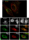
Figure 1. SNX16 is not present on early endosomal membranes.
A) HeLa cells co-expressing EGFP-RAB5 and mRFP-SNX16 were analyzed by fluorescence video microscopy. B) HeLa cells were transfected with EGFP-SNX16 or EGFP-SNX16R144A and then treated or not with 100 nM wortmannin for 30 min at 37°C, as indicated, and analyzed by fluorescence microscopy. C) HeLa cells were transfected with Venus-SNX16, fixed, labeled with antibodies against TFR, and analyzed by fluorescence microscopy. D) HeLa cells were transfected with Venus-SNX16, fixed, labeled with antibodies against EEA1, and analyzed by fluorescence microscopy.
Figure 2
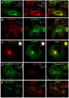
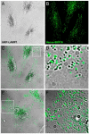
Figure 2. SNX16 is associated to a subset of late endosome membranes.
A) HeLa cells were transfected with Venus-SNX16 and analyzed by immunofluorescence microscopy using antibodies against LAMP1. B) HeLa cells co-expressing mRFP-SNX16 and EGFP-RAB7 were fixed and analyzed by fluorescence microscopy (see also Movie S1). C) HeLa cells co-expressing mRFP-SNX16 and EGFP-RILP were analyzed by fluorescence video microscopy (see Movie S2). D) HeLa cells transfected with Venus-SNX16 were analyzed by immunofluorescence microscopy using antibodies against LBPA. E) HeLa cells were co-transfected with Venus-SNX16 and CD63-mRFP and analyzed by fluorescence video microscopy.
Figure 3
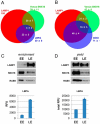
Figure 3. Analysis of SNX16 distribution by fluorescence microscopy and fractionation.
A–B) HeLa cells transfected with Venus-SNX16 were labeled with antibodies against LAMP1 and LBPA (A) or LAMP1 and CD63 (B), and analyzed by confocal microscopy. The distribution of Venus-SNX16 under low expression conditions, LAMP1, and LBPA (A) or Venus-SNX16, LAMP1, and CD63 (B) was quantified after 3D image reconstruction using Imaris software (error bars indicate STDEVA). The data are expressed as the percentage of LAMP1, which co-distributes with the indicated marker. C–D) Untransfected BHK cells were homogenized and a post-nuclear supernatant (PNS) was prepared. The PNS was fractionated by floatation using a well-established step sucrose gradient . Early (EE) and late (LE) endosome fractions were collected and analyzed by SDS gel electrophoresis and western blotting with antibodies against LAMP1, SNX16 or RAB5, or by ELISA with antibodies against LBPA. In (C), the gels were loaded with equal amounts of protein (2.5 µg), as were the wells in the ELISA analysis (5 µg), to visualize enrichment of the corresponding markers in the fractions. RFU: relative fluorescence units. In (D), the gels were loaded with equal volume (1/3 of the total fraction) to visualize the yields of the corresponding markers in the fractions. In the LBPA analysis, yields were calculated from the quantification of the ELISA data (total RFU).
Figure 4
Figure 4. SNX16 distribution after glutaraldehyde fixation, and after brefeldin A treatment.
A–B) HeLa cells transfected with Venus-SNX16 were fixed with paraformaldehyde (A) or glutaraldehyde (0.3%) and paraformaldehyde (3%) for 50 min (B) and analyzed by immunofluorescence microscopy using antibodies against LAMP1. Green arrows point at Venus-SNX16-positive tubules without detectable LAMP1, white arrows point to LAMP1- and SNX16-containing tubules. C) The left panel shows a confocal section of a cell expressing Venus-SNX16 and labeled for LAMP1 (fixation as in B). The middle panel shows a 3D reconstruction of the corresponding confocal stack with Imaris software, and the right panel shows a magnification of the boxed region, displaying only Venus-SNX16 and its colocalization with LAMP1. White arrows point out the presence of LAMP1 at discrete sites of Venus-SNX16-decorated tubules. D) HeLa cells transfected with Venus-SNX16 were treated with brefeldin A (5 µg/ml for 30 min) prior to fixation with paraformaldehyde and analyzed by immunofluorescence microscopy using antibodies against TFR and the cis-Golgi protein p23. The insert in the p23 panel shows the characteristic ribbon-like distribution of p23 in control cells without brefeldin A.
Figure 5
Figure 5. HRP-LAMP1 and SNX16 distribution on tubular and vesicular late endosomes.
A–F) HeLa cells co-transfected with Venus-SNX16 and HRP-LAMP1 were processed as described in , . Briefly, cells were chased with 1 mM DDT for 30 min, to ensure proper HRP-LAMP1 localization . Prior to fixation, HRP-LAMP1 was revealed cytochemically with the DAB reaction and cells were permeabilized; each treatment was for 30 min at 4°C under physiological osmolarity conditions . Samples were analyzed by phase contrast microscopy to reveal HRP-LAMP1 (A) and by fluorescence microscopy to reveal Venus-SNX16 (B). Panel C shows the merged image of A) and B), and panel D a high magnification view of the region boxed in C). In E), an example of a cell is shown where Venus-SNX16 and HRP-LAMP1 colocalize on numerous tubules (magnification in F).
Figure 6
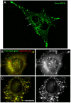
Figure 6. Motility of Venus-SNX16-containing endosomes depends on microtubules.
A) HeLa cells transfected with Venus-SNX16 were analyzed by fluorescence video microscopy. Panel (A) shows a frame of Movie S3, which illustrates the dynamic tubulo-cisternal elements containing SNX16. Arrows point at highly dynamic SNX16-positive tubules. B–C) HeLa cells transfected with EGFP-SNX16 were pretreated (C) or not (B) with 10 µM nocodazole for 2 h, and analyzed by time-lapse video microscopy for 30 sec in the presence (C) or absence (B) of nocodazole. In the left panels, the first (green) and last (red) frames were color-coded and superimposed. The presence of each color indicates that vesicles moved while yellow shows that they remained motionless. All frames superimposed without color-code shown in the right panels feature the trajectories of the corresponding vesicles.
Figure 7
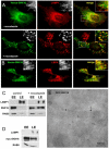
Figure 7. SNX16 distribution in the absence of polymerized microtubules, and re-localization to LBPA-containing multivesicular late endosomes upon overexpression.
A) HeLa cells transfected with Venus-SNX16 were treated or not with 10 µM nocodazole to depolymerize the microtubules as in Fig 6, fixed in 0.3% glutaraldehyde and 3% paraformaldehyde, and analyzed by immunofluorescence microscopy using antibodies against LAMP1. B) HeLa cells overexpressing Venus-SNX16 were analyzed by immunofluorescence microscopy using antibodies against LBPA. C) BHK cells treated or not with nocodazole as in (A) were fractionated as in Fig 3. Early (EE) and late (LE) endosome fractions were analyzed by SDS gel electrophoresis and western blotting using the indicated antibodies. Gels were loaded with equal amounts of protein. D) BHK cells overexpressing myc-SNX16 were fractionated and analyzed as in (C). E) EGFP-SNX16-overexpressing cells were processed for cryosectioning and labeled with anti-GFP antibodies, as described , . Scale bar is 100 nm.
Figure 8
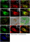
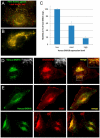
Figure 8. Overexpressed SNX16 inhibits late endosomal dynamics, and interferes with cholesterol and CD81 trafficking.
A–B) HeLa cells expressing either low (A; Movie S4) or high (B; Movie S5) levels of Venus-SNX16 were analyzed by time-lapse video microscopy for 30 sec. The first (green) and last (red) frames were color-coded and superimposed, as in Fig 6B–C. C) The motility of Venus-SNX16-positive structures, depending on SNX16 expression levels (including medium SNX16 expression), was quantified and expressed as a percentage of the motility observed at low Venus-SNX16 expression (error bars indicate SEM). D) After Venus-SNX16 overexpression, HeLa cells were analyzed by fluorescence microscopy using filipin to reveal the distribution of cholesterol. E) CD81 distribution was analyzed by fluorescence microscopy in cells with low Venus-SNX16 (upper panels) or high Venus-SNX16 (lower panels) expression levels.

Similar articles

Cited by

References

    1. Brown MS, Goldstein JL. Cholesterol feedback: from Schoenheimer's bottle to Scap's MELADL. J Lipid Res 50 Suppl. 2009. pp. S15–27. - PMC - PubMed
    1. Lingwood D, Simons K. Lipid rafts as a membrane-organizing principle. Science. 2010;327:46–50. - PubMed
    1. Kobayashi T, Beuchat MH, Lindsay M, Frias S, Palmiter RD, et al. Late endosomal membranes rich in lysobisphosphatidic acid regulate cholesterol transport. Nat Cell Biol. 1999;1:113–118. - PubMed
    1. Di Paolo G, De Camilli P. Phosphoinositides in cell regulation and membrane dynamics. Nature. 2006;443:651–657. - PubMed
    1. Lindmo K, Stenmark H. Regulation of membrane traffic by phosphoinositide 3-kinases. J Cell Sci. 2006;119:605–614. - PubMed

Publication types

MeSH terms