. 2011 Sep;85(18):9527-42.
doi: 10.1128/JVI.00199-11. Epub 2011 Jul 13.

Deletion of the monkeypox virus inhibitor of complement enzymes locus impacts the adaptive immune response to monkeypox virus in a nonhuman primate model of infection

Affiliations

Deletion of the monkeypox virus inhibitor of complement enzymes locus impacts the adaptive immune response to monkeypox virus in a nonhuman primate model of infection

Ryan D Estep et al. J Virol. 2011 Sep.

Abstract

Monkeypox virus (MPXV) is an orthopoxvirus closely related to variola virus, the causative agent of smallpox. Human MPXV infection results in a disease that is similar to smallpox and can also be fatal. Two clades of MPXV have been identified, with viruses of the central African clade displaying more pathogenic properties than those within the west African clade. The monkeypox inhibitor of complement enzymes (MOPICE), which is not expressed by viruses of the west African clade, has been hypothesized to be a main virulence factor responsible for increased pathogenic properties of central African strains of MPXV. To gain a better understanding of the role of MOPICE during MPXV-mediated disease, we compared the host adaptive immune response and disease severity following intrabronchial infection with MPXV-Zaire (n = 4), or a recombinant MPXV-Zaire (n = 4) lacking expression of MOPICE in rhesus macaques (RM). Data presented here demonstrate that infection of RM with MPXV leads to significant viral replication in the peripheral blood and lungs and results in the induction of a robust and sustained adaptive immune response against the virus. More importantly, we show that the loss of MOPICE expression results in enhanced viral replication in vivo, as well as a dampened adaptive immune response against MPXV. Taken together, these findings suggest that MOPICE modulates the anti-MPXV immune response and that this protein is not the sole virulence factor of the central African clade of MPXV.


Figures
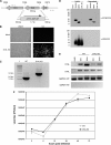
Fig. 1.
Fig. 1.
Construction of D14L KO MPXV-Z. (A) Overview of recombination approach utilized to remove D14L from the MPXV-Z genome. Asterisks indicate the location of recombination within the genomic DNA, small arrows indicate location of primers used in PCR analysis of recombinant virus DNA, and nucleotide numbers of D14L and neighboring ORFs are noted by hash marks. The orientation of the EGFP-GPT cassette is noted by an arrow. (B) EGFP expression in BSC40 cells infected with D14L KO MPXV-Z isolate. (C) PCR analysis of genomic DNA from WT MPXV-Z and D14L KO MPXV-Z demonstrates the complete removal of D14L sequence (650 bp) and the insertion of an EGFP-GPT cassette (∼1.7 kb). (D) Anti-VCP/MOPICE Western blot analysis of lysates and supernatants from vaccinia virus (Western Reserve)-, WT MPXV-Z-, and D14L KO MPXV-Z-infected BSC40 cells, demonstrating production of MOPICE in WT MPXV-Z-infected cells and lack of MOPICE expression in D14L KO MPXV-Z-infected cells. Membranes were probed with anti-VCP antibody and then stripped and reprobed with antibody cross-reactive with vaccinia virus A35R and MPXV-Z A33R as a control for virus infection. (E) RT-PCR analysis of D13L and D15L transcript levels in WT or D14L KO MPXV-Z-infected BSC40 cells, indicating that deletion of D14L and insertion of the EGFP/GPT-expressing cassette in the MPXV-Z genome do not prevent the expression of neighboring genes. Cells were infected at an MOI of 2.5, and total RNA was collected at 6 and 24 h postinfection for analysis using primers specific for D13L or D15L transcripts. RT-PCR for glyceraldehyde-3-phosphate dehydrogenase (GAPDH) serves as a control for equal RNA levels (+RT) and lack of DNA contamination (−RT) in each sample. (F) Single-step growth curve analysis (MOI, 2.5) of WT and D14L KO MPXV-Z in BSC40 cells.
Fig. 2.
Fig. 2.
Development of pox lesions in WT and D14L KO MPXV-Z-infected rhesus macaques. Images of skin lesions demonstrating the development of pox lesions on skin (flank and trunk) of infected RM over the course of infection. (A) WT MPXV-Z-infected animal 24739, and (B) D14L KO MPXV-Z-infected animal 22665. (C) Pox lesions from D14L KO MPXV-infected animals 21965 and 20405, demonstrating EGFP expression within pox lesions due to the presence of virus. Right panels depict the same region shown in left panels exposed to UV light (+UV).
Fig. 3.
Fig. 3.
Viral loads in WT and D14L KO MPXV-Z-infected rhesus macaques. Quantitative real-time PCR analysis was performed using samples collected at the indicated days postinfection (dpi) to determine viral genome copy numbers in whole blood (WB) or peripheral blood mononuclear cells (PBMC) (A and B) and in bronchial alveolar lavage (BAL) fluid cells (C and D). Filled symbols represent WT MPXV-Z-infected animals (A and C), and open symbols depict D14L KO MPXV-Z-infected animals (B and D). DNA from WB was analyzed for all animals except 23358 and 23218, in which cases DNA was obtained from PBMC purified from WB samples. BAL fluid DNA samples for real-time PCR analysis were available only for WT MPXV-Z-infected animals 24739 and 25510. D14L KO MPXV-Z-infected animal 26315 succumbed to disease 17 dpi. BAL fluid samples from D14L KO MPXV-Z-infected animals 21965 and 20405 were not collected 17 and 21 dpi due to low oxygen saturation levels. The highest viral load was detected 7 dpi in BAL fluid of animal 26315 (1.2 × 10⁸), which succumbed to disease on day 17 pi.
Fig. 4.
Fig. 4.
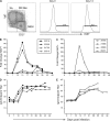
D14L KO MPXV-Z infection results in diminished B-cell response in rhesus macaques. (A) PBMC from infected animals were analyzed by flow cytometry (FCM) to identify three B-cell subsets: naïve (CD27 IgD+), marginal zone-like (CD27+ IgD+), and memory (CD27+ IgD). A representative example from animal 23218 obtained on day 0 is shown in the left panel. B-cell proliferation within the memory cell population was measured by staining for the nuclear protein Ki67, which is upregulated at 14 dpi (right panel) relative to 0 dpi (middle panel). Percentage of Ki67+ memory B cells was converted at every time point to absolute number of Ki67+ memory B cells based on complete blood counts (CBC), and fold increase over baseline (average of three preinfection time points) was calculated. (B) B-cell proliferation in response to WT MPXV-Z infection peaked at 14 dpi. (C) A similar pattern of B-cell proliferation was observed in D14L KO MPXV-Z-infected animals, although only two animals displayed a measurable proliferative burst, which also peaked at 14 dpi. Orthopoxvirus-specific IgG responses in WT (D) and D14L KO MPXV-Z-infected (E) animals were analyzed by endpoint ELISA and were first detectable by 14 dpi in all animals. IgG endpoint titers in D14L KO MPXV-Z-infected animals were on average a log lower than those achieved with WT MPXV-Z infection at 17 dpi.
Fig. 5.
Fig. 5.
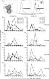
D14L KO MPXV-Z-infected rhesus macaques show decreased T-cell proliferation in peripheral blood compared to WT MPXV-Z-infected animals. PBMC were analyzed by FCM in to delineate CD4 and CD8 T-cell subsets: naïve (CD28+ CD95), central memory (CM) (CD28+ CD95+), and effector memory (EM) (CD28 CD95+). T-cell proliferation was measured by staining for nuclear protein Ki67. (A) A representative example of CD8 T-cell subsets in PBMC from animal 23218 at 0 dpi is shown in the left panel. An example of Ki67 expression within CM and EM population at 0 (middle panel) and 10 dpi (right panel) is shown. Percentage of Ki67+ T cells was converted to numbers of proliferating cells based on CBC values, and fold increase over baseline (average of three preinfection time points) was then calculated. (B, D, F, and H) An initial proliferative burst was detected at 10 dpi in CM and EM CD4 and CD8 subsets in WT MPXV-Z-infected animals, followed by a dramatic decrease in frequency of proliferating cells detected at 14 dpi and a secondary proliferative burst at 17 dpi in most animals. Animals 23218 and 23358 displayed a greater proliferative burst than animals 24739 and 25510. (C, E, G, and I) An increase in Ki67+ T cells was detected at 10 dpi in D14L KO MPXV-Z-infected animals, followed by a gradual return to baseline proliferation levels at 21 dpi, with no consistent secondary proliferative burst occurring. Overall, D14L KO MPXV-Z-infected animals experienced a proliferative burst of lower magnitude than that of WT MPXV-Z-infected animals.
Fig. 6.
Fig. 6.
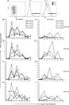
T-cell proliferation is lower in BAL fluid of D14L KO MPXV-Z-infected rhesus macaques compared to WT MPXV-Z-infected animals. In contrast to PBMC, BAL fluid contains only CM and EM T cells. (A) A representative example of CD8 T-cell subsets and frequency of Ki67 cells at 0 (middle panel) and 10 (right panel) dpi from animals 23218 is shown. (B, D, F, and H) In BAL fluid of WT MPXV-Z-infected animals, proliferation in all subsets of T cells followed a pattern similar to that described for PBMC, with an initial proliferative burst first being detected at 10 dpi, a dramatic decrease in frequency of proliferating T cells occurring at 14 dpi, followed by the onset of a second proliferative burst at 17 dpi. The frequency of proliferating T cells returned to baseline at approximately 35 dpi in all animals. (C, E, G, and I) The patterns of proliferation in T-cell subsets from BAL fluid of D14L KO MPXV-Z-infected animals displayed a more sporadic profile, but proliferation was first detected 10 dpi in all subsets and most animals. Data from later time points were not available for animal 26315, which succumbed to disease 17 dpi, or for animals 20405 and 21965 at 17 and 21 dpi.
Fig. 7.
Fig. 7.
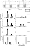
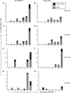
Frequency of MPXV-specific T cells in peripheral blood is higher in WT MPXV-Z-infected animals. MPXV-specific CD4 and CD8 T cells were detected by intracellular cytokine staining (ICCS). PBMC from WT and D14L KO MPXV-Z-infected animals were stimulated with purified MPXV-Z virus (MOI, 1) or simian varicella virus (SVV) (negative control) or anti-CD3 (positive control) as described in Materials and Methods. MPXV-specific T cells were identified based on the production of TNF-α and IFN-γ. (A) Representative example of CD8 CM and EM responses from animal 24739 at 17 dpi, indicating that specific T-cell responses are detected only in response to MPXV-Z stimulation. The average percentage of responding (TNF-α+ IFN-γ+ double-positive or IFN-γ+ only) cells in each subset for all four animals is shown for WT MPXV-Z (B, D, F, and H) and D14L KO MPXV-Z (C, E, G, and I). The T-cell response in all subsets of WT MPXV-Z-infected animals peaked by 21 dpi, while the responses in D14L KO MPXV-Z-infected animals were much lower in most subsets.
Fig. 8.
Fig. 8.
MPXV-Z-specific T cells in BAL fluid of WT and D14L KO MPXV-Z-infected animals. ICCS was performed as described for Fig. 7 to measure frequency of MPXV-specific T cells in BAL fluid of WT and D14L KO MPXV-Z-infected animals. (A, C, E, and G) Average percentage of responding (TNF-α+ IFN-γ+ double-positive or IFN-γ+ only) T-cell subsets in BAL fluid of WT MPXV-Z-infected animals 24739 and 22510. (B, D, F, and H) Frequency of responding T cells from D14L KO MPXV-Z-infected animal 22665.

Similar articles

Cited by

References

    1. Amanna I. J., Slifka M. K., Crotty S. 2006. Immunity and immunological memory following smallpox vaccination. Immunol. Rev. 211:320–337 - PubMed
    1. Blackburn S. D., et al. 2009. Coregulation of CD8+ T cell exhaustion by multiple inhibitory receptors during chronic viral infection. Nat. Immunol. 10:29–37 - PMC - PubMed
    1. Breman J. G., et al. 1980. Human monkeypox, 1970–79. Bull. World Health Organ. 58:165–182 - PMC - PubMed
    1. Brodie R., Smith A. J., Roper R. L., Tcherepanov V., Upton C. 2004. Base-By-Base: single nucleotide-level analysis of whole viral genome alignments. BMC Bioinformatics 5:96. - PMC - PubMed
    1. Cameron C. M., Barrett J. W., Mann M., Lucas A., McFadden G. 2005. Myxoma virus M128L is expressed as a cell surface CD47-like virulence factor that contributes to the downregulation of macrophage activation in vivo. Virology 337:55–67 - PubMed

Publication types

MeSH terms

Associated data

LinkOut - more resources