Review
. 2011 Jul 13;12(8):453-66.
doi: 10.1038/nrn3071.

Gut feelings: the emerging biology of gut-brain communication

Affiliations
Review

Gut feelings: the emerging biology of gut-brain communication

Emeran A Mayer. Nat Rev Neurosci. .

Abstract

The concept that the gut and the brain are closely connected, and that this interaction plays an important part not only in gastrointestinal function but also in certain feeling states and in intuitive decision making, is deeply rooted in our language. Recent neurobiological insights into this gut-brain crosstalk have revealed a complex, bidirectional communication system that not only ensures the proper maintenance of gastrointestinal homeostasis and digestion but is likely to have multiple effects on affect, motivation and higher cognitive functions, including intuitive decision making. Moreover, disturbances of this system have been implicated in a wide range of disorders, including functional and inflammatory gastrointestinal disorders, obesity and eating disorders.


Figures
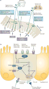
Figure 1
Figure 1. Gut to brain communication
a | Endocrine, immune and neuronal afferent signalling from the gut to the CNS. Information about luminal factors and conditions of the gut are signalled through extrinsic vagal and spinal afferents to the brain stem and spinal cord, respectively. Mechanical stimuli (stretch, pressure, distortion and shearing forces) can activate spinal, vagal and intrinsic primary afferents (IPANs) directly, without intermediary cells such as the enteroendocrine (EE) cells. Although no synaptic connections have been found between IPANs and extrinsic afferents, the latter form networks around myenteric ganglia (intraganglionic laminar endings), many of which receive synaptic input from IPANs. Signalling molecules (including proteases, histamine, serotonin and cytokines) that are produced by immune cells in Peyer's patches and within the gut epithelium can activate their respective receptors on vagal and spinal afferents. Similarly, neuropeptides and hormones (gut peptides) that are released from EE cells in response to other luminal factors, such as nutrients, toxins or antigens, can act both in an endocrine fashion, reaching targets in the brain (area postrema, dorsal vagal complex and hypothalamus), and through receptor activation on spinal and vagal afferents, in a paracrine fashion. Enterochromaffin (EC) cells signal to both IPANs and vagal afferents. b | Encoding of multiple luminal signals by EE cells. Different classes of EE cells are interspersed between gut epithelial cells throughout the gastrointestinal tract. Upon luminal stimulation (or upon activation by postganglionic sympathetic or vagal nerves), these cells can release up to 20 different gut peptides from their basolateral (and possibly luminal) surface. Released peptides can activate closely adjacent vagal afferent nerve terminals in a paracrine fashion, or when released into the circulation they can exert an endocrine effect, signalling to various sites in the brain and other parts of the gastrointestinal tract. Different types of receptors have been identified on the luminal side of EE cells, including G protein-coupled taste receptors (GPCRs) for sweet and bitter tastants, GPCRs that are responsive to fatty acids and toll-like receptors (TLRs). The intestinal taste receptors that are shown are coupled to a specific Gα protein subunit, gustducin (Gαgust), and receptor-induced increases in intracellular calcium result in peptide release from the basolateral membrane. [Ca2+]i, intracellular calcium concentration; DAG, diacylglycerol; GI peptide, gastrointestinal peptide; GPR40, G protein-coupled receptor 40; InsP3, Inositol-1,4,5-trisphosphate;. PIP2, aquaporin PIP2 member; PKC, protein kinase C; PLCβ2, phospholipase Cβ; T1R, taste receptor type 1 member; TRPM5, transient receptor potential cation channel subfamily M member 5 (specifically linked to taste receptor signalling); VSCC, voltage-sensitive Ca2+ channel.
Figure 2
Figure 2. Gut–brain signalling related to food intake
Nutrient-related signals reach the CNS through spinal, vagal and endocrine signalling pathways. Endocrine signalling of gut peptides that are released into the systemic circulation reach the dorsal vagal complex through the area postrema where they modulate the transmission of afferent vagal signals to the dorsal motor nucleus. These gut peptides also reach specialized neurons within the hypothalamus. Paracrine signals activate function-specific vagal afferent fibres that ultimately signal to subregions of the anterior insula (aINS). The sensory aspect of taste is primarily encoded in the aINS, but the multimodal integration of satiety signals with the sensory properties of food (including its flavour, palatability and reward value) as well as the context of food intake (including food related visual and auditory signals) occurs in the orbitofrontal cortex (OFC). Further integration with inputs from the reward system and with interoceptive memories of previous food ingestion generates a multidimensional food-related experience that ultimately determines ingestive behaviour. Prefrontal regions exert cognitive control over ingestive behaviours. Learning about food-related experiences and the formation of interoceptive memories is an important aspect of the cortical circuitry that is involved in this process. ACC, anterior cingulate cortex; AP, area postrema; ARC, arcuate nucleus; cNTS, caudal NTS; HIPP, hippocampus; LH, lateral hypothalamus; NAc, nucleus accumbens; NTS, nucleus tractus solitarius; PeF, pernifornical hypothalamus; OLF, olfaction; PFC, prefrontal cortex; rNTS, rostral NTS; VTA, ventral tegmental area.
Figure 3
Figure 3. Gut signalling systems, gut sensations and meta-representations of such sensations
Gastrointestinal interoceptive signals to the brain play a key part in the maintenance of homeostasis under resting conditions. Whereas signalling under homeostatic conditions is generally associated with pleasant sensations (satiety), signalling under non-homeostatic conditions is typically associated with aversive sensations (pain and nausea). Perception of these signals as gut sensations is associated with activation of the anterior insula (aINS) and the orbitofrontal cortex (OFC), where gustatory, olfactory and viscerosensory signals are integrated and where they are modulated by affective and cognitive input. The generation of such gut sensations can lead to the formation of interoceptive memories of the multidimensional experience associated with the sensation (such as the feeling of nausea associated with a disgusting experience or a particular taste with a pleasant social experience). The interoceptive memory is encoded within a distributed network involving the aINS, OFC, the amygdala (AMYG) and hippocampus (HIPP). Often, environmental stimuli that are unrelated to the gut, such as an image, sound or smell, may trigger the recall of these feeling states as meta-representations of the original gut-triggered sensation. The experience of such memories can result in the engagement of similar brain regions (aINS, anterior cingulate cortex (ACC), OFC and amygdala) in the absence of gut signalling. This recall may be the basis for feelings such as disgust, craving or emotional pain, in the absence of any interoceptive input from the gut, and may occur when watching another person in pain, when hearing about a disgusting experience or when viewing images of rich foods. Recall of such memories without conscious awareness may bias behavioural responses, and such behavioural biases may be a reflection of an internal value map based on ‘gut feelings’. 5-HT, 5-hydroxytryptamine (serotonin); CKK, cholecystokinin; CRF, corticotropin releasing factor; NPY, neuropeptide Y; PY, peptide YY; SP, substance P.
Figure 4
Figure 4. Interoceptive memory and prediction error in chronic disease
According to a theory proposed by Paulus and Stein, the mismatch of actual interoceptive input reaching the anterior insula (aINS) through body to brain signalling, with a falsely predicted interoceptive state (from interoceptive memory and/or influences on the aINS from prefrontal and limbic influences) results in the engagement of anterior cingulate cortex (ACC). Mismatch-related ACC activation is associated with emotional arousal, increased sympathetic and sacral parasympathetic activity and engagement of descending bulbospinal sensory facilitation systems, and may be associated with a conscious feeling of anxiety and worry. When adapted to functional gastrointestinal disorders, the autonomic response to the mismatch is likely to change the state of the gut through modulation of multiple target cells (for example, activation of motor and secretory activity in the distal colon that is associated with stress), which in turn is likely to produce altered interoceptive feedback to the INS. The failure to correct the prediction error by updating the predictive state (as would be expected in a healthy individual) results in chronicity of symptoms through chronic functional and structural dysregulation of the brain–gut axis. Mismatches between actual interoceptive input from nutrient related signals from the gut and interoceptive memories of hedonic experiences of food intake have been implicated in obesity. ACC, anterior cingulate cortex; AMYG amygdala; ANS, autonomic nervous system; HIPP, hippocampus; OFC orbitofrontal cortex; PFC prefrontal cortex.

Similar articles

Cited by

References

    1. Furness JB. The Enteric Nervous System. Blackwell; Oxford: 2006. A comprehensive overview of all aspects of the enteric nervous system.
    1. Furness JB, Kunze WA, Bertrand PP, Clerc N, Bornstein JC. Intrinsic primary afferent neurons of the intestine. Prog Neurobiol. 1998;54:1–18. - PubMed
    1. Costa M, Furness JB, Llewellyn-Smith IJ, Johnson LR. In: Physiology of the Gastrointestinal Tract. Johnson LR, et al., editors. Raven; New York: 1987. pp. 1–40.
    1. Gershon MD, et al. In: Physiology of the Gastrointestinal Tract. Johnson LR, et al., editors. Raven Press; New York: 1994. pp. 381–422.
    1. Gershon MD. The Second Brain. Harper Collins; New York: 1998.

Publication types