. 2011 Aug;138(15):3169-77.
doi: 10.1242/dev.065110.

Mesothelial- and epithelial-derived FGF9 have distinct functions in the regulation of lung development

Affiliations

Mesothelial- and epithelial-derived FGF9 have distinct functions in the regulation of lung development

Yongjun Yin et al. Development. 2011 Aug.

Abstract

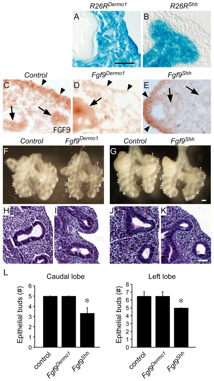
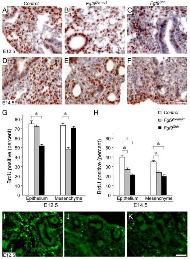
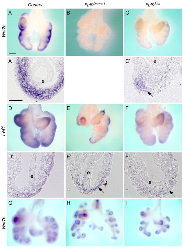
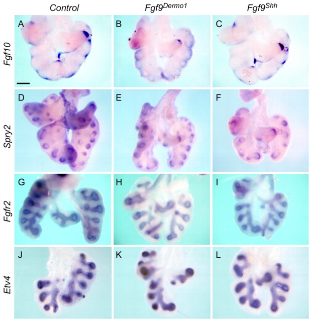
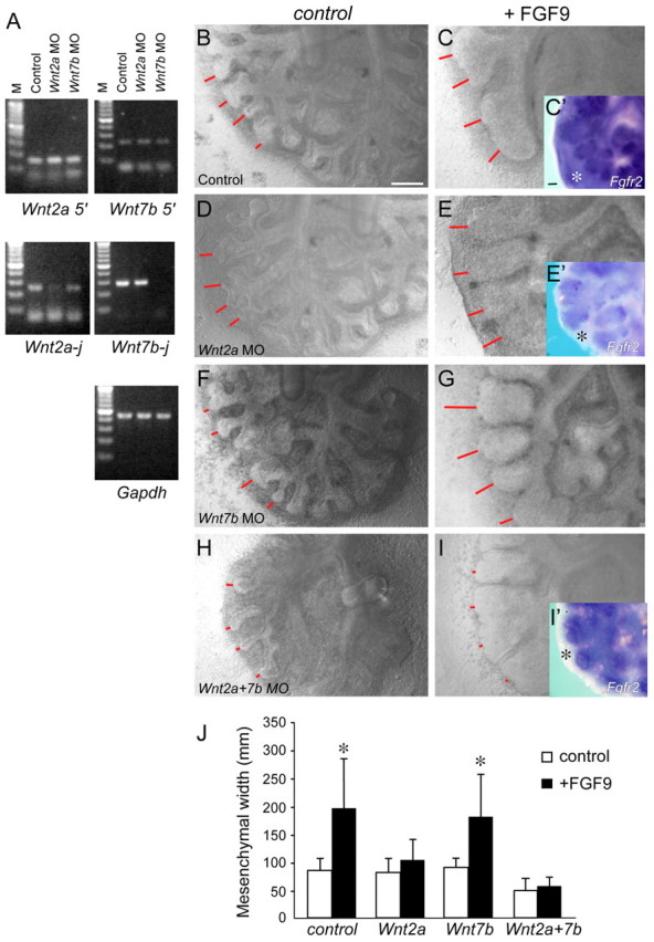
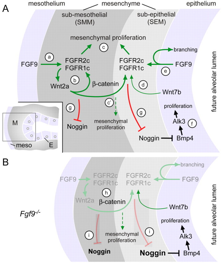
Fibroblast growth factor (FGF) 9 is a secreted signaling molecule that is expressed in lung mesothelium and epithelium and is required for lung development. Embryos lacking FGF9 show mesenchymal hypoplasia, decreased epithelial branching and, by the end of gestation, hypoplastic lungs that cannot support life. Mesenchymal FGF signaling interacts with β-catenin-mediated WNT signaling in a feed-forward loop that functions to sustain mesenchymal FGF responsiveness and mesenchymal WNT/β-catenin signaling. During pseudoglandular stages of lung development, Wnt2a and Wnt7b are the canonical WNT ligands that activate mesenchymal WNT/β-catenin signaling, whereas FGF9 is the only known ligand that signals to mesenchymal FGF receptors (FGFRs). Here, we demonstrate that mesothelial- and epithelial-derived FGF9, mesenchymal Wnt2a and epithelial Wnt7b have unique functions in lung development in mouse. Mesothelial FGF9 and mesenchymal WNT2A are principally responsible for maintaining mesenchymal FGF-WNT/β-catenin signaling, whereas epithelial FGF9 primarily affects epithelial branching. We show that FGF signaling is primarily responsible for regulating mesenchymal proliferation, whereas β-catenin signaling is a required permissive factor for mesenchymal FGF signaling.


Figures

Fig. 1.
Fig. 1.
Mesothelial and epithelial FGF9 have unique functions in developing mouse lung. (A,B) lacZ staining for the Rosa26 reporter (R26R) targeted with Dermo1-Cre (A) and Shh-Cre (B) lungs at E10.5 showing mesenchymal/mesothelial and epithelial cell-specific β-gal staining, respectively. (C-E) Immunohistochemical detection of FGF9 in E12.5 lung showing expression in epithelium (arrows) and mesothelium and submesothelial mesenchyme (arrowheads) in control lungs (C). Fgf9Dermo1 lungs (D) have greatly reduced staining in submesothelial mesenchyme (arrowheads) and Fgf9Shh lungs (E) have reduced expression in epithelium (arrows). (F,G) Anterior views of gross dissections of control (Dermo1-Cre, Fgf9f/+) and Fgf9Dermo1 (F) and control (Shh-Cre, Fgf9f/+) and Fgf9Shh (G) lungs at E12.5. Note the decreased mesenchyme but normal epithelial branch number and orientation of lobes with the Fgf9Dermo1 lung (F) and decreased number of epithelial branches and normal mesenchyme in Fgf9Shh lung (G). (H-K) Hematoxylin and Eosin-stained histological sections of control (H,J), Fgf9Dermo1 (I) and Fgf9Shh (K) lungs at the same stages as shown in F,G. (L) Number of epithelial buds in the caudal and left lobes at E12.5. *P<0.001 (Student's t-test). Error bars represent s.d. Scale bars: 25 μm in A-E,H-L; 200 μm in F,G.
Fig. 2.
Fig. 2.
Mesothelial and epithelial FGF9 differentially affect mesenchymal and epithelial proliferation. (A-F) BrdU labeling of control (A,D), Fgf9Dermo1 (B,E) and Fgf9Shh (C,F) mouse lung showing decreased proliferation in the mesenchyme of the Fgf9Dermo1 lung and the epithelium of the Fgf9Shh lung at E12.5 and E14.5. Note that at E14.5 proliferation was decreased in both the epithelium and mesenchyme in Fgf9Dermo1 and Fgf9Shh lung. (G,H) Quantitation of BrdU labeling at E12.5 (G) and E14.5 (H). *P<0.01 (Student's t-test). Error bars represent s.d. (I-K) TUNEL labeling of control (I), Fgf9Dermo1 (J) and Fgf9Shh (K) lung at E12.5 showing no difference in rates of cell death. Scale bars: 50 μm.
Fig. 3.
Fig. 3.
Mesothelial FGF9 regulates WNT2A expression and mesenchymal WNT/β-catenin signaling. (A-C′) Expression of Wnt2a in control (A,A′), Fgf9Dermo1 (B) and Fgf9Shh (C,C′) mouse lung mesenchyme at E12.5. A′ and C′ are cryosections of the corresponding whole mounts. Note that in Fgf9Dermo1 lung mesenchyme Wnt2a is not detected. In Fgf9Shh lung, Wnt2a is still expressed in the distal mesenchyme (arrow) but not in proximal mesenchyme of epithelium (e). (D-F′) Expression of Lef1 in control (D,D′), Fgf9Dermo1 (E,E′) and Fgf9Shh (F,F′) lung mesenchyme at E12.5. D′, E′ and F′ are cryosections of the corresponding whole mounts. Note that in Fgf9Dermo1 lung, Lef1 expression was detected in the proximal mesenchyme (arrow) and in Fgf9Shh lung Lef1 was detected in the distal mesenchyme (arrow). (G-I) Wnt7b expression in lung epithelium was not significantly changed in control (G), Fgf9Dermo1 (H) and Fgf9Shh (I) lung epithelium at E12.5. Scale bars: 200 μm in A-C,D-E,G-I; 50 μm in A′,C′,D′-F′.
Fig. 4.
Fig. 4.
FGF10 signaling pathways are not affected by mesothelial and epithelial FGF9. (A-C) Expression of Fgf10 in control (A), Fgf9Dermo1 (B) and Fgf9Shh (C) mouse lung mesenchyme at E12.5. (D-F) Expression of Spry2 in control (D), Fgf9Dermo1 (E) and Fgf9Shh (F) lung epithelium at E12.5. No significant differences in Fgf10 or Spry2 expression were observed. (G-I) Expression of Fgfr2 in control (G), Fgf9Dermo1 (H) and Fgf9Shh (I) lung epithelium at E12.5. (J-L) Expression of Etv4 in control (J), Fgf9Dermo1 (K) and Fgf9Shh (L) lung mesenchyme at E12.5. Scale bar: 200 μm.
Fig. 5.
Fig. 5.
Regulation of BMP pathways by mesothelial and epithelial FGF9. (A-C) Expression of Bmp4 in control (A), Fgf9Dermo1 (B) and Fgf9Shh (C) mouse lung mesenchyme at E12.5. (D-F) Expression of Nog in control (D), Fgf9Dermo1 (E) and Fgf9Shh (F) lung epithelium at E12.5. Bmp4 expression was similar but Nog expression was significantly increased in both Fgf9Dermo1 and Fgf9Shh lungs. (G-I) Frozen sections of the whole mounts shown in D-F. (J) Quantitative RT-PCR detection of Nog expression showing increased expression in Fgf9Dermo1 and Fgf9Shh E12.5 lung compared with control. Error bars represent s.d. *P<0.05, **P<0.01, Student's t-test. Scale bar: 200 μm for A-F; 25 μm for G-I.
Fig. 6.
Fig. 6.
WNT2A and WNT7B are required for mesenchymal expression of Fgfr2 and response to FGF9. (A) PCR detection of Wnt2a and Wnt7b mRNA in mouse lung explants treated with control or splice site morpholinos. Top: amplification of the 5′ end of the transcripts showed normal expression in control, Wnt2a MO and Wnt7b MO treated explants. Middle: amplification of the splice site (j) of the transcripts showed reduced or absent mRNA expression in the corresponding treated explant. (B-I′) Wild-type lung explants were untreated (B,C) or exposed to morpholinos targeting a splice site in the Wnt2a (D,E), Wnt7b (F,G) or both Wnt2a and Wnt7b (H,I) transcripts for 48 hours. Explants were also treated with media (B,D,F,H) or FGF9 (C,E,G,I). Expression of Fgfr2 in control (C′), Wnt2a (E′) and Wnt2a and Wnt7b MO (I′) treated explants showed loss of mesenchymal (asterisks) Fgfr2 expression following knockdown of Wnt2a or Wnt2a and Wnt7b. Red lines indicate mesenchymal thickness. (J) Quantification of mesenchymal width. Explants treated with Wnt2a, or Wnt2a and Wnt7b MO showed no mesenchymal growth response to FGF9. (n=3, *P<0.01, Student's t-test). Error bars represent s.d. Scale bars: 200 μm.
Fig. 7.
Fig. 7.
Mesenchymal FGF and WNT/β-catenin signaling requires both FGF9 and WNT inputs to maintain FGF9 responsiveness. (A-L) Lung explant cultures from control (A-C,G-I) and Fgf9–/– (D-F,J-L) mouse embryos were untreated (A,D,G,J) or treated with LiCl (B,E,H,K) or BIO (C,F,I,L). Control and Fgf9–/– explants were also treated with FGF9 (G-L). Red lines indicate mesenchymal thickness. Scale bar: 200 μm. (M) Quantification of mesenchymal growth in response to LiCl, BIO and FGF9 (n=3 explants for each condition). Dashed red line shows mesenchymal thickness of wild-type lung explants. Error bars represent s.d. *P<0.05, **P<0.01, ***P<0.001, Student's t-test.
Fig. 8.
Fig. 8.
Regulatory mechanisms governing mesenchymal FGF-WNT/β-catenin signaling during early pseudoglandular stages of lung development. (A) (a) Mesothelial FGF9 signals to mesenchymal FGFR1C and FGFR2C to regulate mesenchymal proliferation and Wnt2a expression. (b) Mesenchymal β-catenin signaling is required for mesenchymal FGFR expression and mesenchymal response to FGF9. (c) FGFR signaling primarily regulates mesenchymal proliferation but requires WNT/β-catenin signaling to maintain FGFR expression. (c′) In the absence of mesenchymal FGFR signaling, WNT/β-catenin signaling has a limited capacity to induce mesenchymal expansion. (d) Wnt7b, expressed in airway epithelium, is partially redundant with Wnt2a for regulation of mesenchymal β-catenin signaling. (e) Epithelial FGF9 regulates branching either by direct autocrine activation of epithelial FGFRs or indirectly through regulation of mesenchymal FGF-WNT/β-catenin signaling and their downstream targets. (f) BMP4, primarily expressed in epithelium, acts as an autocrine factor to regulate epithelial proliferation and, secondarily, branching, through activation of BMPR1A (ALK3). (g) FGF-WNT/β-catenin signaling in the SEM and SMM negatively regulates Noggin expression. Inset: Diagram showing the region near the distal epithelial bud in which these signaling interactions occur. (B) When Fgf9 is absent, the FGF-WNT/β-catenin feed-forward signaling loop degenerates (gray) and only Wnt7b signaling remains. (h) In the absence of Fgf9, β-catenin is still present but Fgfr1 and Fgfr2 expression is lost. (i) Loss of mesenchymal FGF-WNT/β-catenin signaling results in increased Noggin expression in both the SEM and SMM. Noggin repression of epithelial BMP signaling is proposed to result in decreased epithelial proliferation. E, epithelium; M, mesenchyme.

Similar articles

Cited by

References

    1. Bellusci S., Henderson R., Winnier G., Oikawa T., Hogan B. L. (1996). Evidence from normal expression and targeted misexpression that bone morphogenetic protein (Bmp-4) plays a role in mouse embryonic lung morphogenesis. Development 122, 1693-1702 - PubMed
    1. Bellusci S., Grindley J., Emoto H., Itoh N., Hogan B. L. (1997). Fibroblast growth factor 10 (FGF10) and branching morphogenesis in the embryonic mouse lung. Development 124, 4867-4878 - PubMed
    1. Chen C., Ouyang W., Grigura V., Zhou Q., Carnes K., Lim H., Zhao G. Q., Arber S., Kurpios N., Murphy T. L., et al. (2005). ERM is required for transcriptional control of the spermatogonial stem cell niche. Nature 436, 1030-1034 - PMC - PubMed
    1. Colvin J. S., Feldman B., Nadeau J. H., Goldfarb M., Ornitz D. M. (1999). Genomic organization and embryonic expression of the mouse fibroblast growth factor 9 gene. Dev. Dyn. 216, 72-88 - PubMed
    1. Colvin J. S., White A., Pratt S. J., Ornitz D. M. (2001a). Lung hypoplasia and neonatal death in Fgf9-null mice identify this gene as an essential regulator of lung mesenchyme. Development 128, 2095-2106 - PubMed

Publication types

MeSH terms