Comparative Study
. 2011 Aug 29;52(9):6887-97.
doi: 10.1167/iovs.11-7531.

Spry1 and Spry2 are necessary for lens vesicle separation and corneal differentiation

Affiliations
Comparative Study

Spry1 and Spry2 are necessary for lens vesicle separation and corneal differentiation

Murali R Kuracha et al. Invest Ophthalmol Vis Sci. .

Abstract

Purpose: The studies reported here were performed to analyze the roles of Sproutys (Sprys), downstream targets and negative feedback regulators of the fibroblast growth factor (FGF) signaling pathway, in lens and corneal differentiation.

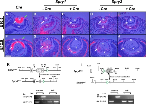
Methods: Spry1 and -2 were conditionally deleted in the lens and corneal epithelial precursors using the Le-Cre transgene and floxed alleles of Spry1 and -2. Alterations in lens and corneal development were assessed by hematoxylin and eosin staining, in situ hybridization, and immunohistochemistry.

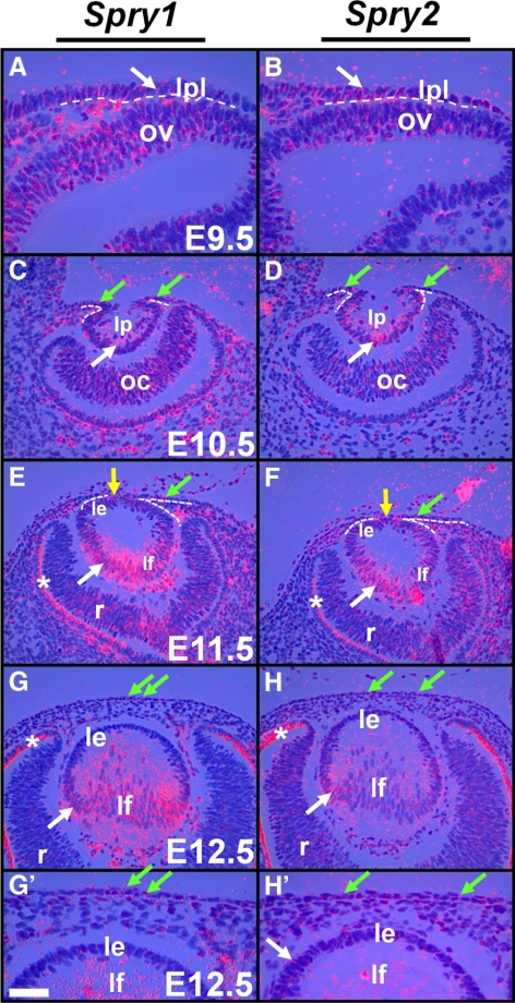
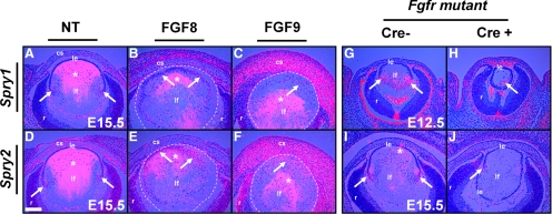
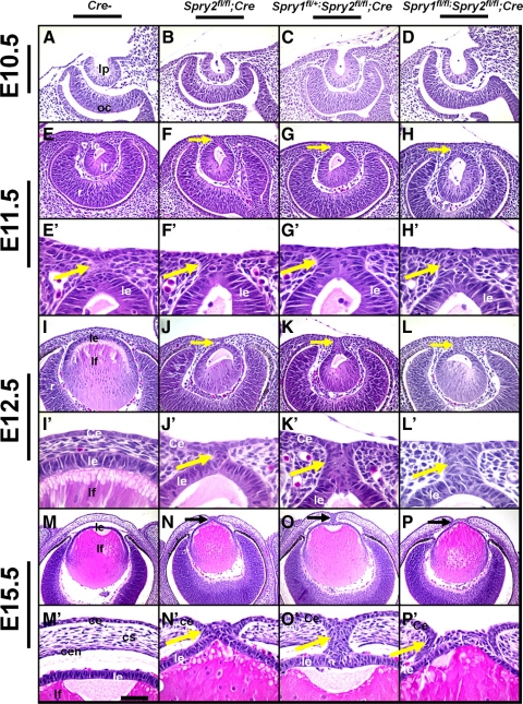
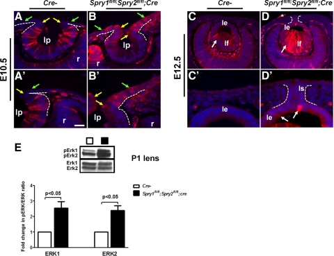
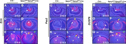
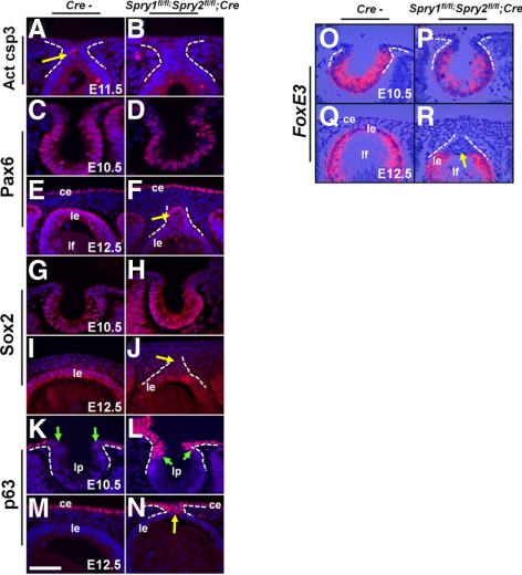
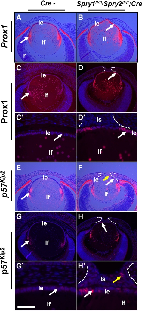
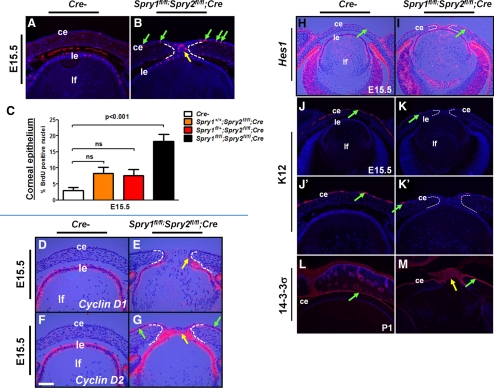
Results: Spry1 and -2 were upregulated in the lens fibers at the onset of fiber differentiation. FGF signaling was both necessary and sufficient for induction of Spry1 and -2 in the lens fiber cells. Spry1 and -2 single- or double-null lenses failed to separate from the overlying ectoderm and showed persistent keratolenticular stalks. Apoptosis of stalk cells, normally seen during lens vesicle detachment from the ectoderm, was inhibited in Spry mutant lenses, with concomitant ERK activation. Prox1 and p57(KIP2), normally upregulated at the onset of fiber differentiation were prematurely induced in the Spry mutant lens epithelial cells. However, terminal differentiation markers such as β- or γ-crystallin were not induced. Corneal epithelial precursors in Spry1 and -2 double mutants showed increased proliferation with elevated expression of Erm and DUSP6 and decreased expression of the corneal differentiation marker K12.

Conclusions: Collectively, the results indicate that Spry1 and -2 (1) through negative modulation of ERKs allow lens vesicle separation, (2) are targets of FGF signaling in the lens during initiation of fiber differentiation and (3) function redundantly in the corneal epithelial cells to suppress proliferation.


Figures

Figure 1.
Figure 1.
Spry expression in the lens. In situ hybridization with 35S-labeled Spry1 and -2 riboprobes was performed on sections of wild-type embryos. Spry1 (A, C, E, G, G′) and Spry2 (B, D, F, H, H′) were expressed initially in the lens placode (A, B, arrows) and later in the lens pit (C, D, arrows) and lens epithelial (E, F) and fiber cells (E, F, G, H, arrows). Spry1 and -2 were weakly expressed in the presumptive cornea (CF, green arrows), in the cells at the junction between the lens and the presumptive cornea (E, F, yellow arrows) that would close to form the lens stalk, and in retinal neuroblasts (CH). (AF, dashed lines) The presumptive lens and corneal epithelium from the periocular mesenchymal cells. (EH) Staining in the retinal pigmented epithelium (RPE, *) is an artifact of dark-field illumination. le, lens epithelium; lf, lens fibers; lp, lens pit; lpl, lens placode; ov, optic vesicle; oc, optic cup; r, retina. Scale bar in (G): (A, B, G′, H′) 10 μm; (CH) 20 μm.
Figure 2.
Figure 2.
Spry1 and -2 expression in FGF transgenic and FGFR mutant mice. In situ hybridization with 35S-labeled Spry1 and -2 riboprobes was performed on sections of nontransgenic (NT) (A, D), FGF8 (B, E), and FGF9 (C, F) transgenic and FGFR mutant (GJ) embryos. Spry1 and -2 were upregulated at the transition zone in the nontransgenic lenses (A, D, arrows). Lens fiber–specific expression of FGF8 (B, E) or FGF9 (C, F), weakly induced Spry1 (B, C, arrows), and strongly induced Spry2 (E, F, arrows) in the lens epithelial cells. (BF, dashed lines) The lens. Spry1 (H, arrow) and Spry2 (J, arrow) expression was reduced in FGFR mutant lenses in contrast to Cre− controls (G, I). Staining within the lens core (*) in (AF) and in the RPE (G) are artifacts of dark-field illumination. True hybridization signals appeared as dots (arrows), and diffraction artifacts had a hazy, less-defined appearance that was visible even in the complete absence of a signal. cs, corneal stroma; le, lens epithelium; lf, lens fibers; r, retina. Scale bar, 40 μm.
Figure 3.
Figure 3.
Conditional deletion of Spry1 and -2 in the lens. In situ hybridization (AJ) was performed with 35S-labeled Cre and Spry1 and -2 riboprobes on sections of Cre transgenic (A, F) and Spry1 (BH) and Spry2 (DJ) mutant embryos. Cre recombinase was expressed in the lens pit (A, white arrow), lens epithelium (F), and presumptive corneal epithelial cells (A, F, green arrows) in the Le-Cre mice, as reported previously. Cre+ embryos showed loss of Spry1 (C, H) and Spry2 (E, J) expression in the lens but not in the optic cup (oc) (BE) or the hyaloid vasculature (hv) (GJ) where Cre recombinase was not expressed. (K, L) Spry1 and -2 floxed alleles (adapted and modified with permission from Basson MA, Akbulut S, Watson-Johnson J, et al. Sprouty1 is a critical regulator of GDNF/RET-mediated kidney induction. Dev Cell 2005;8:229–239. © Elsevier, and Shim K, Minowada G, Coling DE, Martin GR. Sprouty2, a mouse deafness gene, regulates cell fate decisions in the auditory sensory epithelium by antagonizing FGF signaling. Dev Cell 2005;8:553–564. © Elsevier). Cre-mediated recombination in the cornea was determined by PCR using primers (P1, P2, and P3) flanking the loxP sequences (gray triangles) of Spry1 and -2 genes. Tail genomic DNA was used as negative controls. Open rectangles: exons. Green open triangles: frt sequences. After recombination, P2 and P3 amplicons were seen in Cre+ corneas but not in Cre− corneas, as they are too large to be amplified. ce, corneal epithelium; le, lens epithelium; lf, lens fibers; lp, lens pit; ORF, open reading frame; r, retina. Scale bar, 20 μm.
Figure 4.
Figure 4.
Failure of lens detachment in Spry mutants. Sections of E10.5 (AD), E11.5 (EH′), E12.5 (IL′), and E15.5 (MP′), control (Cre−, A, E, E′, I, I′, M, M′), Spry2fl/fl; Cre (B, F, F′, J, J′, N, N′), Spry1fl/+;Spry2 fl/fl;Cre (C, G, G′, K, K′, O, O′), and Spry1fl/fl;Spry2fl/fl;Cre (D, H, H′, L, L′, P, P′) embryos were analyzed by hematoxylin and eosin staining. (E′–P′) Higher magnifications of (EP). Lens placode invagination (BD) was unaffected in Spry1 and -2 mutants. At E11.5, Spry mutants showed prominent stalks (FH′) in contrast to control embryos (E′). At E12.5 and at E15.5, Spry mutant lenses failed to separate and displayed persistent lens stalks (JL′, NP′, arrows) in contrast to controls (I, I′, M, M′). Corneal stroma (cs) and endothelium (cen) were discontinuous in Spry mutants at E15.5 (NP′). ce, corneal epithelium; cs, corneal stroma; cen, corneal endothelium; le, lens epithelium; lf, lens fibers; lp, lens pit; oc, optic cup; r, retina. Scale bar in (M): (AH) 30 μm; (IL,MP) 60 μm; (E′–H′, I′–L′) 10 μm; (M′–P′) 15 μm.
Figure 5.
Figure 5.
ERK activation in Spry mutants. Immunohistochemistry was performed on sections of E10.5 (AB′) and E12.5 (CD′) control and Spry1fl/fl;Spry2fl/fl;Cre embryos using an anti-pERK1/2 antibody. (A′–D′) Higher magnifications of (AD). Elevated pErk1/2 levels were detected at the anterior margins of the lens pit (compare B to A, B′ to A′, yellow arrows) and corneal epithelial precursors (B, B′, green arrows) in the Spry mutant eyes. Similarly, pERK1/2 levels were elevated in the Spry mutant lens epithelial and fiber cells at E12.5 (D, D′, arrows). (D, *) pERK1/2 staining in the incompletely dissected amnion. (E) Western blot analysis of lens lysates from postnatal day 1 (P1) control (Cre−) and Spry mutants. The blots were probed either with the anti-ERK1/2 (bottom) or the anti-pERK1/2 (top) antibody. pERK/ERK ratios were quantified and normalized to Cre− controls. Error bars, SEM. pERK1 and -2 levels were significantly increased in Spry1fl/fl;Spry2fl/fl;Cre mutant lenses. ce, corneal epithelium; le, lens epithelium; lf, lens fibers; ls, lens stalk; r, retina. Scale bar in (A′): (A, B) 15 μm; (C, D) 30 μm; (E, F) 60 μm; (A′–D′) 10 μm.
Figure 6.
Figure 6.
Induction of FGF targets in the Spry mutants. In situ hybridizations were performed on sections of control (Cre−) and Spry mutant embryos using 35S-labeled Erm, Pea3, and DUSP6 riboprobes. Erm and DUSP6 were induced in the Spry mutant lens epithelial (D, F, R, white arrows), stalk (F, P, R, yellow arrow), and corneal epithelial cells (F, R, green arrows). Pea3 expression was seen in the Spry mutant lenses (H, J, L, white arrows) and stalks (J, L, yellow arrows). The stalks were, in some cases, more ventrally placed and in these cases, peripheral sections were chosen (D, J) to include the stalks. The lenses in these sections therefore, appear smaller. ce, corneal epithelium; le, lens epithelium; lf, lens fibers. Scale bar in (E): (AE, GR) 30 μm; (E, F) 60 μm.
Figure 7.
Figure 7.
Early lens differentiation in Spry mutant embryos. Immunohistochemistry (AN) and in situ hybridizations (OR) were performed on control (Cre−) and Spry mutant embryos, to detect expression of activated caspase 3 (A, B), Pax6 (CF), Sox2 (GJ), p63 (KN), and FoxE3 (OR). In situ hybridizations were performed using 35S-labeled riboprobes (OR). Activated caspase 3 was seen in the stalks of Cre− controls (A, arrow) but not in Spry mutants. Pax6 (D, F) and Sox2 (H, J) were expressed in their normal spatial pattern in Spry mutants. Spry mutant lens stalks, however, showed reduced Sox2 expression compared to lens epithelial cells (J, arrow). p63, normally excluded from the lens placodal cells (K, green arrows), was expressed at the anterior margins of the lens pit (L, arrows) and in the stalks (N, arrow) of Spry mutants. FoxE3 expression in the invaginating lens placodal cells at E10.5 (O) and epithelial cells (Q) was similar to controls (O). Spry mutant stalk cells did not express FoxE3 (R, arrow). ce, corneal epithelium; le, lens epithelium; lf, lens fibers; lp, lens pit. Scale bar in (M): (AD, M, N) 10 μm; (E, F, IL, OR) 15 μm.
Figure 8.
Figure 8.
Lens fiber differentiation in Spry mutant embryos. In situ hybridizations (A, B, E, F) and immunohistochemistry (CD′, GH′) were performed on E15.5 control (Cre−) and Spry mutant embryos to detect expression of Prox1 (AD′) and p57KIP2 (EH′). In situ hybridizations were performed using 35S-labeled riboprobes (A, B, E, F). Prox1 (B, D, D′, arrows), and p57KIP2 (F, H, H′, arrows) were upregulated in Spry mutant lens epithelial cells. Some of the stalk cells also expressed p57KIP2 (H′, yellow arrow). ce, corneal epithelium; le, lens epithelium; lf, lens fibers; r, retina. Scale bar in (G′): (AD, EH) 60 μm; (C′, D′, G′, H′) 10 μm.
Figure 9.
Figure 9.
Cell proliferation and differentiation in Spry mutant corneal epithelial cells. (AC) BrdU incorporation assay. Immunohistochemistry was performed on E15.5 Spry mutant (B) and control (A) embryos. BrdU proliferation index in the corneal epithelial cells (C) was quantified. Each genotype was compared to Cre− controls. Error bars, SEM. BrdU incorporation was significantly increased in the Spry mutant corneal epithelial cells (B, green arrows, C). (DG) Cell cycle targets in the lens and cornea. In situ hybridizations were performed on E15.5 control (Cre−) and Spry mutant embryos to detect expression of cyclin D1 (D, E) and cyclin D2 (F, G). Cyclin D2 (G, green arrows) but not cyclin D1 (E) was upregulated in the Spry mutant corneas. Both cyclin D1 (E, arrow) and D2 (G, yellow arrow) were expressed in the stalk cells of Spry mutants. (HM) Corneal epithelial differentiation. In situ hybridization (H, I) and immunohistochemistry (JM) were performed on sections of control (Cre−) and Spry mutant embryos. In situ hybridization was performed with a 35S-labeled Hes1 riboprobe. (J′, K′) Higher magnifications of (J) and (K). Increased Hes1 expression in the Spry mutant corneal epithelial cells (I, green arrows) suggests an expansion of progenitor cells. Expression of K12, but not 14-3-3σ, a corneal epithelial differentiation marker, was reduced in Spry mutant corneas (K, K′, M). ce, corneal epithelium; le, lens epithelium; ir, iris; lf, lens fibers; r, retina. Scale bar in (F): (A, B, DG, J′, K′) 20 μm; (HM) 40 μm.

Similar articles

Cited by

References

    1. Coulombre JL, Coulombre AJ. Lens development: fiber elongation and lens orientation. Science. 1963;142:1489–1490 - PubMed
    1. Lovicu FJ, Overbeek PA. Overlapping effects of different members of the FGF family on lens fiber differentiation in transgenic mice. Development. 1998;125:3365–3377 - PubMed
    1. McAvoy JW, Chamberlain CG. Fibroblast growth factor (FGF) induces different responses in lens epithelial cells depending on its concentration. Development. 1989;107:221–228 - PubMed
    1. Robinson ML, Ohtaka-Maruyama C, Chan CC, et al. Disregulation of ocular morphogenesis by lens-specific expression of FGF-3/int-2 in transgenic mice. Dev Biol. 1998;198:13–31 - PubMed
    1. Robinson ML, Overbeek PA, Verran DJ, et al. Extracellular FGF-1 acts as a lens differentiation factor in transgenic mice. Development. 1995;121:505–514 - PubMed

Publication types

MeSH terms