. 2011 Oct;10(10):M111.009936.
doi: 10.1074/mcp.M111.009936. Epub 2011 Jul 8.

Quantitative proteomic analyses of human cytomegalovirus-induced restructuring of endoplasmic reticulum-mitochondrial contacts at late times of infection

Affiliations

Quantitative proteomic analyses of human cytomegalovirus-induced restructuring of endoplasmic reticulum-mitochondrial contacts at late times of infection

Aiping Zhang et al. Mol Cell Proteomics. 2011 Oct.

Abstract

Endoplasmic reticulum-mitochondrial contacts, known as mitochondria-associated membranes, regulate important cellular functions including calcium signaling, bioenergetics, and apoptosis. Human cytomegalovirus is a medically important herpesvirus whose growth increases energy demand and depends upon continued cell survival. To gain insight into how human cytomegalovirus infection affects endoplasmic reticulum-mitochondrial contacts, we undertook quantitative proteomics of mitochondria-associated membranes using differential stable isotope labeling by amino acids in cell culture strategy and liquid chromatography-tandem MS analysis. This is the first reported quantitative proteomic analyses of a suborganelle during permissive human cytomegalovirus infection. Human fibroblasts were uninfected or human cytomegalovirus-infected for 72 h. Heavy mitochondria-associated membranes were isolated from paired unlabeled, uninfected cells and stable isotope labeling by amino acids in cell culture-labeled, infected cells and analyzed by liquid chromatography-tandem MS analysis. The results were verified by a reverse labeling experiment. Human cytomegalovirus infection dramatically altered endoplasmic reticulum-mitochondrial contacts by late times. Notable is the increased abundance of several fundamental networks in the mitochondria-associated membrane fraction of human cytomegalovirus-infected fibroblasts. Chaperones, including HSP60 and BiP, which is required for human cytomegalovirus assembly, were prominently increased at endoplasmic reticulum-mitochondrial contacts after infection. Minimal translational and translocation machineries were also associated with endoplasmic reticulum-mitochondrial contacts and increased after human cytomegalovirus infection as were glucose regulated protein 75 and the voltage dependent anion channel, which can form an endoplasmic reticulum-mitochondrial calcium signaling complex. Surprisingly, mitochondrial metabolic enzymes and cytosolic glycolytic enzymes were confidently detected in the mitochondria-associated membrane fraction and increased therein after infection. Finally, proapoptotic regulatory proteins, including Bax, cytochrome c, and Opa1, were augmented in endoplasmic reticulum-mitochondrial contacts after infection, suggesting attenuation of proapoptotic signaling by their increased presence therein. Together, these results suggest that human cytomegalovirus infection restructures the proteome of endoplasmic reticulum-mitochondrial contacts to bolster protein translation at these junctions, calcium signaling to mitochondria, cell survival, and bioenergetics and, thereby, allow for enhanced progeny production.


Figures

Fig. 1.
Fig. 1.
Experimental design for quantitative proteomics of enriched heavy MAM fraction from uninfected and HCMV-infected HFFs. A, Forward (left) and reverse (right) SILAC approaches. HFFs were grown in light (12C6-Arg and 12C6, 14N2-Lys) or heavy (13C6-Arg and 15N2, 13C6-Lys) medium for four cell doublings. HFFs, grown in heavy medium, were HCMV (strain BAD wt)-infected for 72 h; whereas, HFFs, grown in light medium, were uninfected for 72 h. Prior to subcellular fractionation, two roller bottles of SILAC-labeled, HCMV-infected HFFs were mixed (1:1) with two roller bottles of unlabeled, uninfected HFFs (forward experiment). In the reverse experiment, SILAC-labeled, uninfected HFFs were mixed (1:1) with unlabeled, HCMV-infected HFFs at 72 hpi. The mixed cells were fractionated to obtain heavy MAM as previously described (28, 47). Heavy MAM proteins (100 μg), obtained from mixed unlabeled and SILAC-labeled cells, were resolved by SDS-PAGE. Fifty sequential slices of each lane were in-gel digested with trypsin and analyzed by LC-MS/MS analysis. B, Percoll gradients of uninfected and HCMV infected HFFs. Uninfected HFFs and HCMV-infected HFFs (∼5 × 10⁷ cells/each) were gently lysed by homogenization, pelleted by centrifugation at 10,300 × g for 10 min at 4 °C. The supernatant was removed and alternately processed, whereas the pellet was resuspended in 300 μl of ice-cold mannitol buffer A, briefly homogenized, layered over 10 ml of a 30% Percoll suspension in mannitol buffer B, and subjected to ultracentrifugation at 95,000 × g for 65 min at 4 °C. Banded MAM and mitochondria were recovered from the gradient by needle extraction from above (MAM, 200 μl) and below (Mito, 2 ml) the indicated points. C, SDS-PAGE of forward and reverse MAM samples. Heavy MAM from the paired forward or reverse paired HFFs were resolved by SDS-PAGE and stained by BioSafe Coomassie.
Fig. 2.
Fig. 2.
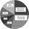
The subcellular distribution of cellular proteins identified in the heavy MAM fractions from uninfected and HCMV-infected HFFs. The known subcellular localization of 991 human proteins identified in heavy MAM proteome in both forward and reverse experiments (with identified peptide number cutoff of at least 2) using UniProt are represented by percent of total. Identified HCMV proteins were excluded from the analysis. The portion designated microsomes includes proteins, which localize in the ER, Golgi apparatus, endosomes, lysosomes, melanosomes, and vesicles using Uniprot.
Fig. 3.
Fig. 3.
Validation of subcellular fractionation using Western analyses and induction of MAM components during HCMV infection. A, Total protein (TP, 30 μg) or fractions were isolated from uninfected or HCMV (BADwt) HFFs at 72 hpi. Fractionated microsomes (Mi), cytosol (Cy), heavy MAM (M) and mitochondria (Mt) (10 μg) were resolved by SDS-PAGE, blotted, and probed with antibodies against markers of ER-mitochondria (FACL4, COX2, and Prohibitin), the secretory apparatus (Membrin and Golgin 97), and glycolytic enzymes (PGM 5 and Hexokinase 1). B, The relative abundances of cellular markers in uninfected HFFs. The relative abundance of each marker tested in Panel A was determined in each fraction by densitometry of the Western blots. The values were normalized to the relative abundance of α-tubulin in the same fraction. ND, not detected. C, Fold induction of cellular markers during HCMV infection of HFFs. The induction of each protein tested in Panel A was determined by comparing the normalized values in HCMV-infected cell fractions to the normalized abundances of the corresponding fractions from uninfected cells. NQ, not quantifiable.
Fig. 4.
Fig. 4.
A, Scatter Plot showing correlation in ratio distribution of proteins detected in forward and reverse SILAC experiments. Log ratio distribution of all proteins detected in the MAM fraction of uninfected and unlabeled HFFs versus HCMV-infected and SILAC-labeled HFFs were plotted versus log ratios of the same set of proteins detected in HCMV-infected and unlabeled HFF cells versus uninfected and SILAC-labeled HFF cells. This plot shows strong correlation between the two experiments with an R2 = -0.8. B, Example of MS spectra of labeled and unlabeled peptide pairs detected for an MAM marker protein, Ero1 α, in both forward and reverse experiments. The peptide was detected as doubly charged species at m/z of 753.4 and 757.4 corresponding to the unlabeled and labeled peptide, respectively. The peptide intensity was consistently increased in HCMV-infected versus uninfected cells.
Fig. 5.
Fig. 5.
A, Alteration of the MAM proteome by HCMV at late times of infection. Total cellular proteins (991 proteins) associated with the MAM fraction in uninfected and in HCMV-infected HFFs at 72 hpi were identified using quantitative proteomics. The number of proteins (at least two identified unique peptides) whose expression was increased (>twofold), showed discordant change (inconsistent values in forward and reverse experiments), had no change (0.5 to twofold), or was decreased (<0.5) after HCMV infection is shown. B, The fold induction levels of MAM-associated cellular proteins after HCMV infection. The number of MAM-associated cellular proteins showing low (>two- to fivefold), intermediate (>five- to eightfold), intermediate high (>eight- to 10-fold), or high (>10-fold) induction is shown.
Fig. 6.
Fig. 6.
A, Verification of quantitative MAM proteomic results using Western analyses. HFFs were uninfected (left) or HCMV-infected (right). At 72 hpi, microsomal (Mi), cytosol (Cy), heavy MAM (M), mitochondria (Mt) were isolated as previously described (28, 47). Proteins in total (T) lysates (30 μg) or indicated fractions (10 μg) were resolved by SDS-PAGE and analyzed by Western analyses using ER chaperones (anti-GRP78/BiP, -calnexin, -calreticulin), MAM junction (anti-PACS-2, -Mfn 1, -Mfn 2), MAM lipid rafts (anti-erlin 2, Sig-1R), MAM Ca2+ signaling complex (anti-GRP75, -VDAC) and mitochondrial chaperones (anti-HSP60, -Prohibitin). The presence of pUL37×1 was verified by anti-UL37×1 (DC35) and α-tubulin was used as a loading control. B, Comparison of HCMV induction of key proteins associated with the MAM calculated by Western analyses (top) or quantitative proteomic analyses (bottom). Top right: The HCMV induction for each protein was calculated for each fraction in the Western analyses (Panel A) by comparing the indicated protein levels in HCMV-infected cells normalized to α-tubulin levels and then divided by the protein levels in uninfected cells normalized to α-tubulin levels. Bottom right: The range of HCMV induction of each protein was determined by quantitative proteomics as in Tables I and III. ND: Proteins were not detected by MS/MS analyses. Prohibitin data from quantitative proteomics were not shown.
Fig. 7.
Fig. 7.
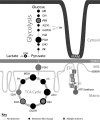
Increased levels of the glycolytic, TCA cycle, and ETC pathways associated with ER-mitochondrial contacts at late times of HCMV infection. The relative fold-inductions for proteins involved in glycolysis, TCA cycle, and ETC detected in association with the MAM fraction from HCMV-infected HFFs above uninfected cells (numeric values are shown in supplementary Table S2) are represented by the areas of corresponding filled black circles. The open circles represent glycolytic enzymes not detected by this quantitative proteomic analysis. Proteins with multiple isoforms are also represented by a smaller, gray-filled circle (isoform with lowest induction) and surrounded by a darker outline (isoform with the largest induction). Constituent members of the ETC and ATP synthase complexes are represented only by solid blocks, as these proteins were observed to have increased expression levels of at least sevenfold, but typically over 10-fold higher than in uninfected cells. Glycolysis hexokinase (HK), glucose-6-phosphate isomerase (GPI), phosphofructokinase (PFK), fructose-biphosphate aldolase (ALDO), glyceraldehyde-3-phosphate dehydrogenase (GAPDH), phosphoglycerate mutase (PGM), alpha-enolase (ENO), pyruvate kinase (PK), lactate dehydrogenase (LDH), pyruvate dehydrogenase (PDH), citrate synthase (CS), aconitase hydratase (ACO), isocitrate dehydrogenase (IDH), 2-oxoglutarate dehydrogenase (OGDH), succinyl-CoA ligase (SUCL), succinate dehydrogenase (SDH), fumarate hydratase (FH), malate dehydrogenase (MDH).
Fig. 8.
Fig. 8.
Relocalization of Sig-1R during HCMV infection of HFFs. HFFs were uninfected or HCMV infected at a multiplicity of 1 and harvested at 72 hpi. A, Induction of HSP60 during HCMV infection. Uninfected and HCMV-infected HFFs were probed with anti-HSP60 (green), anti-HCMV IE 1/2 (blue), and human anti mitochondria (red) antibodies and the corresponding secondary antibodies. The stained cells were imaged by confocal microscopy. The three leftmost panels are greyscale and the right panel shows the overlay of confocal sections. The insets show enlarged regions of interest in the cells. B, Induction of Hexokinase 1 at late times of HCMV infection. Cells were probed with anti-Hexokinase (green), anti-HCMV IE 1/2 (blue), and human anti mitochondria (red) and the corresponding secondary antibodies and imaged as above. The three leftmost panels are greyscale and the right panel shows the overlay of confocal sections. The insets show enlarged regions of interest in the cells. C, Relocalization of Sig-1R during HCMV infection of HFFs. Cells were probed with anti-Sig 1R (green) and anti HCMV IE 1/2 (blue) antibodies. The left and middle panels are greyscale and the right shows the overlay of confocal sections. The insets show enlarged regions of interest in the cells.

Similar articles

Cited by

References

    1. Mocarski E. S., Shenk T., Pass R. F. (2007) Cytomegaloviruses. In: Knipe D. M., Howley P. M. eds. Fields Virology, 5th Ed., pp. 2701–2772, Wolters Kluwer Health, Lippincott Williams & Wilkins, Philadelphia
    1. Gerna G., Baldanti F., Revello M. G. (2004) Pathogenesis of human cytomegalovirus infection and cellular targets. Hum. Immunol. 65, 381–386 - PubMed
    1. Boeckh M., Nichols W. G. (2004) The impact of cytomegalovirus serostatus of donor and recipient before hematopoietic stem cell transplantation in the era of antiviral prophylaxis and preemptive therapy. Blood. 103, 2003–2008 - PubMed
    1. Khanna R., Diamond D. J. (2006) Human cytomegalovirus vaccine: time to look for alternative options. Trends Mol. Med. 12, 26–33 - PubMed
    1. Munger J., Bajad S. U., Coller H. A., Shenk T., Rabinowitz J. D. (2006) Dynamics of the cellular metabolome during human cytomegalovirus infection. PLoS Pathog. 2, e132. - PMC - PubMed

Publication types