. 2011 Sep;85(17):8738-51.
doi: 10.1128/JVI.00798-11. Epub 2011 Jun 29.

Autocatalytic activity of the ubiquitin-specific protease domain of herpes simplex virus 1 VP1-2

Affiliations

Autocatalytic activity of the ubiquitin-specific protease domain of herpes simplex virus 1 VP1-2

M Bolstad et al. J Virol. 2011 Sep.

Abstract

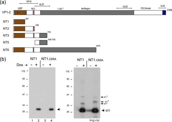
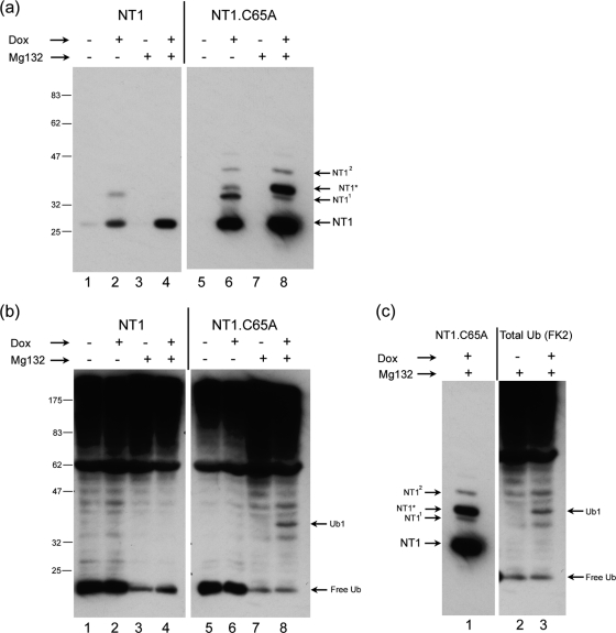
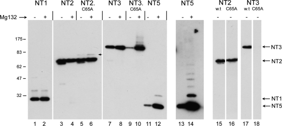
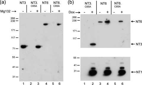
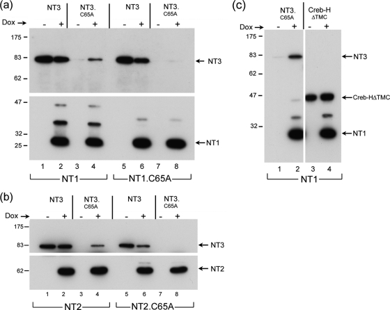
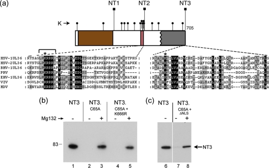
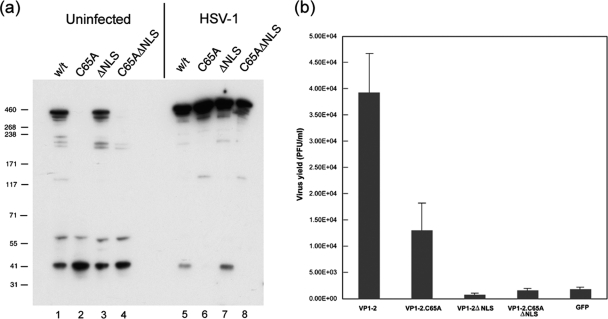
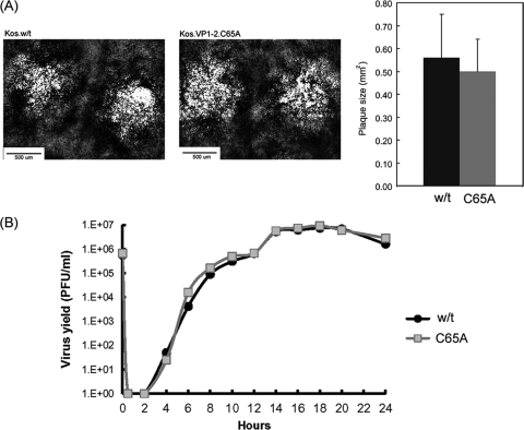
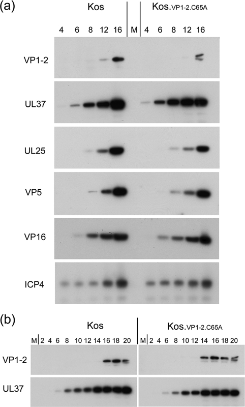
The herpes simplex virus (HSV) tegument protein VP1-2 is essential for virus entry and assembly. VP1-2 also contains a highly conserved ubiquitin-specific protease (USP) domain within its N-terminal region. Despite conservation of the USP and the demonstration that it can act on artificial substrates such as polyubiquitin chains, identification of the relevance of the USP in vivo to levels or function of any substrate remains limited. Here we show that HSV VP1-2 USP can act on itself and is important for stability. VP1-2 N-terminal variants encompassing the core USP domain itself were not affected by mutation of the catalytic cysteine residue (C65). However, extending the N-terminal region resulted in protein species requiring USP activity for accumulation. In this context, C65A mutation resulted in a drastic reduction in protein levels which could be stabilized by proteosomal inhibition or by the presence of normal C65. The functional USP domain could increase abundance of unstable variants, indicating action at least in part, in trans. Interestingly, full-length variants containing the inactive USP, although unstable when expressed in isolation, were stabilized by virus infection. The catalytically inactive VP1-2 retained complementation activity of a VP1-2-negative virus. Furthermore, a recombinant virus expressing a C65A mutant VP1-2 exhibited little difference in single-step growth curves and the kinetics and abundance of VP1-2 or a number of test proteins. Despite the absence of a phenotype for these replication parameters, the USP activity of VP1-2 may be required for function, including its own stability, under certain circumstances.


Figures

Fig. 1.
Fig. 1.
Doxycycline-inducible expression of the VP1-2 USP domain. (a) Summary of various N-terminal constructs of VP1-2, NT1, NT2, NT3, NT5, and NT6 in relation to general features of VP1-2 organization. Numbering refers to the final residue in each construct. The N-terminal core USP is indicated by brown shading, the NLS in pink shading, the central core of VP1-2 in gray, and the C-terminal highly conserved region in blue. Various alignments within each of the alpha, beta, and gamma classes indicate this overall organization, as summarized in Fig. S1 in the supplemental material. Previously characterized regions involved in UL37, VP16, and UL25 binding are indicated (7, 14, 24, 26, 30, 31, 40). (b) Induction of NT1 and NT1.C65A after transfection in 293-Tet On cells. Cells were transfected with the appropriate vectors, Dox was added 24 h later, and cells were harvested 48 h after Dox addition. The primary NT1 species are indicated by an arrow. The panel in the right hand side shows a longer exposure revealing additional lower-abundance species and the presence of NT1* in NT1.C65A, a species never observed for NT1.
Fig. 2.
Fig. 2.
The USP domain can deubiquitinate itself. (a) Cell lines containing inducible NT1 (lanes 1 to 4) and NT1.C65A (lanes 5 to 8) were established and accumulation measured after Dox induction with or without the addition of MG132 (added for 4 h before harvesting) as indicated. The species NT11 and NT12 were readily observed for NT1.C65A and could be seen for NT1 (Fig. 1). NT1* was specific for NT1.C65A and its accumulation was significantly increased by MG132 addition. (b) The same samples as in panel a were probed for the presence of total ubiquitinated species using the anti-ubiquitin antibody FK2. We did not observe major changes in the total ubiquitinated species upon NT1 (or NT1.C65A) induction. However, MG132 treatment resulted in the loss of free ubiquitin and a general increase in higher-molecular-weight species. A novel ubiquitinated species appeared upon Dox induction of NT1.C65A. This species labeled Ub1 (lane 8) was not observed for NT1 induction. (c) Alignment of the same blots as in panels a and b. The Dox-induced NT1.C65A band NT1* (lane 1) comigrates with the novel Dox-induced ubiquitinylated band, Ub1 (lane 3).
Fig. 3.
Fig. 3.
Dependence of N-terminal variants on USP activity for inhibition of ubiquitination and degradation. N-terminal variants of VP1-2 containing wt C65 or the C65A substitution as indicated (Fig. 1) were transfected into Cos cells and treated without or with addition of MG132 (added 4 h prior to harvesting). Mutation in the active site of the USP domain in NT2 resulted in a novel low-abundance species (arrowed lane 6) but had very little overall effect on NT2 levels (cf. lanes 3 and 5). Proteosome inhibition had little effect on total levels of either NT2 or NT2.C65A (lanes 3 to 6). In contrast, mutation in the USP active site resulted in a profound reduction in NT3 levels (cf. lanes 7 and 9 to shorter-exposure lanes 17 and 18). Moreover, proteosome inhibition restored levels of NT3.C65A and induced a series of poorly resolved higher-molecular-weight species (cf. lanes 9 and 10). NT5 was also poorly expressed with proteosome inhibition inducing a significant increase in levels and higher-molecular-weight species (cf. lanes 11 and 12) and longer exposure (lanes 13 and 14).
Fig. 4.
Fig. 4.
The USP domain expressed in trans stabilizes VP1-2 C65A mutants. (a) Variant encoding approximately 60% of the protein NT6 was expressed in comparison to NT3, each containing C65 or C65A as indicated. As for NT3, substitution of C65A in NT6 resulted in a profound decrease in levels of the protein (cf. lanes 4 and 5) which could be stabilized by proteosome inhibition (cf. lanes 5 and 6). (b) Cells were transfected with constructs for constitutive expression of NT3.C65A, NT6, or NT6.C65A as indicated, together with the vector for the Dox-inducible expression of NT1. Twenty-four hours after transfection, Dox was added to one set of duplicate transfections and cells were incubated for a further 24 h. Samples were then probed simultaneously for expression of the various species. Dox induction resulted in expression of NT1 and a concomitant increase in NT3.C65A and NT6.C65A. An increase was seen also for NT6 itself.
Fig. 5.
Fig. 5.
Active USP is required for stabilization in trans. (a) Cells were transfected as for Fig. 4b with constructs for constitutive expression of NT3 (lanes 1, 2, and 5, 6) or NT3.C65A (lanes 3, 4 and 7, 8), together with vectors for the Dox-inducible expression of NT1 or NT1.C65A as indicated. Twenty-four hours after transfection, Dox was added to one set of duplicate transfections and cells were incubated for a further 24 h. Samples were then probed simultaneously for expression of the various species. Dox induction of NT1 resulted in expression of NT3.C65A (lanes 3 and 4), while no increase was observed after induction of NT1.C65A (cf. lanes 7 and 8). (b) Exactly as for panel a, but comparing the effect of NT2 versus NT2.C65A on NT3 and NT3.C65A levels. (c) Cellular protein subject to proteosome-mediated turnover shows little increase upon induction of NT1. Cells were transfected as for panel a, with vectors for expression of NT3.C65A or CREBHΔTMC and processed as described. Despite significant increase in NT3.C65A (lanes 1 and 2), little effect was observed for CREBHΔTMC (lanes 3 and 4).
Fig. 6.
Fig. 6.
Single conserved lysine residues within the VP1-2 N terminus are not sufficient to mediate degradation of catalytically inactive variants. (a) Schematic of the N-terminal constructs NT1, NT2, and NT3, whose C-terminal endpoints are indicated (square lollipops). Lysine residues are indicated by small round lollipops. Sequence alignments of the N terminus of VP1-2 reveal a highly conserved lysine residue within the boundary between NT2 and NT3, indicated by an asterisk above the expanded section of sequence. Numerous lysine residues are present between the boundaries of NT1 and NT2, which also includes a demonstrated functional NLS (2). Within the NLS is located a conserved lysine, as indicated by an asterisk above the expanded sequence. (b) Substitution of these individual lysines does not result in significant stabilization of NT3.C65A. NT3, NT3.C65A, variants of NT3.C65A containing K666R, and a 7-residue deletion at the NLS (indicated by bracket above sequence alignment) were analyzed in the absence or presence of MG132 (added 8 h before harvesting). The double mutants behaved like NT3.C65A, being virtually undetectable in the absence of proteosome inhibition and stabilized by proteosome inhibition to approximately similar levels.
Fig. 7.
Fig. 7.
Relative instability of full-length VP1-2.C65A is rescued by replication-defective virus. (a and b) Cells were transiently transfected with expression vectors for full-length VP1-2 or mutant versions as indicated. Duplicate sets were either mock infected or superinfected with the VP1-2-negative deletion mutant, KΔUL36 (MOI 5), and further incubated for 16 h, after which time cells were harvested and processed for expression levels of VP1-2 (a) or recovery of virus growth by plaque titration in HS30 cells (b). Virus infection resulted in a significant increase in all species of VP1-2 but in particular altered the ratio of the C65A mutants, whose levels were now overall similar to the wt VP1-2, unlike what was seen for uninfected cells. As described previously, VP1-2 substantially complemented growth of KΔUL36. Background levels seen with the control vector expressing green fluorescent protein (GFP) are due to the low level of revertant virus obtained in KΔUL36 preparation (11). VP1-2.ΔNLS showed no complementation above background level. However, VP1-2.C65A exhibited significant levels of complementation above background and only 2- to 3-fold lower than that for wt VP1-2.
Fig. 8.
Fig. 8.
Growth characteristics of a recombinant virus expressing VP1-2.C65A. (A) KOS.wt and KOS.VP1-2.C65A viruses were constructed as described in Materials and Methods and grown in RSC cells. Typical examples of plaque formation after 72 h are shown. No significant difference in average plaque size was observed counting approximately 50 plaques from each strain (right panel). (B) RSC cells were infected with KOS.wt or KOS.VP1-2.C65A at an MOI of 5 and incubated for 2 h, after which time inoculum virus was inactivated by a low-pH wash and the cells were incubated and harvested every 2 h at the times indicated. Cultures were harvested by pooling medium and cell-associated virus, and the pooled lysate was subjected to 3 freeze-thaw cycles. Debris was then pelleted, and production of infectious virus was titrated in RSC cells.
Fig. 9.
Fig. 9.
Accumulation of virus proteins after infection with KOS.wt and KOS.VP1-2.C65A. (a) RSC cells were infected as in Fig. 8 and at the times indicated (h) lysed in SDS lysis buffer; accumulation of candidate proteins was then analyzed by gel electrophoresis and Western blotting. (b) More extended comparison showing accumulation of VP1-2 and UL37 at 2-h intervals at the times indicated.
Fig. 10.
Fig. 10.
Summary illustration of results, as described in the text, indicating the following. (A) The USP domain is ubiquitinated with relatively low efficiency and is able to deubiquitinate itself. (B) Determinants within the N-terminal region of NT3 confer efficient ubiquitin targeting, and protein accumulation is now dependent upon an active USP, removing ubiquitin in trans and potentially in cis. The inactive form of NT3 accumulates ubiquitin (yellow circles) and is targeted for degradation. (C) In virus-infected cells, alteration in processes or the presence of additional factors, e.g., known partners such as UL25, UL37, or VP16, antagonize ubiquitination, and as a consequence the protein is stabilized.

Similar articles

Cited by

References

    1. Abaitua F., Daikoku T., Crump C. M., Bolstad M., O'Hare P. 2011. A single mutation responsible for temperature-sensitive entry and assembly defects in the VP1-2 protein of HSV. J. Virol. 85:2024–2036 - PMC - PubMed
    1. Abaitua F., O'Hare P. 2008. Identification of a highly conserved, functional nuclear localization signal within the N-terminal region of herpes simplex virus type 1 VP1-2 tegument protein. J. Virol. 82:5234–5244 - PMC - PubMed
    1. Bailey D., Barreca C., O'Hare P. 2007. Trafficking of the bZIP transmembrane transcription factor CREB-H into alternate pathways of ERAD and stress-regulated intramembrane proteolysis. Traffic 8:1796–1814 - PubMed
    1. Batterson W., Roizman B. 1983. Characterization of the herpes simplex virion-associated factor responsible for the induction of alpha genes. J. Virol. 46:371–377 - PMC - PubMed
    1. Bonifacino J. S., Weissman A. M. 1998. Ubiquitin and the control of protein fate in the secretory and endocytic pathways. Annu. Rev. Cell Dev. Biol. 14:19–57 - PMC - PubMed

Publication types