. 2011 Aug 26;286(34):29973-83.
doi: 10.1074/jbc.M111.270785. Epub 2011 Jun 28.

The tumor suppressor hamartin enhances Dbl protein transforming activity through interaction with ezrin

Affiliations

The tumor suppressor hamartin enhances Dbl protein transforming activity through interaction with ezrin

Marzia Ognibene et al. J Biol Chem. .

Abstract

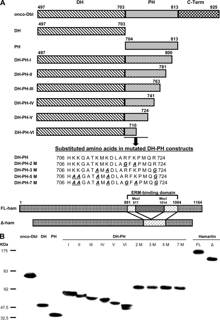
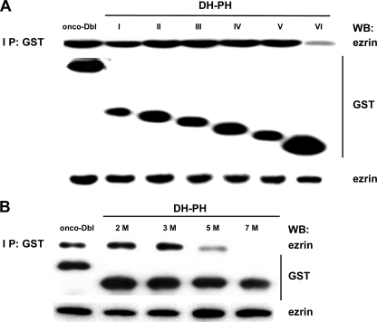
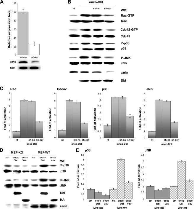
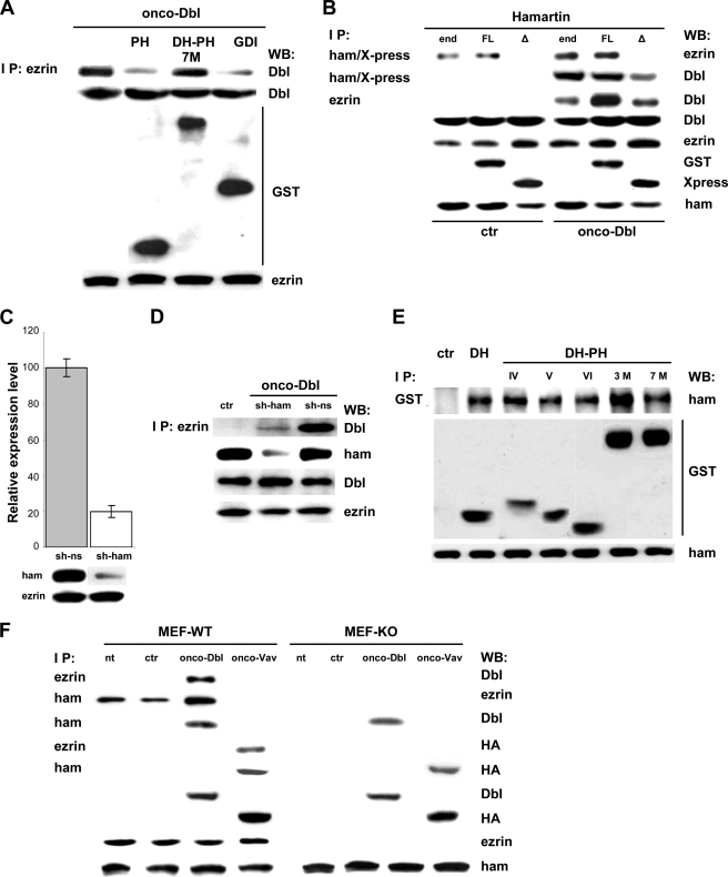
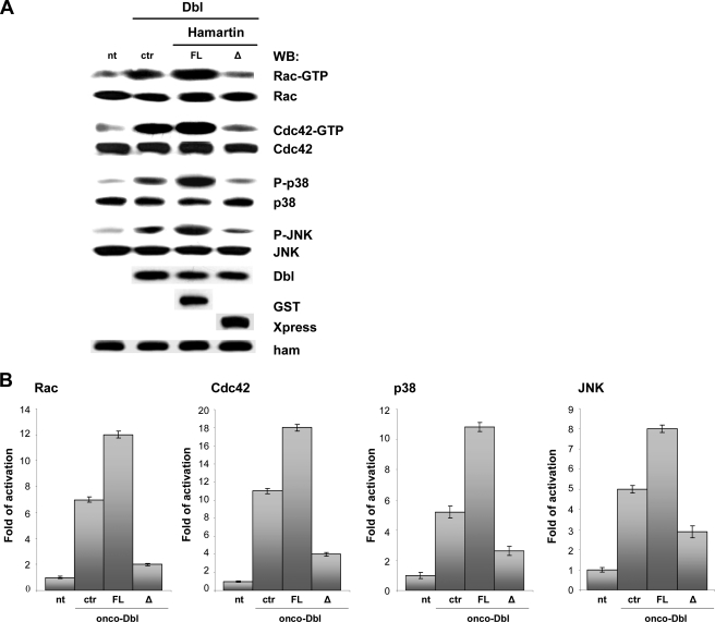
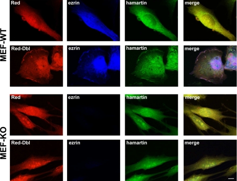
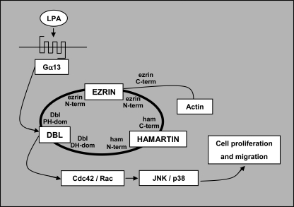
The Rho guanine nucleotide exchange factor (GEF) Dbl binds to the N-terminal region of ezrin, a member of the ERM (ezrin, radixin, moesin) proteins known to function as linkers between the plasma membrane and the actin cytoskeleton. Here we have characterized the interaction between ezrin and Dbl. We show that binding of Dbl with ezrin involves positively charged amino acids within the region of the pleckstrin homology (PH) domain comprised between β1 and β2 sheets. In addition, we show that Dbl forms a complex with the tuberous sclerosis-1 (TSC-1) gene product hamartin and with ezrin. We demonstrate that hamartin and ezrin are both required for activation of Dbl. In fact, the knock-down of ezrin and hamartin, as well as the expression of a mutant hamartin, unable to bind ezrin, inhibit Dbl transforming and exchange activity. These results suggest that Dbl is regulated by hamartin through association with ezrin.


Figures

FIGURE 1.
FIGURE 1.
Schematic representation and expression of Dbl and hamartin mutants. A, Dbl oncogene lacks the N-terminal fragment of proto-Dbl; PH represents the isolated pleckstrin homology domain; DH represents the isolated Dbl homology domain; the DH-PH deletion mutants DH-PH-I to DH-PH-VI were derived from the DH-PH fragment by progressive truncations. The amino acid substitutions in the DH-PH mutants, DH-PH-2 M to DH-PH-7 M, were introduced between positions 707 and 724 in the PH domain of the DH-PH fragment. FL-ham represents the full-length hamartin. Hamartin deletion mutant, Δ-ham, lacks 98 amino acids within the C-terminal ERM-binding domain. B, expression of various Dbl and hamartin constructs. Transient transfectants of Dbl and hamartin were generated in COS7 cells. Cell lysates were subjected to Western blotting. Dbl products were visualized with anti-GST antibody, while FL-ham and Δ-ham were detected using anti-hamartin and anti-Xpress antibody, respectively.
FIGURE 2.
FIGURE 2.
Mapping of the binding site for ezrin on Dbl PH domain. COS7 cells were transiently transfected with (A) onco-Dbl, DH-PH-I, DH-PH-II, DH-PH-III, DH-PH-IV, DH-PH-V, or DH-PH-VI and with (B) onco-Dbl, DH-PH-2M, DH-PH-3M, DH-PH-5M, or DH-PH-7M. Cells lysates were subjected to anti-GST immunoprecipitation followed by anti-ezrin Western blot. The amount of GST constructs and endogenous ezrin was determined using anti-GST and anti-ezrin antibodies, respectively. The results shown are representative of three independent experiments.
FIGURE 3.
FIGURE 3.
Knockdown of ezrin expression inhibits activation of Rho GTPases, JNK, and p38 in Dbl-expressing cells. A, quantitative Western blot evaluation of ezrin expression level after silencing. Stable transfectants of sh-ezr or sh-ns were generated in NIH 3T3 cells by puromycin selection. Cell lysates of the puromycin-resistant clones were subjected to anti-ezrin Western blotting. The blot was re-probed with anti-hamartin antibody as loading control. A 75% reduction in total ezrin level was achieved in comparison with the non-silenced control, considered as 100%. Data are mean ± S.D., n = 3. B, untransfected NIH-3T3 cells (nt), and NIH-3T3 cells stably transfected with onco-Dbl alone or together with shRNA-ns (sh-ns) or shRNA-ezrin (sh-ezr) were lysed. Whole cell lysates were subjected to GST-PAK pull-down assay and anti-Rac or anti-Cdc42 Western blot analysis. Alternatively, cell lysates were subjected to SDS-PAGE, transferred to PVDF membrane and probed with anti-phospho-p38 and anti-phospho-JNK antibodies. The amount of Rac, Cdc42, p38, JNK, ezrin, and onco-Dbl in total cell lysates was determined by Western blotting with the respective antibodies. The results shown are representative of three independent experiments. C, amount of activated Rac and Cdc42 and the phosphorylation of p38 and JNK were quantified by densitometry and normalized to the content of each total protein in cell extracts. The optical density of the scanned film was measured with Quantity One v. 2–3 Image software (Versa Doc, Bio-Rad). Results represent the mean values ± S.D. from three different experiments. D, MEF-WT and MEF-KO were transiently transfected with onco-Dbl, onco-Vav, or with the empty vector, as control (ctr). Whole cell lysates were subjected to SDS-PAGE, transferred to PVDF membrane and probed with anti-phospho-p38 and anti-phospho-JNK antibodies. The amount of p38, JNK, onco-Dbl, and endogenous ezrin in cell lysates was determined by Western blotting with the respective antibodies, while the amount of onco-Vav was determined with anti-HA antibody. The results shown are representative of three independent experiments. E, phosphorylation of p38 and JNK was quantified by densitometry and normalized to the content of each total protein in cell extracts. The optical density of the scanned film was measured with Quantity One v. 2–3 Image software (Versa Doc, Bio-Rad). Results represent the mean values ± S.D. from three different experiments.
FIGURE 4.
FIGURE 4.
Overexpression of GDI and hamartin induces opposite effects on Dbl GEF activity. A, COS7 cells were transiently transfected with onco-Dbl or cotransfected with onco-Dbl and PH, DH-PH-7M, or GDI. Cells lysates were subjected to anti-ezrin immunoprecipitation followed by anti-onco-Dbl Western blot. Protein expression level was determined by Western blot analysis with specific anti-Dbl, anti-GST, and anti-ezrin antibodies using total cell lysates. The results shown are representative of three independent experiments. B, COS7 cells were transiently cotransfected with the empty vector (ctr) or onco-Dbl, together with FL-ham (FL) or Δ-ham (Δ). The effects of the endogenous hamartin (end) on Dbl activity was evaluated by transfecting COS7 cells with onco-Dbl alone. Cells lysates were subjected to anti-hamartin, to visualize FL-ham, to anti-Xpress, to visualize Δ-ham, or to anti-ezrin immunoprecipitation followed by anti-ezrin or anti-Dbl Western blot. Onco-Dbl, endogenous ezrin, FL-ham, Δ-ham, and endogenous hamartin expression level was determined by Western blot analysis with anti-Dbl, anti-ezrin, anti-GST, anti-Xpress, and anti-hamartin specific antibodies. The results shown are representative of three independent experiments. C, quantitative Western blot evaluation of hamartin expression level after silencing. Stable transfectants of sh-ham or sh-ns were generated in NIH 3T3 cells by puromycin selection. Cell lysates of the puromycin-resistant clones were subjected to anti-hamartin Western blotting. The blot was re-probed with anti-ezrin antibody as loading control. A 80% reduction in total hamartin level was achieved in comparison with the non-silenced control, considered as 100%. Data are means ± S.D., n = 3. D, NIH-3T3 cells were stably transfected with the empty vector (ctr) or cotransfected with onco-Dbl together with shRNA-hamartin (sh-ham) or with shRNA-ns (sh-ns). Cell lysates were subjected to anti-ezrin immunoprecipitation followed by anti-Dbl Western blot. Protein expression level of onco-Dbl, endogenous hamartin and endogenous ezrin was determined by Western blot analysis with specific antibodies. E, binding of hamartin with Dbl constructs. COS7 cells were transiently transfected with the empty vector (ctr), DH, DH-PH-IV, DH-PH-V, DH-PH-VI, DH-PH-3M, or DH-PH-7M. Cells lysates were subjected to anti-GST immunoprecipitation followed by anti-hamartin Western blot. The amount of GST-constructs and endogenous hamartin was determined using anti-GST and anti-hamartin antibodies, respectively. The results shown are representative of three independent experiments. F, in vivo interaction of hamartin with Dbl. MEF-WT and MEF-KO were left untreated (nt) or transiently transfected with the empty vector (ctr), onco-Dbl, or onco-Vav. Cells lysates were subjected to anti-ezrin or anti-hamartin immunoprecipitation followed by anti-Dbl, anti-ezrin, or anti-HA Western blot. Protein expression level of onco-Dbl, onco-Vav, endogenous ezrin, and endogenous hamartin was determined by Western blot analysis with anti-Dbl, anti-HA, anti-ezrin, and anti-hamartin specific antibodies. The results shown are representative of three independent experiments.
FIGURE 5.
FIGURE 5.
Hamartin enhances activation of JNK and p38 in Dbl-expressing cells. A, COS7 cells were used untransfected (nt) or transiently cotransfected with onco-Dbl and the empty vector (ctr), FL-ham (FL), or Δ-ham (Δ). Whole cell lysates were subjected to GST-PAK pull-down assay and anti-Rac or anti-Cdc42 Western blot analysis. Alternatively, cell lysates were subjected to SDS-PAGE, transferred to PVDF membrane and probed with anti-phospho-p38 and anti-phospho-JNK antibodies. Expression level of Rac, Cdc42, p38, JNK, onco-Dbl, FL-ham, Δ-ham, and endogenous hamartin was determined by Western blot analysis with specific antibodies using total cell lysates. The results shown are representative of three independent experiments. B, amount of activated Rac and Cdc42 and phosphorylated of p38 and JNK were quantified by densitometry and normalized to the content of each total protein in cell extracts. The optical density of the scanned film was measured with Quantity One v. 2–3 Image software (Versa Doc, Bio-Rad). Results represent the mean values ± S.D. from three different experiments.
FIGURE 6.
FIGURE 6.
Knock-out of ezrin expression prevents the translocation of Dbl and hamartin to the plasma membrane. MEF-WT and MEF-KO were infected with the lentivirus expressing onco-Dbl fused to the fluorescence Red protein (Red-Dbl) or with the control lentivirus (Red). Cells were double immunolabeled for ezrin (blue) and hamartin (green), as described under “Experimental Procedures.” MEF-WT infected with Red-Dbl show the typical Dbl phenotype and localization (red) along the plasma membrane. Colocalization of Dbl with ezrin and hamartin is shown (merge). Staining of Red-Dbl, hamartin, and ezrin appears diffuse in the cytoplasm of the elongated MEF-KO cells, and no localization along the plasma membrane is evident. Cells infected with Red appear elongated, and the red fluorescence is diffuse in the cytoplasm of both MEF-WT and MEF-KO, with no plasma membrane localization of either ezrin or hamartin (bar, 10 μm).
FIGURE 7.
FIGURE 7.
Schematic model of Dbl activation by ezrin and hamartin. Activation of Gα13 by stimulation of Gα13-associated G-protein-coupled receptors by the agonist LPA (48) induces Dbl activation (14). Hamartin associates with its N-terminal domain to Dbl DH domain and with its C-terminal domain to ezrin N-terminal domain (31). Ezrin associates with Dbl PH domain through its N-terminal domain (30) and with actin through its C-terminal domain, inducing Dbl activity on Rho GTPases by inhibition of Rho GDI. Activation of Cdc42 and Rac by Dbl induces cell proliferation and migration.

Similar articles

Cited by

References

    1. Bretscher A., Edwards K., Fehon R. G. (2002) Nat. Rev. Mol. Cell Biol. 3, 586–599 - PubMed
    1. Takahashi K., Sasaki T., Mammoto A., Takaishi K., Kameyama T., Tsukita S., Takai Y. (1997) J. Biol. Chem. 272, 23371–23375 - PubMed
    1. Ng T., Parsons M., Hughes W. E., Monypenny J., Zicha D., Gautreau A., Arpin M., Gschmeissner S., Verveer P. J., Bastiaens P. I., Parker P. J. (2001) EMBO J. 20, 2723–2741 - PMC - PubMed
    1. Poullet P., Gautreau A., Kadaré G., Girault J. A., Louvard D., Arpin M. (2001) J. Biol. Chem. 276, 37686–37691 - PubMed
    1. Perez O. D., Kinoshita S., Hitoshi Y., Payan D. G., Kitamura T., Nolan G. P., Lorens J. B. (2002) Immunity. 16, 51–65 - PubMed

Publication types