. 2011 Jun 24:12:29.
doi: 10.1186/1471-2121-12-29.

Digging deeper into lymphatic vessel formation in vitro and in vivo

Affiliations

Digging deeper into lymphatic vessel formation in vitro and in vivo

Benoit Detry et al. BMC Cell Biol. .

Abstract

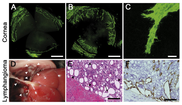
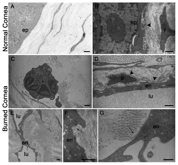
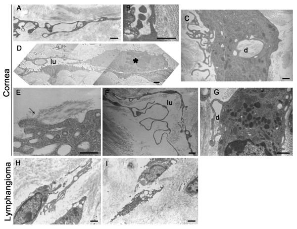
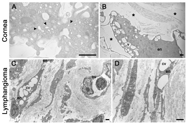
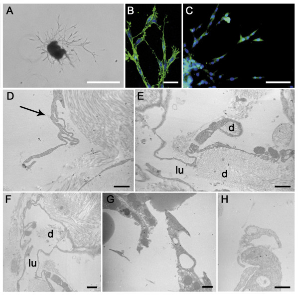
Background: Abnormal lymphatic vessel formation (lymphangiogenesis) is associated with different pathologies such as cancer, lymphedema, psoriasis and graft rejection. Lymphatic vasculature displays distinctive features than blood vasculature, and mechanisms underlying the formation of new lymphatic vessels during physiological and pathological processes are still poorly documented. Most studies on lymphatic vessel formation are focused on organism development rather than lymphangiogenic events occurring in adults. We have here studied lymphatic vessel formation in two in vivo models of pathological lymphangiogenesis (corneal assay and lymphangioma). These data have been confronted to those generated in the recently set up in vitro model of lymphatic ring assay. Ultrastructural analyses through Transmission Electron Microscopy (TEM) were performed to investigate tube morphogenesis, an important differentiating process observed during endothelial cell organization into capillary structures.

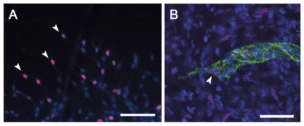
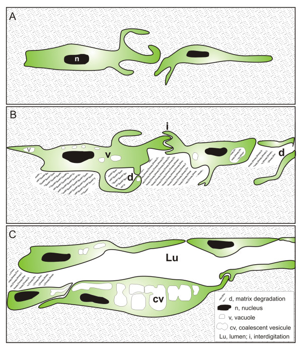
Results: In both in vivo models (lymphangiogenic corneal assay and lymphangioma), migrating lymphatic endothelial cells extended long processes exploring the neighboring environment and organized into cord-like structures. Signs of intense extracellular matrix remodeling were observed extracellularly and inside cytoplasmic vacuoles. The formation of intercellular spaces between endothelial cells led to tube formation. Proliferating lymphatic endothelial cells were detected both at the tips of sprouting capillaries and inside extending sprouts. The different steps of lymphangiogenesis observed in vivo are fully recapitulated in vitro, in the lymphatic ring assay and include: (1) endothelial cell alignment in cord like structure, (2) intracellular vacuole formation and (3) matrix degradation.

Conclusions: In this study, we are providing evidence for lymphatic vessel formation through tunneling relying on extensive matrix remodeling, migration and alignment of sprouting endothelial cells into tubular structures. In addition, our data emphasize the suitability of the lymphatic ring assay to unravel mechanisms underlying lymphangiogenesis.


Figures

Figure 1
Figure 1
Lymphangiogenesis in vivo. (A-C): Corneal lymphangiogenesis induced by thermal cauterization. Corneal flatmounts are labeled in green with an anti-LYVE-1 antibody and observed on fluorescent (A, B) or confocal (C) microscope. (A): In normal condition, cornea is avascular and the limbal vascular arcade is positive for LYVE-1 staining. (B, C): Seven days after an inflammatory stimulus, lymphatic vessels outgrow from the limbus towards the central cornea. (C): Migrating LEC display filopodia-like structures. (D-F): Mouse lymphangioma induced by intraperitoneal injection of incomplete Freund's adjuvant. (D): Lymphangiomas appear as white masses at the surface of the diaphragm (white arrowheads). (E): Hematoxylin-eosin staining of a histological section of the diaphragm (black arrowheads: lymph vessels) (F): Lymphatic vessels are evidenced by LYVE-1 immunostaining. The arrow delineated the process of fusion leading to increased lumen size. Scale bars in (A, B): 1 mm, in (C): 20 μm, and in (E, F): 100 μm.
Figure 2
Figure 2
Electron microscopy of normal or burned mouse cornea. (A, B): Normal cornea reveals epithelial cells (ep) apposed on a regular basement membrane (arrowhead in B). Flattened fibroblastic cells (f) are surrounded by collagen fibrils in 2 perpendicular orientations. (C-G): burned cornea. (C): A neutrophil is seen in a remodeled collagen matrix. (D): A blood capillary lined by an endothelial cell (en) is surrounded by a continuous basal lamina (arrowhead) in which is incorporated a pericyte (p). (E): A lymphatic capillary is shown with a narrow irregular lumen (lu). (F, G): LEC (en) are associated with bundles of thin anchoring filaments into collagen fibrils (arrows). en = endothelial cell; lu = lumen. Scale bars in (A, E): 2 μm, in (B-D): 1 μm, and in (F, G): 0.5 μm.
Figure 3
Figure 3
Electron microscopy pictures of lymphangiogenesis in vivo. Lymphangiogenesis was observed after thermal cauterization of the cornea (A-G) and in lymphangioma (H, I). (A): Lymphatic endothelial cells (LEC) form long processes containing vesicles and delimitating extracellular spaces devoid of matrix or with reminiscent matrix fragments. Note the presence of intracellular vesicles in endothelial processes. (B): Endothelial cells are joined by interdigitations. (C): Intracellular vesicle contains matrix fragments (d). (D): Aligned endothelial cells form a tubular structure that delimits a narrow lumen (lu). The luminal surface of endothelial cells is ruffled with small cell processes. A mitotic endothelial cell is visible (*). (E): LEC are anchored to the matrix through anchoring filaments (arrow). (F): Tubular structures containing a lumen (lu) are lined by long cytoplasmic extensions of LEC. (G): Connection of two cell extensions delineates an extracellular space containing degradation products of the matrix that are reminiscent of collagen fibrils. (H, I): LEC are aligned and surrounded by matrix-free extracellular spaces. Note the presence of coalescent vacuoles. Scale bars in (A-C; F-H): 1 μm, in (D, I): 2 μm, and in (E): 0.5 μm.
Figure 4
Figure 4
Vacuolization and lumen formation during lymphangiogenesis in vivo. Lymphangiogenesis was observed after thermal cauterization of the cornea (A, B) and in lymphangioma (C, D). (A): Prominent pinocytic activity (arrowhead) is visible along the plasma membrane and at cell junction. (B) Endothelial cells (en) containing intracellular vesicles are aligned and surrounded by matrix-free extracellular spaces (*). (C): Aligned elongated endothelial cells surrounded by extracellular spaces. Note the coalescence of intracellular vesicles (cv) and the presence of a blood vessel (bv) containing a white cell. (D) Vesicle coalescence (cv) into an intracellular luminal space is visible through a process similar to that depicted in B. bv = blood vessel; cv = coalescent vesicle; en: endothelial cell. Scale bars: 1 μm.
Figure 5
Figure 5
Lymphatic ring assay. (A): Visualization by optical microscopy of LEC spreading from mouse thoracic duct fragment embedded for 11 days in a 3D-type I collagen gel. (B): Outgrowing LEC display filopodia-like structures visible upon phalloidin staining under confocal microscopy. (C) LEC are labeled in green with an anti-LYVE-1 antibody and observed under a fluorescent microscope. Nuclei are counterstained in blue (Dapi). (D-H) Electron microscopy micrographs of 3D-cultures of lymphatic thoracic duct rings. (D): Long extended processes of LEC. Note the invagination process (arrow). (E, F): Establishment of an irregular lumen in tubular structures lined by long thin endothelial cell processes. (G, H): Sprouting endothelial cells display processes and intracellular vesicles. d = matrix degradation. d = matrix degradation; lu = lumen. Scale bars in (A): 500 μm, in (B): 40 μm, in (C): 100 μm, and in (D-H): 1 μm.
Figure 6
Figure 6
LEC proliferation during lymphangiogenesis. Cell proliferation was visualised by immunostaining following BrdU incorporation (red) in vitro, in the lymphatic ring assay (A) and in vivo, in whole mounted burned cornea (B). Proliferating LEC at the tip of capillary bud (arrowhead) are detected both in vitro (A) and in vivo (B). LEC are labeled in green with an anti-LYVE-1 antibody (B). Nuclei are counterstained in blue with Dapi (A) or TO-PRO3 (B). The whole mounted samples are observed under fluorescent (A) or confocal (B) microscope. Scale bars in (A): 100 μm and in (B): 40 μm.
Figure 7
Figure 7
Modulation of lymphatic vessel outgrowth by protease inhibitor. Lymphatic rings cultured in the presence of serum were treated with a broad spectrum MMP inhibitor (GM6001) at 0.1 μg/ml and 0.001 μg/ml. Quantification of LEC sprouting was measured by determining the number of intersections between capillaries and a grid obtained by the dilatation of the ring boundary, as previously described [38]. The number of intersections (Ni) is plotted as a function of the distance to the ring. * = P < 0.05.
Figure 8
Figure 8
Tunneling model of lymphatic vessel formation. The model is based on ultrastructural observations performed in in vitro and in vivo models of lymphangiogenesis. (A): LEC alignment. Elongated LEC migrate and extend long cytoplasmic protrusions. (B): Vacuolization and matrix degradation. The continuity of LEC lining is mediated by interdigitations (i). Vesicle invaginations lead to the formation of intracellular vacuoles (v) in the cytoplasm and in protrusions. Matrix degradation (d) occurs intracellularly and extracellularly generating space between cells. (C): Luminogenesis. The lumen (lu) is formed de novo in the intercellular space. The intracellular vacuoles coalesce (cv) and likely fuse with the cytoplasmic membrane to increase the lumen.

Similar articles

Cited by

References

    1. Cursiefen C, Chen L, Dana MR, Streilein JW. Corneal lymphangiogenesis: evidence, mechanisms, and implications for corneal transplant immunology. Cornea. 2003;22:273–281. doi: 10.1097/00003226-200304000-00021. - DOI - PubMed
    1. Ellenberg D, Azar DT, Hallak JA, Tobaigy F, Han KY, Jain S, Zhou Z, Chang JH. Novel aspects of corneal angiogenic and lymphangiogenic privilege. Prog Retin Eye Res. 2010;29:208–248. doi: 10.1016/j.preteyeres.2010.01.002. - DOI - PMC - PubMed
    1. Karpanen T, Alitalo K. Molecular biology and pathology of lymphangiogenesis. Annu Rev Pathol. 2008;3:367–397. doi: 10.1146/annurev.pathmechdis.3.121806.151515. - DOI - PubMed
    1. Tammela T, Alitalo K. Lymphangiogenesis: Molecular mechanisms and future promise. Cell. 2010;140:460–476. doi: 10.1016/j.cell.2010.01.045. - DOI - PubMed
    1. Thiele W, Sleeman JP. Tumor-induced lymphangiogenesis: a target for cancer therapy? J Biotechnol. 2006;124:224–241. doi: 10.1016/j.jbiotec.2006.01.007. - DOI - PubMed

Publication types

MeSH terms

Substances