Androgen receptor driven transcription in molecular apocrine breast cancer is mediated by FoxA1
- PMID: 21701558
- PMCID: PMC3160190
- DOI: 10.1038/emboj.2011.216
Androgen receptor driven transcription in molecular apocrine breast cancer is mediated by FoxA1
Erratum in
- EMBO J. 2012 Mar 21;31(6):1617
Abstract
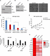
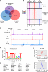
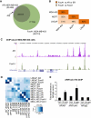
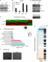
Breast cancer is a heterogeneous disease and several distinct subtypes exist based on differential gene expression patterns. Molecular apocrine tumours were recently identified as an additional subgroup, characterised as oestrogen receptor negative and androgen receptor positive (ER- AR+), but with an expression profile resembling ER+ luminal breast cancer. One possible explanation for the apparent incongruity is that ER gene expression programmes could be recapitulated by AR. Using a cell line model of ER- AR+ molecular apocrine tumours (termed MDA-MB-453 cells), we map global AR binding events and find a binding profile that is similar to ER binding in breast cancer cells. We find that AR binding is a near-perfect subset of FoxA1 binding regions, a level of concordance never previously seen with a nuclear receptor. AR functionality is dependent on FoxA1, since silencing of FoxA1 inhibits AR binding, expression of the majority of the molecular apocrine gene signature and growth cell growth. These findings show that AR binds and regulates ER cis-regulatory elements in molecular apocrine tumours, resulting in a transcriptional programme reminiscent of ER-mediated transcription in luminal breast cancers.
Conflict of interest statement
The authors declare that they have no conflict of interest.
Figures


Similar articles
-
Overexpression of androgen receptor and forkhead-box A1 protein in apocrine breast carcinoma.Anticancer Res. 2014 Mar;34(3):1261-7. Anticancer Res. 2014. PMID: 24596370
-
Androgen receptor and FOXA1 coexpression define a "luminal-AR" subtype of feline mammary carcinomas, spontaneous models of breast cancer.BMC Cancer. 2019 Dec 30;19(1):1267. doi: 10.1186/s12885-019-6483-6. BMC Cancer. 2019. PMID: 31888566 Free PMC article.
-
Identification of molecular apocrine breast tumours by microarray analysis.Oncogene. 2005 Jul 7;24(29):4660-71. doi: 10.1038/sj.onc.1208561. Oncogene. 2005. PMID: 15897907
-
Apocrine lesions of the breast: part 2 of a two-part review. Invasive apocrine carcinoma, the molecular apocrine signature and utility of immunohistochemistry in the diagnosis of apocrine lesions of the breast.J Clin Pathol. 2019 Jan;72(1):7-11. doi: 10.1136/jclinpath-2018-205485. Epub 2018 Nov 13. J Clin Pathol. 2019. PMID: 30425121 Review.
-
FOXA1: a transcription factor with parallel functions in development and cancer.Biosci Rep. 2012 Apr 1;32(2):113-30. doi: 10.1042/BSR20110046. Biosci Rep. 2012. PMID: 22115363 Free PMC article. Review.
Cited by
-
Human Epidermal Growth Factor Receptor-3 Expression Is Regulated at Transcriptional Level in Breast Cancer Settings by Junctional Adhesion Molecule-A via a Pathway Involving Beta-Catenin and FOXA1.Cancers (Basel). 2021 Feb 19;13(4):871. doi: 10.3390/cancers13040871. Cancers (Basel). 2021. PMID: 33669586 Free PMC article.
-
Metastatic colon cancer of the small intestine diagnosed using genetic analysis: a case report.Diagn Pathol. 2020 Aug 31;15(1):106. doi: 10.1186/s13000-020-01019-6. Diagn Pathol. 2020. PMID: 32867793 Free PMC article.
-
A Combination of RNA-Seq Analysis and Use of TCGA Database for Determining the Molecular Mechanism and Identifying Potential Drugs for GJB1 in Ovarian Cancer.Onco Targets Ther. 2021 Apr 14;14:2623-2633. doi: 10.2147/OTT.S303589. eCollection 2021. Onco Targets Ther. 2021. PMID: 33883906 Free PMC article.
-
Prognostic Role of Androgen Receptor in Triple Negative Breast Cancer: A Multi-Institutional Study.Cancers (Basel). 2019 Jul 17;11(7):995. doi: 10.3390/cancers11070995. Cancers (Basel). 2019. PMID: 31319547 Free PMC article.
-
Androgen Receptor: A Complex Therapeutic Target for Breast Cancer.Cancers (Basel). 2016 Dec 2;8(12):108. doi: 10.3390/cancers8120108. Cancers (Basel). 2016. PMID: 27918430 Free PMC article. Review.
References
-
- Benjamini Y, Hochberg Y (1995) Controlling the false discovery rate: a practical and powerful approach to multiple testing. J Royal Stat Soc B 57: 289–300
-
- Birney E, Stamatoyannopoulos JA, Dutta A, Guigo R, Gingeras TR, Margulies EH, Weng Z, Snyder M, Dermitzakis ET, Thurman RE, Kuehn MS, Taylor CM, Neph S, Koch CM, Asthana S, Malhotra A, Adzhubei I, Greenbaum JA, Andrews RM, Flicek P et al. (2007) Identification and analysis of functional elements in 1% of the human genome by the ENCODE pilot project. Nature 447: 799–816 - PMC - PubMed
-
- Birrell SN, Bentel JM, Hickey TE, Ricciardelli C, Weger MA, Horsfall DJ, Tilley WD (1995) Androgens induce divergent proliferative responses in human breast cancer cell lines. J Steroid Biochem Mol Biol 52: 459–467 - PubMed
-
- Birrell SN, Hall RE, Tilley WD (1998) Role of the androgen receptor in human breast cancer. J Mammary Gland Biol Neoplasia 3: 95–103 - PubMed
Publication types
MeSH terms
Substances
Grants and funding
LinkOut - more resources
Full Text Sources
Other Literature Sources
Medical
Molecular Biology Databases
Research Materials
Miscellaneous

