Converging pathways lead to overproduction of IL-17 in the absence of vitamin D signaling
- PMID: 21697289
- PMCID: PMC3139478
- DOI: 10.1093/intimm/dxr045
Converging pathways lead to overproduction of IL-17 in the absence of vitamin D signaling
Abstract
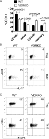
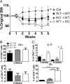
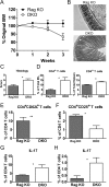
Multiple pathways converge to result in the overexpression of T(h)17 cells in the absence of either vitamin D or the vitamin D receptor (VDR). CD4(+) T cells from VDR knockout (KO) mice have a more activated phenotype than their wild-type (WT) counterparts and readily develop into T(h)17 cells under a variety of in vitro conditions. Vitamin D-deficient CD4(+) T cells also overproduced IL-17 in vitro and 1,25 dihydroxyvitamin D(3) inhibited the development of T(h)17 cells in CD4(+) T-cell cultures. Conversely, the induction of inducible (i) Tregs was lower in VDR KO CD4(+) T cells than WT and the VDR KO iTregs were refractory to IL-6 inhibition. Host-specific effects of the VDR were evident on in vivo development of naive T cells. Development of naive WT CD4(+) T cells in the VDR KO host resulted in the overexpression of IL-17 and more severe experimental inflammatory bowel disease (IBD). The increased expression of T(h)17 cells in the VDR KO mice was associated with a reduction in tolerogenic CD103(+) dendritic cells. The data collectively demonstrate that T(h)17 and iTreg cells are direct and indirect targets of vitamin D. The increased propensity for development of T(h)17 cells in the VDR KO host results in more severe IBD.
Figures


Similar articles
-
Failure of T cell homing, reduced CD4/CD8alphaalpha intraepithelial lymphocytes, and inflammation in the gut of vitamin D receptor KO mice.Proc Natl Acad Sci U S A. 2008 Dec 30;105(52):20834-9. doi: 10.1073/pnas.0808700106. Epub 2008 Dec 18. Proc Natl Acad Sci U S A. 2008. PMID: 19095793 Free PMC article.
-
Vitamin D receptor expression controls proliferation of naïve CD8+ T cells and development of CD8 mediated gastrointestinal inflammation.BMC Immunol. 2014 Feb 7;15:6. doi: 10.1186/1471-2172-15-6. BMC Immunol. 2014. PMID: 24502291 Free PMC article.
-
Mechanisms underlying the effect of vitamin D on the immune system.Proc Nutr Soc. 2010 Aug;69(3):286-9. doi: 10.1017/S0029665110001722. Epub 2010 Jun 2. Proc Nutr Soc. 2010. PMID: 20515520 Free PMC article.
-
Vitamin D status, 1,25-dihydroxyvitamin D3, and the immune system.Am J Clin Nutr. 2004 Dec;80(6 Suppl):1717S-20S. doi: 10.1093/ajcn/80.6.1717S. Am J Clin Nutr. 2004. PMID: 15585793 Review.
-
Vitamin D regulation of immune function in the gut: why do T cells have vitamin D receptors?Mol Aspects Med. 2012 Feb;33(1):77-82. doi: 10.1016/j.mam.2011.10.014. Epub 2011 Nov 6. Mol Aspects Med. 2012. PMID: 22079836 Free PMC article. Review.
Cited by
-
Metabolic control of the Treg/Th17 axis.Immunol Rev. 2013 Mar;252(1):52-77. doi: 10.1111/imr.12029. Immunol Rev. 2013. PMID: 23405895 Free PMC article. Review.
-
Vitamin D inhibited endometriosis development in mice model through interleukin-17 modulation.Open Vet J. 2022 Nov-Dec;12(6):956-964. doi: 10.5455/OVJ.2022.v12.i6.23. Epub 2022 Dec 9. Open Vet J. 2022. PMID: 36650872 Free PMC article.
-
Peripheral Neuropathies Derived from COVID-19: New Perspectives for Treatment.Biomedicines. 2022 May 2;10(5):1051. doi: 10.3390/biomedicines10051051. Biomedicines. 2022. PMID: 35625788 Free PMC article. Review.
-
The Role of Vitamin D in Immune System and Inflammatory Bowel Disease.J Inflamm Res. 2022 May 28;15:3167-3185. doi: 10.2147/JIR.S363840. eCollection 2022. J Inflamm Res. 2022. PMID: 35662873 Free PMC article. Review.
-
Anti-Inflammatory Effects of Vitamin D on Human Immune Cells in the Context of Bacterial Infection.Nutrients. 2016 Dec 12;8(12):806. doi: 10.3390/nu8120806. Nutrients. 2016. PMID: 27973447 Free PMC article.
References
-
- Podolsky DK. Inflammatory bowel disease. N. Engl. J. Med. 2002;347:417. - PubMed
Publication types
MeSH terms
Substances
Grants and funding
LinkOut - more resources
Full Text Sources
Other Literature Sources
Medical
Molecular Biology Databases
Research Materials

