

Gastric tuft cells express DCLK1 and are expanded in hyperplasia
- PMID: 21688022
- PMCID: PMC3570962
- DOI: 10.1007/s00418-011-0831-1
Gastric tuft cells express DCLK1 and are expanded in hyperplasia
Abstract
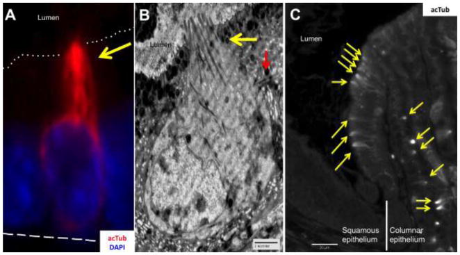
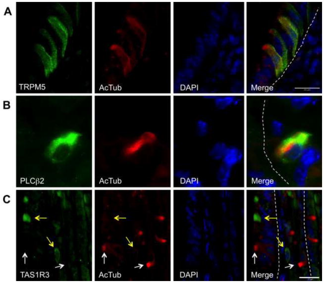
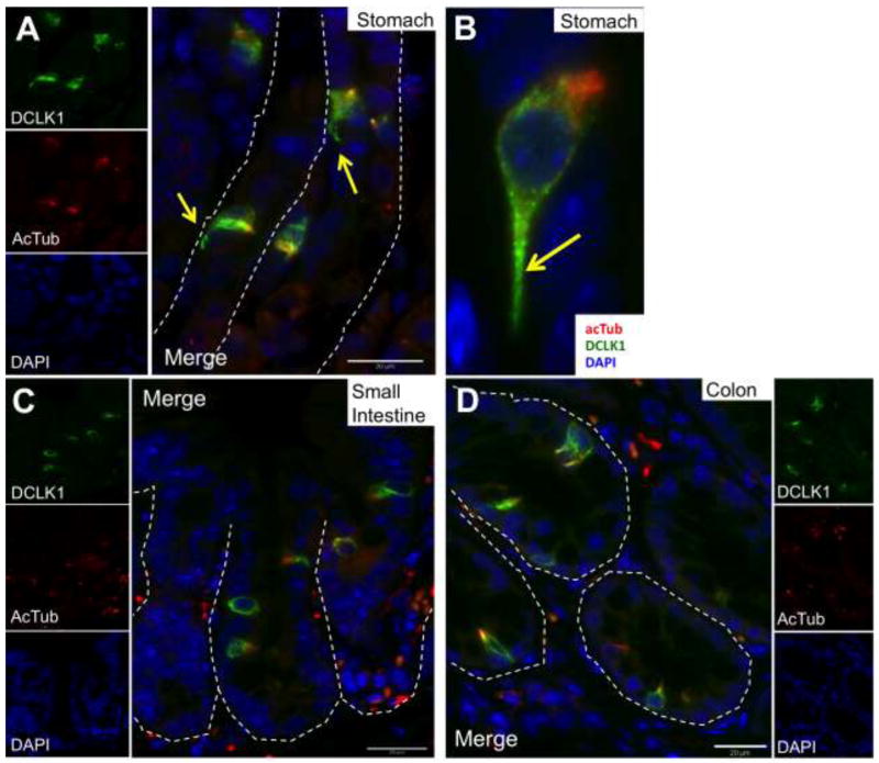
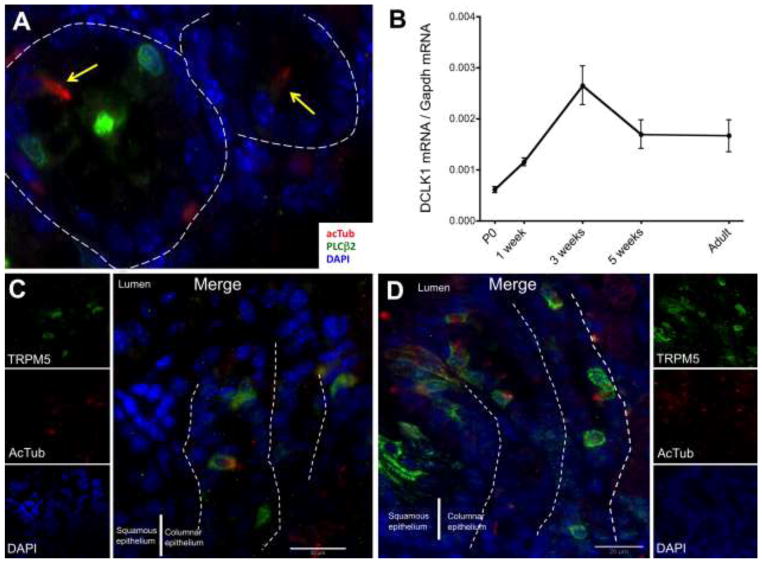
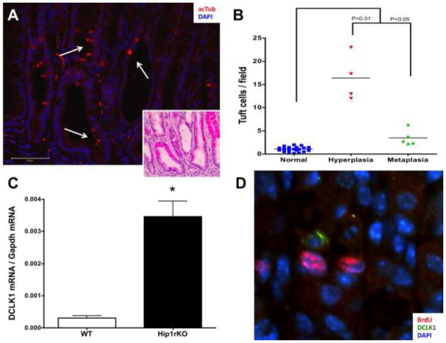
Epithelial tuft cells are named after their characteristic microtubule bundles located at the cell apex where these are exposed to the luminal environment. As such, tuft cells are found in multiple organs, including the gastrointestinal (GI) tract where the apical "tuft" is hypothesized to detect and transmit environmental signals. Thus, the goal of our study was to characterize gastric tuft cells during GI tract development, then subsequently in the normal and metaplastic adult stomach. GI tracts from mouse embryos, and newborn and postnatal mice were analyzed. Tuft cells were identified by immunohistochemistry using acetylated-α-tubulin (acTub) antibody to detect the microtubule bundle. Additional tuft cell markers, e.g., doublecortin-like kinase 1 (DCLK1), were used to co-localize with acTub. Tuft cells were quantified in human gastric tissue arrays and in mouse stomachs with or without inflammation. In the developing intestine, tuft cells in both the crypts and villi expressed all markers by E18.5. In the stomach, acTub co-localized with DCLK1 and other established tuft cell markers by E18.5 in the antrum, but not until postnatal day 7 in the corpus, with the highest density of tuft cells clustered at the forestomach ridge. Tuft cell numbers increased in hyperplastic human and mouse stomachs. In the adult GI tract, the tuft cell marker acTub co-expressed with DCKL1 and chemosensory markers, e.g.,TRPM5. In summary, tuft cells appear in the gastric antrum and intestine at E18.5, but their maximal numbers in the corpus are not achieved until after weaning. Tuft cell numbers increase with inflammation, hyperplasia, and metaplasia.
Figures
Similar articles
-
Dclk1-expressing tuft cells: critical modulators of the intestinal niche?Am J Physiol Gastrointest Liver Physiol. 2017 Oct 1;313(4):G285-G299. doi: 10.1152/ajpgi.00073.2017. Epub 2017 Jul 6. Am J Physiol Gastrointest Liver Physiol. 2017. PMID: 28684459 Free PMC article. Review.
-
Dynamic tuft cell expansion during gastric metaplasia and dysplasia.J Pathol Clin Res. 2024 Jan;10(1):e352. doi: 10.1002/cjp2.352. Epub 2023 Dec 20. J Pathol Clin Res. 2024. PMID: 38117182 Free PMC article.
-
Dynamic expansion of gastric mucosal doublecortin-like kinase 1-expressing cells in response to parietal cell loss is regulated by gastrin.Am J Pathol. 2015 Aug;185(8):2219-31. doi: 10.1016/j.ajpath.2015.04.009. Epub 2015 Jun 12. Am J Pathol. 2015. PMID: 26073039 Free PMC article.
-
A novel serotonin-containing tuft cell subpopulation in mouse intestine.Cell Tissue Res. 2019 May;376(2):189-197. doi: 10.1007/s00441-018-02988-3. Epub 2019 Jan 21. Cell Tissue Res. 2019. PMID: 30666535
-
Advances in tuft cells, a chemosensory cell in sequential diseases of the pancreas.Biochim Biophys Acta Rev Cancer. 2023 Jul;1878(4):188911. doi: 10.1016/j.bbcan.2023.188911. Epub 2023 May 12. Biochim Biophys Acta Rev Cancer. 2023. PMID: 37182665 Review.
Cited by
-
Group 2 Innate Lymphoid Cells Coordinate Damage Response in the Stomach.Gastroenterology. 2020 Dec;159(6):2077-2091.e8. doi: 10.1053/j.gastro.2020.08.051. Epub 2020 Sep 4. Gastroenterology. 2020. PMID: 32891625 Free PMC article.
-
Thymic tuft cells: potential "regulators" of non-mucosal tissue development and immune response.Immunol Res. 2023 Aug;71(4):554-564. doi: 10.1007/s12026-023-09372-6. Epub 2023 Mar 24. Immunol Res. 2023. PMID: 36961668 Free PMC article. Review.
-
Gastric cancer and Hedgehog signaling pathway: emerging new paradigms.Genes Cancer. 2018 Jan;9(1-2):1-10. doi: 10.18632/genesandcancer.168. Genes Cancer. 2018. PMID: 29725500 Free PMC article. Review.
-
Evaluation of Lineage Changes in the Gastric Mucosa Following Infection With Helicobacter pylori and Specified Intestinal Flora in INS-GAS Mice.J Histochem Cytochem. 2019 Jan;67(1):53-63. doi: 10.1369/0022155418785621. Epub 2018 Jul 3. J Histochem Cytochem. 2019. PMID: 29969055 Free PMC article.
-
Dclk1-expressing tuft cells: critical modulators of the intestinal niche?Am J Physiol Gastrointest Liver Physiol. 2017 Oct 1;313(4):G285-G299. doi: 10.1152/ajpgi.00073.2017. Epub 2017 Jul 6. Am J Physiol Gastrointest Liver Physiol. 2017. PMID: 28684459 Free PMC article. Review.
References
-
- Barker N, Huch M, Kujala P, van de Wetering M, et al. Lgr5(+ve) stem cells drive self-renewal in the stomach and build long-lived gastric units in vitro. Cell Stem Cell. 2010;6:25–36. - PubMed
-
- Elia G, Chinery R, Hanby AM, Poulsom R, et al. The production and characterization of a new monoclonal antibody to the trefoil peptide human spasmolytic polypeptide. Histochem J. 1994;26:644–647. - PubMed
-
- Friis-Hansen L, Sundler F, Li Y, Gillespie PJ, et al. Impaired gastric acid secretion in gastrin-deficient mice. Am J Physiol. 1998;274:G561–568. - PubMed
-
- Gerbe F, Brulin B, Makrini L, Legraverend C, et al. DCAMKL-1 expression identifies Tuft cells rather than stem cells in the adult mouse intestinal epithelium. Gastroenterology. 2009;137:2179–2180. author reply 2180–2171. - PubMed
Publication types
MeSH terms
Substances
Grants and funding
LinkOut - more resources
Full Text Sources
Molecular Biology Databases
Research Materials
Miscellaneous
