. 2011 Jun 19;13(7):838-45.
doi: 10.1038/ncb2267.

Inhibition of glycogen synthase kinase-3 alleviates Tcf3 repression of the pluripotency network and increases embryonic stem cell resistance to differentiation

Affiliations

Inhibition of glycogen synthase kinase-3 alleviates Tcf3 repression of the pluripotency network and increases embryonic stem cell resistance to differentiation

Jason Wray et al. Nat Cell Biol. .

Erratum in

  • Nat Cell Biol. 2012 May;14(5):555

Abstract

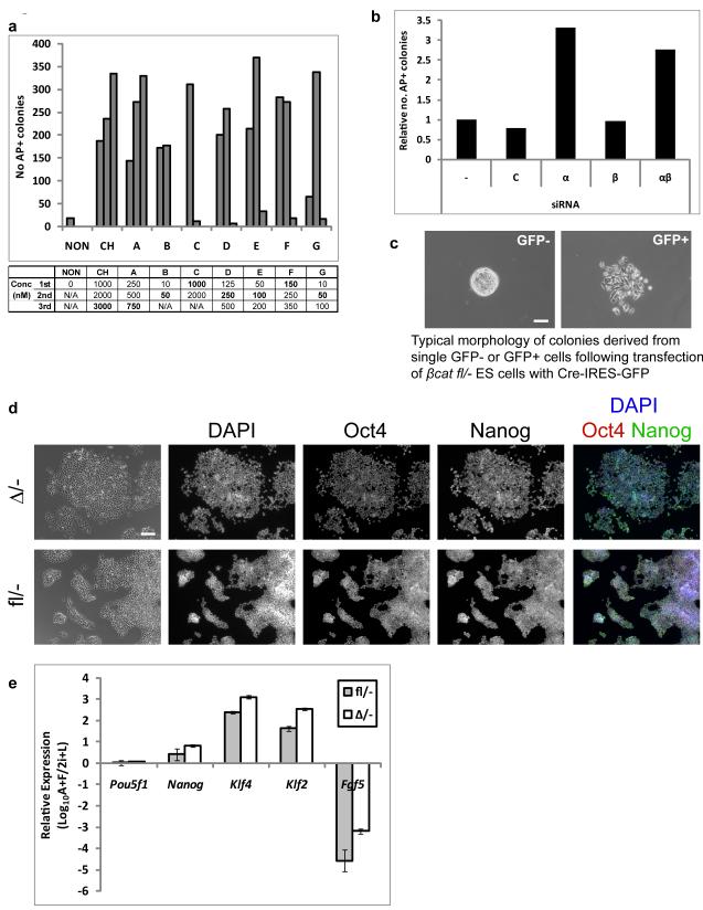
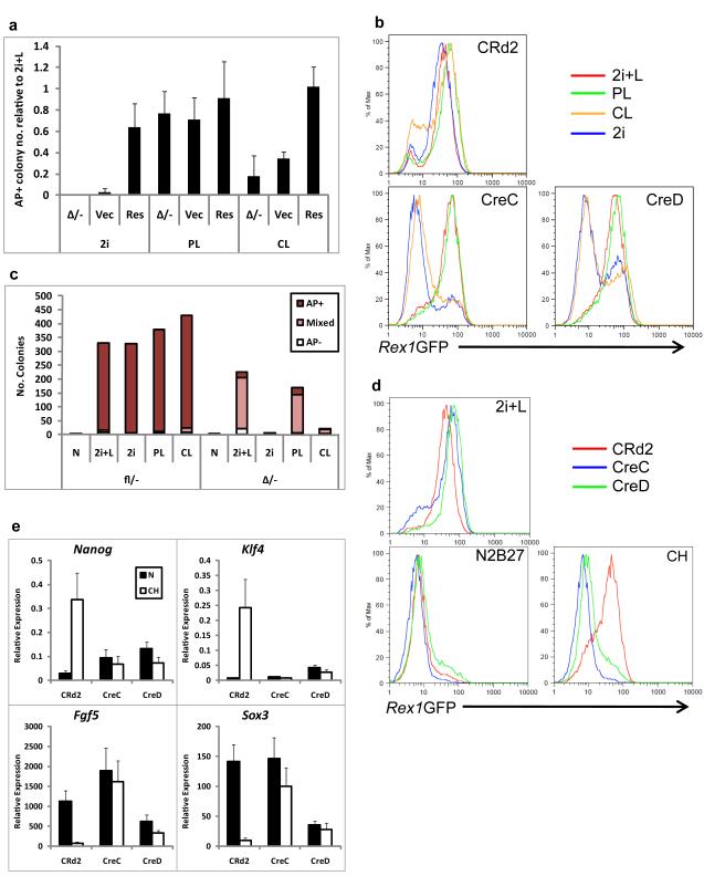
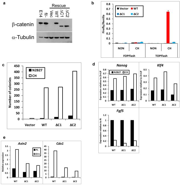
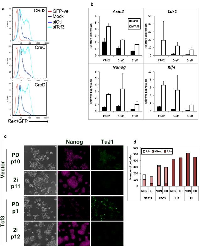
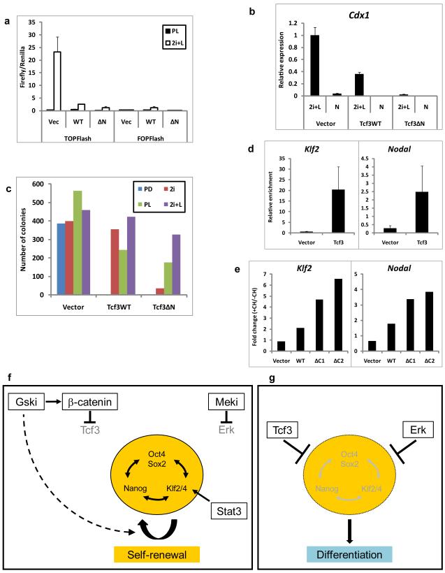
Self-renewal of rodent embryonic stem cells is enhanced by partial inhibition of glycogen synthase kinase-3 (Gsk3; refs 1, 2). This effect has variously been attributed to stimulation of Wnt signalling by β-catenin, stabilization of Myc protein and global de-inhibition of anabolic processes. Here we demonstrate that β-catenin is not necessary for embryonic stem cell identity or expansion, but its absence eliminates the self-renewal response to Gsk3 inhibition. Responsiveness is fully restored by truncated β-catenin lacking the carboxy-terminal transactivation domain. However, requirement for Gsk3 inhibition is dictated by expression of T-cell factor 3 (Tcf3) and mediated by direct interaction with β-catenin. Tcf3 localizes to many pluripotency genes in embryonic stem cells. Our findings confirm that Tcf3 acts as a transcriptional repressor and reveal that β-catenin directly abrogates Tcf3 function. We conclude that Gsk3 inhibition stabilizes the embryonic stem cell state primarily by reducing repressive influence on the core pluripotency network.


Figures

Figure 1
Figure 1. Suppression of Gsk3 mediates enhanced ES cell self-renewal but β-catenin is dispensable for ES cell maintenance
(a). Histogram showing number of undifferentiated (alkaline phosphatase positive, AP+), colonies formed from 600 E141VC ES cells plated in N2B27 with Mek inhibitor PD0325901 (PD, 1 μM) plus CHIRON99021 (CH); or alternative Gsk3 inhibitors A, B, C, D, E, F, and G (see methods for details). Concentrations (Conc) of the inhibitors (nM) are indicated in the table beneath the graph. 1st, 2nd and 3rd correspond to the bars from left to right. Optimum concentrations are shown in bold print. (b). Histogram showing relative number of undifferentiated (alkaline phosphatase positive, AP+), colonies formed from 600 3/4KO ES cells plated in N2B27 plus PD. Cells were untreated (−) or transfected with control siRNA (C) or siRNAs against Gsk3α (α), Gsk3β (β), or both (αβ). Mean of two biological replicates. (c). Phase contrast images showing typical morphology of primary colonies isolated from GFP-negative (left) and –positive (right) fractions of βcatfl/− ES cells transiently transfected with Cre-IRES-GFP. Note the lack of cell-cell contacts in colonies from the GFP-positive fraction. Scale bar, 200μm (d). Phase contrast and fluorescent images showing immunostaining of βcatfl/− and βcatΔ/− ES cells for Oct4 and Nanog. Scale bar, 100μm. (e). Histogram showing gene expression in βcatfl/− and βcatΔ/− ES cells cultured in 2i+LIF relative to EpiSC-like cells derived by culture in activin+FGF2 (A+F) for 7 passages. Klf4 and Klf2 are specific for naive ES cells while Fgf5 is up-regulated and Nanog down-regulated in EpiSCs. Mean ± s.d. of three biological replicates.
Figure 2
Figure 2. βcatΔ/− ES cells do not resist differentiation upon Gsk3 inhibition
(a). Histogram showing number of undifferentiated (alkaline phosphatase positive, AP+), colonies formed by parental βcatΔ/− or βcatΔ/− ES cells expressing a β-catenin transgene (Res) or the corresponding vector control (Vec) plated in N2B27 plus 2i, PD+LIF (PL) or CH+LIF (CL). Data are expressed relative to the number of colonies in 2i+LIF. Mean ± s.d. of three biological replicates. (b). Flow cytometry analysis of Rex1GFP expression in βcatfl/− (CRd2) or βcatΔ/− (CreC and CreD) Rex1GFP reporter ES cells cultured in 2i+LIF, PD+LIF (PL), CH+LIF (CL) or 2i for 96 hours. (c). Histogram showing number of undifferentiated (alkaline phosphatase positive, AP+), mixed and differentiated (AP−) colonies formed from 600 βcatfl/− or βcatΔ/− ES cells plated in 2i+LIF following 48 hours culture in N2B27 alone or plus 2i+LIF (2i+L), 2i, PD+LIF (PL) or CH+LIF (CL).(d). Flow cytometry analysis of Rex1GFP expression in βcatfl/− (CRd2) or βcatΔ/− (CreC and CreD) Rex1GFP reporter ES cells cultured in 2i+LIF, N2B27 alone or CH for 48 hours. (e). Histogram showing relative expression of pluripotency markers Nanog and Klf4 and early differentiation markers Fgf5 and Sox3 in βcatfl/− (CRd2) or βcatΔ/− (CreC and CreD) Rex1GFP reporter ES cells cultured in 2i+LIF, or N2B27 alone or plus CH for 48 hours. Mean ± s.d. of three biological replicates.
Figure 3
Figure 3. β-catenin inhibits differentiation independent of its transcriptional activation domain
(a). Western blot showing β-catenin and α-Tubulin (loading control) expression in wild-type (E14), βcatfl/− or “Rescue” βcatΔ/− ES cells expressing randomly integrated wild-type (WT) or C-terminal-deleted β-catenin (ΔC) transgenes or the corresponding vector control (Vec). ΔC1 and ΔC2 are independent clones. Uncropped images of blots are shown in Supplementary Fig. S9. (b). Histogram showing TOPFlash and FOPFlash reporter activity in βcatΔ/− cells expressing randomly integrated wild-type (WT) or C-terminal-deleted (ΔC) β-catenin transgenes in PD+LIF with or without CH. ΔC1 and ΔC2 are independent clones. Mean ± s.d. of three biological replicates. (c). Histogram showing number of undifferentiated colonies formed from 600 βcatΔ/− cells expressing randomly integrated wild-type (WT) or C-terminal-deleted (ΔC) β-catenin transgenes plated in 2i+LIF after 48 hours in N2B27 alone or plus CH. (d) Histograms showing relative expression of Nanog, Klf4, and Fgf5 in βcatΔ/− cells expressing randomly integrated wild-type (WT) or C-terminal-deleted β-catenin (ΔC) transgenes cultured in 2i+LIF or in N2B27 alone or plus CH for 24 hours. Expression is shown relative to levels in 2i+LIF. ΔC1 and ΔC2 are independent clones. (e). Histogram showing relative expression of Axin2 and Cdx1 in βcatΔ/− cells expressing randomly integrated wild-type (WT) or C-terminal-deleted (ΔC) β-catenin transgenes cultured in PD+LIF or 2i+LIF for 48 hours. Data in (c)-(e) are mean of two biological replicates.
Figure 4
Figure 4. β-catenin functions by abrogating Tcf3 repression
(a). Flow cytometry analysis showing the profile of Rex1GFP expression in βcatfl/− (CRd2) or βcatΔ/− (CreC and CreD) Rex1GFP reporter ES cells mock transfected (Mock) or transfected with control siRNAs (siCtl) or siRNA against Tcf3 (siTcf3) and cultured for 48 hours in N2B27 alone. GFP-ve: wild-type ES cells that do not express GFP. (b). Histograms showing relative expression of Axin2, Cdx1, Nanog and Klf4 in βcatfl/− (CRd2) or βcatΔ/− (CreC and CreD) Rex1GFP reporter ES cells. Cells were cultured for 24 hours in N2B27 alone and were transfected with control siRNAs (siCtl) or siRNAs against Tcf3 (siTcf3). Expression is shown relative to untreated. Mean ± s.d. of three biological replicates. (c). Phase contrast and fluorescent images showing Nanog and βIII-tubulin (TuJ1) expression in Tcf3-null ES cells stably expressing a randomly integrated Tcf3 transgene or the corresponding vector control cultured in N2B27 plus PD or plus 2i for the indicated number of passages (p). Scale bar, 100μm. (d). Histogram showing number of undifferentiated (AP+), mixed and differentiated (AP-) colonies formed from 600 Tcf3-null ES cells in N2B27 alone or plus PD, LIF or PD+LIF in the presence or absence of CH
Figure 5
Figure 5. Gsk3 Inhibition Relieves the Core Pluripotency Network from Repression by Tcf3 and Complements Mek Inhibition and/or Stat3 Activation to Stabilise ES Cell Self-Renewal
a) Histogram showing TOPFlash and FOPFlash activation in Tcf3-null ES cells stably expressing randomly integrated wild-type (WT) or N-terminal-deleted (ΔN) Tcf3 transgenes or the corresponding vector control (Vec) in PD+LIF(PL) or 2i+LIF (2i+L). Mean ± s.d. of three biological replicates is shown b) Histogram showing relative expression of Cdx1 in Tcf3-null ES cells stably expressing randomly integrated wild-type (Tcf3WT) or N-terminal-deleted (Tcf3ΔN) Tcf3 transgenes or the corresponding vector control in 2i+LIF (2i+L) or N2B27 alone. Mean ± s.d. of three biological replicates is shown c) Histogram showing number of undifferentiated colonies formed from 600 Tcf3-null ES cells stably expressing randomly integrated wild-type (Tcf3WT) or N-terminal-deleted (Tcf3ΔN) Tcf3 transgenes or the corresponding vector control in N2B27 plus PD, 2i, PD+LIF (PL) or 2i+LIF (2i+L). Mean of two biological replicates. d) Histogram showing relative enrichment of Klf2 and Nodal promoter regions following chromatin immunoprecipitation for Tcf3 in Tcf3-null ES cells stably expressing a randomly integrated Tcf3 transgene or the corresponding vector control. Mean ± sd of three replicates is shown e) Histogram showing response to CH of Klf2 and Nodal in βcatΔ/− cells expressing randomly integrated wild-type (WT) or C-terminal-deleted (ΔC) β-catenin transgenes cultured in N2B27 alone for 24 hours followed by 8 hours in N2B27 plus PD in the presence or absence of CH. Mean ratio (+CH/−CH) of two biological replicates. f) In the presence of Gsk-3 inhibitor (Gski) and a mitogen activated protein kinase (Erk) kinase inhibitor (Meki), repressive effects on the pluripotent gene regulatory network are abolished. The pluripotent circuitry is also positively regulated by Stat3 acting primarily through Klf4. Any two of these three effects are sufficient to stabilise the network and sustain ES cell self-renewal. Gski generates intracellular β-catenin which interacts with Tcf3 and abolishes its repressor effect on multiple genes in the pluripotent network. Gski additionally supports ES cell propagation through stimulatory effects on metabolic and biosynthetic processes (dashed arrow). g) In the absence of inhibitors, Tcf3 repression and activated Erk drive ES cells into differentiation. When ES cells are maintained in serum using LIF without inhibitors, cultures are heterogeneous and metastable due to co-existence of states (f) and (g).

Comment in

Similar articles

Cited by

References

    1. Sato N, Meijer L, Skaltsounis L, Greengard P, Brivanlou AH. Maintenance of pluripotency in human and mouse embryonic stem cells through activation of Wnt signaling by a pharmacological GSK-3-specific inhibitor. Nat Med. 2004;10:55–63. - PubMed
    1. Ying QL, et al. The ground state of embryonic stem cell self-renewal. Nature. 2008;453:519–23. - PMC - PubMed
    1. Cartwright P, et al. LIF/STAT3 controls ES cell self-renewal and pluripotency by a Myc-dependent mechanism. Development. 2005;132:885–96. - PubMed
    1. Doble BW, Woodgett JR. GSK-3: tricks of the trade for a multi-tasking kinase. J Cell Sci. 2003;116:1175–86. - PMC - PubMed
    1. Hsu SC, Galceran J, Grosschedl R. Modulation of transcriptional regulation by LEF-1 in response to Wnt-1 signaling and association with beta-catenin. Mol Cell Biol. 1998;18:4807–18. - PMC - PubMed

Publication types

MeSH terms

Substances