. 2011 Jun 17;30(15):3134-46.
doi: 10.1038/emboj.2011.203.

Molecular basis of positive allosteric modulation of GluN2B NMDA receptors by polyamines

Affiliations

Molecular basis of positive allosteric modulation of GluN2B NMDA receptors by polyamines

Laetitia Mony et al. EMBO J. .

Abstract

NMDA receptors (NMDARs) form glutamate-gated ion channels that have central roles in neuronal communication and plasticity throughout the brain. Dysfunctions of NMDARs are involved in several central nervous system disorders, including stroke, chronic pain and schizophrenia. One hallmark of NMDARs is that their activity can be allosterically regulated by a variety of extracellular small ligands. While much has been learned recently regarding allosteric inhibition of NMDARs, the structural determinants underlying positive allosteric modulation of these receptors remain poorly defined. Here, we show that polyamines, naturally occurring polycations that selectively enhance NMDARs containing the GluN2B subunit, bind at a dimer interface between GluN1 and GluN2B subunit N-terminal domains (NTDs). Polyamines act by shielding negative charges present on GluN1 and GluN2B NTD lower lobes, allowing their close apposition, an effect that in turn prevents NTD clamshell closure. Our work reveals the mechanistic basis for positive allosteric modulation of NMDARs. It provides the first example of an intersubunit binding site in this class of receptors, a discovery that holds promise for future drug interventions.


Conflict of interest statement

The authors declare that they have no conflict of interest.

Figures

Figure 1
Figure 1
Properties of the glycine-independent and voltage-independent spermine potentiation. (A) The extent of spermine potentiation is greatly magnified by decreasing extracellular pH (pHext). Typical current traces obtained at three different pHext from oocytes expressing wild-type (wt) GluN1/GluN2B receptors. Spermine was applied at 200 μM. The bars above the current traces indicate the duration of agonists and spermine applications. Inset: Mean relative current amplitudes. Values are 9.3±0.7 (n=4), 1.33±0.06 (n=5) and 0.6±0.1 (n=5) at pHext of 6.3, 7.3 and 8.3, respectively. (B) Spermine potentiation is GluN2B specific. Typical current traces obtained at pHext=6.5 from oocytes expressing NMDARs incorporating wt GluN1 and either one of the four wt GluN2 subunits. Spermine was applied at 200 μM. Inset: mean relative current amplitudes. Values are 0.90±0.04 (n=17), 8.0±1.6 (n=50), 0.88±0.04 (n=7) and 0.86±0.02 (n=7) for GluN2A-, GluN2B-, GluN2C- and GluN2D-containing receptors, respectively. The dashed line in the bar graphs indicates the lack of spermine effect (Ispermine/I0=1).
Figure 2
Figure 2
Both GluN1 and GluN2B NTDs are required for spermine potentiation. (A) Typical current traces from oocytes expressing receptors incorporating wild-type GluN1 and different chimeric GluN2 subunits, or YFP-GluN2B and a NTD-deleted GluN1 subunit (GluN1-ΔNTD) (lower trace; see Materials and methods). Spermine was applied at 200 μM; pHext=6.5. (B) Summary of the spermine-induced effects. Values are (from left to right): 8.0±1.6 (n=50), 1.00±0.01 (n=5), 0.99±0.04 (n=8), 5.5±0.3 (n=5), 1.0±0.1 (n=11), 0.94±0.05 (n=16), 1.00±0.04 (n=7), 2.3±0.2 (n=8), 5.1±1.1 (n=8) and 0.90±0.04 (n=17). (C) Spermine dose–response curves of receptors containing GluN2B wt, GluN2A-2B(NTD) or GluN2A-2B(NTD+L) subunits. The spermine EC50, maximal potentiation and Hill coefficient are, respectively: 127±5 μM, 11.4±0.3 and 1.40±0.08 (n=6) for wt GluN1/GluN2B receptors; 260±30 μM, 11.0±1.0 and 1.33±0.07 (n=5) for GluN1 wt/GluN2A-2B(NTD+L) receptors and 370±30 μM, 4.9±0.2 and 1.03±0.05 (n=5) for GluN1 wt/GluN2A-2B(NTD) receptors.
Figure 3
Figure 3
The β6–β8 region in the lower lobe of GluN2B NTD is critical for spermine potentiation. (A) Sequence alignment of the β6–β8 regions of GluN1, GluN2A and GluN2B subunits. Indicated α-helices (coils) and β-strands (arrows) are from GluN2B NTD crystal structure (pdb 3JPW; Karakas et al, 2009). Secondary structures are numbered according to the secondary structures of the AMPA GluA2 NTD (Jin et al, 2009). Red boxes correspond to acidic residues previously shown to influence spermine sensitivity (Masuko et al, 1999). Other acidic residues located in the β6–β8 region are highlighted by orange boxes. Limits of the different GluN2A/GluN2B NTD chimeras are represented by brackets and coloured arrows. (B) CPK (Corey–Pauling–Koltun) representation of GluN2B NTD (pdb 3JPW; Karakas et al, 2009) viewed from its putative dimerization face. The star indicates the position of the domain interlobe cleft. The green, red and blue represent the different regions exchanged between GluN2A and GluN2B in the chimeras (see corresponding coloured arrows in (A)). (C) (Top) Schematic representation of a dimer of LIVBP-like domains. The area corresponding to the β6–β8 regions of GluN1 and GluN2B NTDs is squared. (Bottom) Electrostatic potential surface of GluN1 (pdb: 3Q41; Farina et al, 2011) and GluN2B NTDs (pdb: 3JPW; Karakas et al, 2009) viewed from their putative dimerization face. Stars indicate the position of the domain interlobe cleft. Note the high density of negative charges in the lobe 2 dimerization face of both GluN1 and GluN2B NTDs. Dashed circles represent the clusters of residues highlighted in red in (A). UL, upper lobe; LL, lower lobe. (D) Spermine sensitivities of the different β6–β8 chimeras. Values are, from top to bottom: 3.4±0.4 (n=8), 2.6±0.2 (n=7), 2.1±0.2 (n=8), 0.94±0.03 (n=10) and 1.28±0.05 (n=11). ***P<0.001 (Student's t-test). (E) Spermine dose–response curve of GluN1 wt/GluN2A-2B(175–227) receptors. The spermine EC50, maximal potentiation and Hill coefficient are, respectively: 1.36±0.08 mM, 4.2±0.2 and 1.54±0.05 (n=5). The dashed and dotted curves represent spermine dose–response curves for GluN1 wt/GluN2A-2B(NTD) and GluN1 wt/GluN2A-2B(NTD+L) receptors, respectively (replotted from Figure 2C).
Figure 4
Figure 4
GluN1 and GluN2B NTDs assemble as heterodimers. (A) (Top) Chemical structure and length of M2M. (Bottom) Schematic depiction of the cross-linking experiment. (B) Western blots using an anti-GluN1 antibody on membrane fractions of Xenopus oocytes expressing four combinations of wt and mutated GluN1 and GluN2B subunits. Samples were run in non-reducing conditions after incubation of the intact oocytes in either 1% DMSO (−) or 2 mM M2M (+). The arrowheads indicate non-specific bands (also seen in non-injected oocytes). (C) Functional effects of M2 M applications (0.2 mM, pHext=6.5). Values are (from left to right): 0.97±0.09 (n=4), 1.3±0.3 (n=6), 1.7±0.2 (n=8) and 3.0±0.3 (n=6). ***P<0.001 (Student's t-test). (D) Spermine sensitivities before and after treatment with 200 μM M2M. The dashed line represents the absence of spermine effect. Note the complete abolition of spermine potentiation after M2M treatment of GluN1-E181C/GluN2B-E201C receptors. Each bar corresponds to the mean value from 4 to 6 different cells. ***P<0.001; NS, non-significant (Student's t-test).
Figure 5
Figure 5
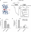
Mimicking spermine potentiation by introducing basic residues. (A) Typical current traces from oocytes expressing receptors in which acidic residues located in the putative lower lobe dimer interface between GluN1 and GluN2B NTDs were substituted by positively charged (arginine or lysine) or neutral (alanine) residues. Spermine was applied at 200 μM; pHext=6.5. (B) Examples of spermine sensitivities of a series of mutant receptors substituted with positively charged residues at different GluN1 and GluN2B positions. Values are, from left to right: 8.0±1.6 (n=50), 1.43±0.08 (n=9), 1.9±0.1 (n=4), 0.83±0.07 (n=9), 0.86±0.03 (n=4), 1.10±0.01 (n=5), 0.93±0.02 (n=6) and 0.91±0.01 (n=3). The numbers in bold indicate the number of charge inversions (i) or charge neutralizations (n). (C) MK-801 inhibition kinetics of the mutant receptors described in (B). Onset time constants (τon) were normalized to the value obtained for wild-type GluN1/GluN2B receptors. Values are, from left to right: 1.0±0.1 (n=86), 0.38±0.07 (n=8), 0.33±0.06 (n=4), 0.27±0.04 (n=15), 0.28±0.04 (n=4), 0.34±0.03 (n=6), 0.40±0.06 (n=6) and 1.1±0.1 (n=6).
Figure 6
Figure 6
Spermine stabilizes an open-cleft conformation of GluN2B NTD. (A) Ribbon representation of GluN2B NTD (pdb: 3JPW; Karakas et al, 2009). The interlobe cleft residue Y282 is highlighted in red CPK. UL, upper lobe; LL, lower lobe. (B) Typical current traces from oocytes expressing GluN1wt/GluN2B-Y282C receptors during treatment with MTSPtrEA (200 μM) with or without spermine (200 μM). Inset: MTSPtrEA modification kinetics are faster in the presence of spermine (normalized currents). (C) Time constants of MTSPtrEA-induced potentiation (τon MTSPtrEA) of GluN1 wt/GluN2B-Y282C receptors. Mean values are 18±2 s (n=16) in the presence of spermine and 9±3 s (n=17) in its absence. ***P<0.001 (Student's t-test). (D) Mean amplitudes of MTSPtrEA-induced potentiations on GluN1 wt/GluN2B-Y282C and GluN1 wt/GluN2B-Y282S receptors in the presence or absence of 200 μM spermine. Values are (from left to right): 6.3±0.6 (n=16), 0.89±0.01 (n=3), 1.6±0.2 (n=16) and 0.67±0.04 (n=3). ***P<0.001 (Student's t-test).
Figure 7
Figure 7
Proposed mechanism for allosteric control of GluN2B-containing NMDARs by the NTDs. (A) Schematic view of a heterodimer of GluN1 and GluN2B subunits. GluN1 NTD is represented in orange and GluN2B NTD in blue. GluN1 and GluN2B NTDs likely dimerize primarily through upper lobe interactions (see Text). (B) Intrinsic control of receptor activity. GluN1/GluN2B NTD dimer assembly undergoes spontaneous (ligand-independent) interconversions between conformation A, in which the NTDs are in an open state with their lower lobes closely apposed, and conformation D, in which the lower lobes have swung apart because of domain closure. The A conformation favours channel opening (‘high Po’ mode) while the D conformation promotes channel closure (i.e. receptor inhibition) by disrupting the ABD dimer interface (‘low Po’ mode; Gielen et al, 2008, 2009). In GluN1/GluN2B receptors, electrostatic repulsion (red arrows) between the lower lobes of GluN1 and GluN2B NTDs favours entry into the D state thus accounting for the low Po of this receptor subtype. UL, upper lobe; LL, lower lobe. (C) Pharmacological control of NMDAR activity. Polyamines such as spermine and spermidine shield the negative charges of the lower lobe NTD dimer interface and convert electrostatic repulsion into attraction (red arrows), thus stabilizing the dimer assembly in the high Po A state. In contrast, negative allosteric modulators like ifenprodil or zinc, which bind the GluN2B NTD cleft (Rachline et al, 2005; Karakas et al, 2009; Mony et al, 2009b), promote cleft closure and NTD lower lobes separation, thus favouring entry in the ‘low Po’ D state. (D) Energetic diagram summarizing the different conformational states of a GluN1/GluN2B NTD dimer in presence or in absence of negative allosteric modulators (NAM) and positive allosteric modulators (PAM). ≠, transition state.

Similar articles

Cited by

References

    1. Armstrong N, Jasti J, Beich-Frandsen M, Gouaux E (2006) Measurement of conformational changes accompanying desensitization in an ionotropic glutamate receptor. Cell 127: 85–97 - PubMed
    1. Benveniste M, Mayer ML (1993) Multiple effects of spermine on N-methyl-D-aspartic acid receptor responses of rat cultured hippocampal neurones. J Physiol 464: 131–163 - PMC - PubMed
    1. Bertrand D, Gopalakrishnan M (2007) Allosteric modulation of nicotinic acetylcholine receptors. Biochem Pharmacol 74: 1155–1163 - PubMed
    1. Chesler M, Kaila K (1992) Modulation of pH by neuronal activity. Trends Neurosci 15: 396–402 - PubMed
    1. Choi YB, Lipton SA (1999) Identification and mechanism of action of two histidine residues underlying high-affinity Zn2+ inhibition of the NMDA receptor. Neuron 23: 171–180 - PubMed

Publication types

LinkOut - more resources