. 2011 Jun 13;193(6):1101-14.
doi: 10.1083/jcb.201103132.

Cbp3-Cbp6 interacts with the yeast mitochondrial ribosomal tunnel exit and promotes cytochrome b synthesis and assembly

Affiliations

Cbp3-Cbp6 interacts with the yeast mitochondrial ribosomal tunnel exit and promotes cytochrome b synthesis and assembly

Steffi Gruschke et al. J Cell Biol. .

Erratum in

  • J Cell Biol. 2011 Jul 11;194(1):155
  • J Cell Biol. 2011 Jun 13;193(6):1101

Abstract

Mitochondria contain their own genetic system to express a small number of hydrophobic polypeptides, including cytochrome b, an essential subunit of the bc(1) complex of the respiratory chain. In this paper, we show in yeast that Cbp3, a bc(1) complex assembly factor, and Cbp6, a regulator of cytochrome b translation, form a complex that associates with the polypeptide tunnel exit of mitochondrial ribosomes and that exhibits two important functions in the biogenesis of cytochrome b. On the one hand, the interaction of Cbp3 and Cbp6 with mitochondrial ribosomes is necessary for efficient translation of cytochrome b transcript [corrected]. On the other hand, the Cbp3-Cbp6 complex interacts directly with newly synthesized cytochrome b in an assembly intermediate that is not ribosome bound and that contains the assembly factor Cbp4. Our results suggest that synthesis of cytochrome b occurs preferentially on those ribosomes that have the Cbp3-Cbp6 complex bound to their tunnel exit, an arrangement that may ensure tight coordination of cytochrome b synthesis and assembly.


Figures

Figure 1.
Figure 1.
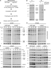
Cbp3 binds to mitochondrial ribosomes in proximity to the polypeptide tunnel exit. (A) Identification of Cbp3 as a cross-linking partner of Mrpl4. Mitochondria containing Mrpl4His7 were incubated with the cross-linker MBS or mock treated (DMSO). Mrpl4His7 and cross-linked proteins were purified on Ni-NTA beads and analyzed by Western blotting with antibodies against Mrpl4 (left) and Cbp3 (right). E, elution after Ni-NTA purification; T, 10% total before Ni-NTA purification. (B) Verification of the cross-link between Mrpl4 and Cbp3. The analysis depicted in A was repeated with mitochondria containing Cbp3His7. Fractions were analyzed by Western blotting using antibodies against Cbp3 (left) and Mrpl4 (right). (C) Cbp3 binds to mitochondrial ribosomes in a salt-sensitive manner. (left) Experimental setup. Ribosomes and co-migrating proteins (P, pellet) are separated from the soluble fraction (S, supernatant) by high-speed centrifugation through a sucrose cushion. (right) Triton X-100 lysates of wild-type mitochondria were fractionated in the presence of 50 or 150 mM KCl. The fractions were analyzed by Western blotting using antibodies against Aco1 (a soluble protein), Mrp20 (a ribosomal component), and Cbp3. T, 100% total before high-speed centrifugation. (D) Cbp3 and ribosomes are present in similar quantities in mitochondria. Increasing amounts of proteins of wild-type, mrpl4his7, and cbp3his7 cells were analyzed by Western blotting using the indicated antibodies. MM, molecular mass.
Figure 2.
Figure 2.
Cbp3 stabilizes newly synthesized cytochrome b. (A) An experimental procedure to analyze the contact of Cbp3 to mitochondrial translation products. (B) Cbp3 can be cross-linked to newly synthesized cytochrome b. Translation products of mitochondria isolated from wild-type or cbp3his7 cells were labeled with [35S]methionine in the presence of the cleavable cross-linking reagent DSP and purified on Ni-NTA beads. The samples were analyzed by autoradiography. The black arrow indicates the newly synthesized cytochrome b that is copurified with Cbp3His7. E, elution after Ni-NTA purification; T, 1% total before Ni-NTA purification. (C) Mitochondrial translation products of wild-type or Δcbp3 cells were labeled with [35S]methionine in the presence of cycloheximide to block cytosolic protein synthesis. Samples were taken after the indicated time points and subjected to alkaline lysis followed by SDS-PAGE and autoradiography. A Western blot using anti-Mrpl40 antibody served as a loading control. (D) Newly synthesized cytochrome b is destabilized in the absence of Cbp3. Radiolabeled translation products of the indicated strains were followed after the labeling had been stopped by addition of unlabeled methionine. (E) Cytochrome b is less efficiently labeled in Δcbp4 cells but is stable for up to 2.5 h. The experiments described in C and D were repeated with Δcbp4 cells. (F) The absence of Cbp3 and Cbp4 reduces the levels of components of the bc1 complex. Increasing amounts of proteins of wild-type, Δcbp3, and Δcbp4 cells were analyzed by Western blotting using the indicated antibodies. MM, molecular mass.
Figure 3.
Figure 3.
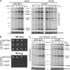
Cbp3, but not Cbp4, is critical for efficient translation of the cob::ARG8m mRNA. (A) Growth test of wild-type, Δcbp3, or Δcbp4 yeast harboring mitochondrial genomes where a recoded version of Arg8 was inserted into the mitochondrial genome replacing the ORF of Cytb (cob::ARG8m) or Cox2 (cox2::ARG8m). Cells were plated in serial 10-fold dilutions onto media containing or lacking arginine and incubated for 2 d. SD, synthetic medium supplemented with glucose. (B) Western blot analysis of steady-state levels of proteins from strains described in A. (C and D) The absence of Cbp3 in cells harboring the cob::ARG8m mitochondrial genome leads to a strong decrease in the synthesis of Arg8 (C) but does not affect its stability (D). Mitochondrial translation products from cob::ARG8m wild-type or Δcbp3 cells were labeled with [35S]methionine as described in Fig. 2 C. Western blotting using anti-Mrpl40 antibody served as a loading control. MM, molecular mass.
Figure 4.
Figure 4.
Cbp3 forms a complex with Cbp6. (A) Native purification of Cbs1, Cbs2, and Cbp6 complexes. Cbs1 and Cbs2 were equipped with C-terminal ProteinAHis7 tags to allow sensitive detection as well as purification of the proteins via metal affinity chromatography, whereas Cbp6 was expressed with a C-terminal His7 tag. Triton X-100 lysates of the indicated mitochondria were subjected to Ni-NTA chromatography, and the resulting fractions were analyzed by Western blotting. E, elution after Ni-NTA purification; NB, 20% of unbound material after Ni-NTA purification; PAHis7 tag, ProteinA-heptahistidine tag; T, 20% total before Ni-NTA purification. (B) Cbp6 is efficiently purified with Cbp3His7. Native purification of the Cbp3 complex from cbp3his7 mitochondria was performed as described in A. (C) Cbp3 and Cbp6 can be cross-linked to each other. Mitochondria harboring Cbp3His7 were incubated with the cross-linker MBS or a control (DMSO). Cbp3 and cross-linked proteins were purified as described in Fig. 1 B. The elution fractions were analyzed by Western blotting using antibodies against Cbp3 (left) and Cbp6 (right). The asterisk indicates the cross-link of Mrpl4 to Cbp3His7. (D) Cbp6 co-migrates with mitochondrial ribosomes in a salt-sensitive manner. Triton X-100 lysates of wild-type mitochondria were fractionated as described in Fig. 1 C. MM, molecular mass; P, pellet; S, supernatant; T, 100% total before ribosome fractionation.
Figure 5.
Figure 5.
Deletion of CBP6 provokes a phenotype similar to that of Δcbp3 cells. (A) Cytochrome b is destabilized in the absence of Cbp6. Mitochondrial translation products from wild-type or Δcbp6 cells were analyzed as described in Fig. 2 (C and D). (B) Cbp6 is critical for efficient translation of a cob::ARG8m reporter. Cells of wild type, Δcbp3, or Δcbp6 harboring the modified mitochondrial genome described in Fig. 3 A were plated in serial 10-fold dilutions onto synthetic media (SD) containing or lacking arginine and incubated for 2 d. (C) The absence of Cbp6 impairs translation of COB-ARG8m-COB mRNA. Mitochondrial translation products of wild-type and Δcbp6 cob::ARG8m cells were labeled with [35S]methionine and analyzed by autoradiography. MM, molecular mass.
Figure 6.
Figure 6.
Cbp3 and Cbp6 act in one complex. (A) Cbp3 is destabilized when Cbp6 is absent and vice versa. Cell lysates of the indicated yeast strains were analyzed by Western blotting using the indicated antibodies. The asterisk indicates an unspecific cross-reaction of the Cbp6 antibody. (B) The levels of the other translational activators of the COB mRNA, Cbs1 and Cbs2, are not affected when CBP3 or CBP6 is deleted. Cells expressing ProteinAhis7-tagged variants of Cbs1 or Cbs2 were lysed and analyzed by Western blotting. (C and D) Overexpression of either CBP3 or CBP6 cannot complement deletion of the other. The indicated yeast strains harboring the wild-type (C) or the cob::ARG8m mitochondrial DNA (D) were transformed with 2µ plasmids encoding CBP3 or CBP6 under control of the TPI promoter or an empty vector. Cell lysates were analyzed by Western blotting using the indicated antibodies. MM, molecular mass.
Figure 7.
Figure 7.
Interaction of Cbp3 and Cbp6 with the ribosome requires formation of a Cbp3–Cbp6 complex and is influenced by cytochrome b. (A) Cbp3 and Cbp6 cannot bind to mitochondrial ribosomes in the absence of the other factor. Triton X-100 lysates of mitochondria from the indicated strains were fractionated as described in Fig. 1 C in the presence of 50 mM KCl. Fractions were analyzed by Western blotting. Because Cbp6 is not detectable in the Δcbp3 strain, mitochondria from Δcbp3 cells overexpressing CBP6 were used. (B) COB mRNA is absent in Δcbp1 but not in Δcbp3 and Δcbp6 cells. Total RNA was isolated from mitochondria of the indicated strains and analyzed by Northern blotting. 15S ribosomal RNA served as a loading control. (C) The Cbp3–Cbp6 complex can bind to mitochondrial ribosomes in the absence of cytochrome b but is mainly present in a non–ribosome-bound form in the wild type (WT). Mitochondria of the indicated strains were lysed with digitonin in the presence of 50 mM KCl and fractionated as described in Fig. 1 C. P, pellet; S, supernatant; T, 100% total before ribosome fractionation. (D) Cbp3 is more tightly bound to ribosomes of mitochondria harboring a cob::ARG8m mitochondrial genome and is released upon treatment with increasing salt concentrations. Mitochondria of wild-type and cob::ARG8m cells were lysed with digitonin and increasing KCl concentrations as indicated on the x axis and fractionated as in Fig. 1 C. The percentage of Cbp3 that was found in the pellet fraction was determined densitometrically from three independently performed experiments. The error bars represent the standard deviation of these experiments. MM, molecular mass.
Figure 8.
Figure 8.
The Cbp3–Cbp6 complex is released from ribosomes upon cytochrome b binding. (A and B) Cbp4 interacts with the Cbp3–Cbp6 complex only in the presence of cytochrome b. Mitochondria of the indicated strains were lysed with Triton X-100 (A) or digitonin (B), and Cbp3His7 was purified under native conditions as described in Fig. 4 A. The asterisks indicate a nonspecific cross-reaction of the antibody against cytochrome b. E, elution after Ni-NTA purification; NB, 20% of unbound material after Ni-NTA purification; T, 20% total before Ni-NTA purification. (C) Cbp6 and Cbp4 are tightly associated with the inner mitochondrial membrane (IMM). Mitochondrial membranes were extracted with high salt (left) or carbonate (right) as described in the Materials and methods section. M, membrane fraction. (D) Cbp3 and Cbp6 are located in the mitochondrial matrix, whereas Cbp4 faces the intermembrane space (IMS). Wild-type mitochondria were incubated in isoosmotic or hypoosmotic buffers (swelling) and treated with proteinase K (PK) or left untreated. The arrow points to a fragment of Oxa1 (f-27) that confirms efficient swelling. (right) A model of the topology of Cbp3, Cbp6, and Cbp4. (E) The Cbp3–Cbp6 complex can bind newly synthesized cytochrome b in the absence of Cbp4. Translation products from wild-type and Δcbp4 mitochondria were labeled with [35S]methionine. The organelles were reisolated, lysed with digitonin, and subjected to coimmunoprecipitation with antibodies against Cbp3 or preimmune serum. The samples were analyzed by autoradiography. T, 5% total before immunoprecipitation (IP). (F) The Cbp3–Cbp6 complex is mainly present in a non–ribosome-bound state in the absence of Cbp4, and newly synthesized cytochrome b is liberated from ribosomes independent of Cbp4. Translation products from wild-type and Δcbp4 mitochondria were labeled with [35S]methionine. Mitochondria were reisolated, lysed with digitonin, and fractionated as described in Fig. 1 C in the presence of 50 mM KCl. Samples were analyzed by autoradiography (left) and Western blotting using the indicated antibodies (right). MM, molecular mass; P, pellet; S, supernatant.
Figure 9.
Figure 9.
Hypothetical model for the role of the Cbp3–Cbp6 complex during biogenesis of cytochrome b. In the absence of an mRNA, the Cbp3–Cbp6 complex is bound to mitochondrial ribosomes (state 0). COB mRNA binds to the ribosome, and translation can only be initiated efficiently when the Cbp3–Cbp6 complex is ribosome bound (state 1). Cbp3 and Cbp6 then interact with the newly synthesized cytochrome b. Cbp4 is recruited (state 2), and this complex mediates the assembly of cytochrome b into a functional bc1 complex. IMM, inner mitochondrial membrane; IMS, intermembrane space.

Similar articles

Cited by

References

    1. Barrientos A., Zambrano A., Tzagoloff A. 2004. Mss51p and Cox14p jointly regulate mitochondrial Cox1p expression in Saccharomyces cerevisiae. EMBO J. 23:3472–3482 10.1038/sj.emboj.7600358 - DOI - PMC - PubMed
    1. Bingel-Erlenmeyer R., Kohler R., Kramer G., Sandikci A., Antolic´ S., Maier T., Schaffitzel C., Wiedmann B., Bukau B., Ban N. 2008. A peptide deformylase-ribosome complex reveals mechanism of nascent chain processing. Nature. 452:108–111 10.1038/nature06683 - DOI - PubMed
    1. Bonnefoy N., Fox T.D. 2000. In vivo analysis of mutated initiation codons in the mitochondrial COX2 gene of Saccharomyces cerevisiae fused to the reporter gene ARG8m reveals lack of downstream reinitiation. Mol. Gen. Genet. 262:1036–1046 10.1007/PL00008646 - DOI - PubMed
    1. Bonnefoy N., Fox T.D. 2007. Directed alteration of Saccharomyces cerevisiae mitochondrial DNA by biolistic transformation and homologous recombination. Methods Mol. Biol. 372:153–166 10.1007/978-1-59745-365-3_11 - DOI - PMC - PubMed
    1. Borst P., Grivell L.A. 1978. The mitochondrial genome of yeast. Cell. 15:705–723 10.1016/0092-8674(78)90257-X - DOI - PubMed

MeSH terms