. 2011 Aug 4;118(5):1329-39.
doi: 10.1182/blood-2011-01-327197. Epub 2011 Jun 9.

Distribution of Bim determines Mcl-1 dependence or codependence with Bcl-xL/Bcl-2 in Mcl-1-expressing myeloma cells

Affiliations

Distribution of Bim determines Mcl-1 dependence or codependence with Bcl-xL/Bcl-2 in Mcl-1-expressing myeloma cells

Alejo A Morales et al. Blood. .

Abstract

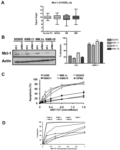
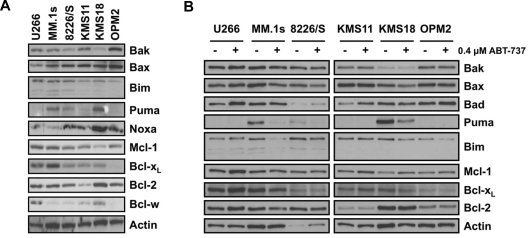
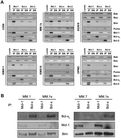
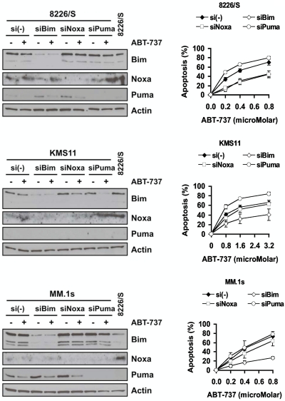
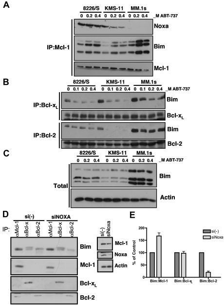
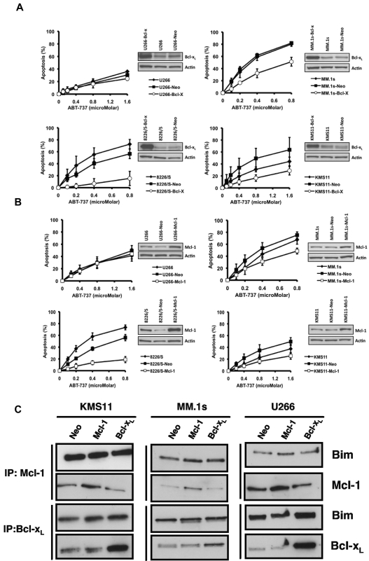
Dependence on Bcl-2 proteins is a common feature of cancer cells and provides a therapeutic opportunity. ABT-737 is an antagonist of antiapoptotic Bcl-2 proteins and therefore is a good predictor of Bcl-x(L)/Bcl-2 dependence. Surprisingly, analysis of Mcl-1-dependent multiple myeloma cell lines revealed codependence on Bcl-2/Bcl-x(L) in half the cells tested. Codependence is not predicted by the expression level of antiapoptotic proteins, rather through interactions with Bim. Consistent with these findings, acquired resistance to ABT-737 results in loss of codependence through redistribution of Bim to Mcl-1. Overall, these results suggest that complex interactions, and not simply expression patterns of Bcl-2 proteins, need to be investigated to understand Bcl-2 dependence and how to better use agents, such as ABT-737.


Figures

Figure 1
Figure 1
All MM cells are dependent on Mcl-1, although they differ in their sensitivity to ABT-737. (A) mRNA levels for Mcl-1 were derived from the analysis of normalized gene expression profile data deposited at GEO from CD138-selected plasma cells from healthy donors: normal PC (n = 22), monoclonal gammopathy of undetermined significance (n = 44), smoldering myeloma (SM, n = 12), and newly diagnosed MM (MM, n = 538). The data are taken from the 214056_at probe on the affymetrix Hu133 2.0 plus array. Similar results were obtained from the 214057_at probe (not shown). (B) Four myeloma cell lines were transfected with siRNA for Mcl-1 and efficiency of knock-down was determined by Western blot (left panel), whereas the apoptosis of each cell line 16 hours after transfection was measured by annexin PI assays. P values compare with si(-), P < .005 (KMS11, MM.1s), P < .001 (8226, KMS18). (C) Six myeloma cell lines were treated with indicated doses of ABT-737 for 24 hours, and apoptosis was measured by annexin V/PI staining. Data are mean ± SD of 4 independent experiments. (D) CD138+ cells purified from bone marrow aspirates of 6 MM patients were incubated with the indicated concentrations of ABT-737 for 24 hours and apoptosis determined by annexin V/PI staining. Each sample was run in parallel with MM.1s (●), which is presented as the mean ± SD of the 6 experiments. The 50% inhibitory concentration values for each sample are as follows (μM): MM.1s, 0.65; MM1-0.3, MM6-0.58, MM7-0.92, MM23-0.1, MM24, and MM26, < 0.1.
Figure 2
Figure 2
Sensitivity to ABT-737 does not correlate with the expression pattern of Bcl-2 family proteins. (A) Protein expression of Bcl-2 family proteins was determined by Western blot analysis using lysates from untreated myeloma cell lines. Membranes were probed with specific pAbs against the indicated Bcl-2 family members as described in “Western blot analysis” and “Antibodies.” Actin was used as a loading control. (B) Change in the expression of Bcl-2 proteins after treatment with 0.4μM ABT-737 for 24 hours was determined by Western blot analysis.
Figure 3
Figure 3
Binding of Bim to Bcl-xL and Bcl-2 in myeloma cell correlates with codependence and sensitivity to ABT-737. (A) Coimmunoprecipitation of proapoptotic proteins with Mcl-1, Bcl-xL, and Bcl-2 was determined by Western blot analysis after lysing all 6 cell lines in 2% 3(3-cholamidopropyl) dimethylammonio-1-propane sulfonate buffer and immunoprecipitating with specific monoclonal antibodies as described in “Antibodies.” and “Coimmunoprecipitation studies.” Lysates and supernatant contain 7%, whereas the immunoprecipitated proteins contain 60% of the input. (B) CD138+ myeloma cells were isolated, and 1 million cells were subjected to lysis and immunoprecipitation with anti–Bcl-x and anti–Mcl-1 antibodies as described in “Antibodies” and “Coimmunoprecipitation studies.” Immunoprecipitates were subjected to SDS-PAGE and Western blot analysis performed for Bcl-xL, Mcl-1, and Bim. Two of 6 patient samples tested are shown, and each experiment was performed in parallel with MM.1s as a reference.
Figure 4
Figure 4
Effect of silencing Bim, Noxa, and Puma on ABT-737–induced apoptosis. Knock-down efficiency was analyzed by Western blotting of each protein in untreated and 0.4μM ABT-737 treated samples (left panels). Activity of ABT-737 was analyzed by annexin/PI staining after knock-down of each protein (right panels). Data are mean ± SD of at least 3 independent experiments. P values: 8226, silencing of all 3 BH3 proteins are significantly different from [si(−)] control (P < .005), with the exception of siPuma at 0.8μM ABT-737 (not significant). KMS11, all values are significantly different from control (P < .005), with the exception of siNoxa at 1.6 and 3.2μM (not significant). MM.1s, siBim significantly different from control (P < .005).
Figure 5
Figure 5
Noxa expression inhibits Mcl-1 binding to Bim displaced from Bcl-xL and Bcl-2. 8226/S, MM.1s, and KMS11 cell lines were treated with indicated concentrations of ABT-737 for 24 hours and lysates generated as described in “Western blot analysis.” (A) Mcl-1 and (B) Bcl-xL and Bcl-2 were immunoprecipitated using specific monoclonal antibodies. Immunoprecipitates were sequentially probed with pAbs for Bim and Noxa as well as for Bcl-2, Bcl-xL, and Mcl-1. Immunoprecipitates contain 60% of input. (C) The total amount of Bim was assayed by Western blot in lysates from control and treated samples of 8226/S, KMS-11, MM.1S. (D) Twenty-four hours after transfection with si(-) or siNoxa, expression of Noxa and Mcl-1 was determined by Western blot (right panel). The remaining lysate was subjected to immunoprecipitation with Mcl-1, Bcl-xL, or Bcl-2 followed by Western blot analysis of Bim, Mcl-1, Bcl-xL, and Bcl-2 (left panel). (E) The bands from 2 independent experiments were quantified using ImageJ software Version 1.45r, and the graph represents the average ratio of relative intensity of indicated bands.
Figure 6
Figure 6
Bcl-xL and Mcl-1 overexpression protect codependent cell lines from ABT-737–induced apoptosis. U266, MM.1s, 8226/S, and KMS-11 cell lines were stably transfected with pcDNA3.1 (Neo) and (A) pcDNA–Bcl-xL or (B) pcDNA–Mcl-1 vectors. Cell lines were treated with indicated concentrations of ABT-737 for 24 hours, and viability was determined by annexin V/PI staining. The data presented are the mean ± SD of at least 3 independent experiments. U266–Bcl-xL is significantly different from U266-Neo (P < .05) at 0.1μM ABT-737. MM.1s-Bcl-xL is significantly different from MM.1s-Neo at all concentrations of ABT-737 (0.1μM, P < .05; 0.2μM, P < .001; 0.4-0.8μM, P < .005). 8226-Bcl-xL is significantly different from 8226-Neo at all concentrations of ABT-737 (P < .005). MM.1s-Mcl-1 is significantly different from MM.1s-Neo (P < .005) at 0.4 to 0.8μM ABT-737. KMS11-Mcl-1 is statistically different from KMS11-Neo (P < .05) at all concentrations of ABT-737. 8226-Mcl-1 is significantly different from 8226-Neo (P < .001) at all concentrations of ABT-737. (C) Protein lysates were obtained using 2% 3(3-cholamidopropyl) dimethylammonio-1-propane sulfonate buffer for the indicated transfectants of KMS11, MM.1s, U266. Antiapoptotic proteins Mcl-1 and Bcl-xL were immunoprecipitated using specific monoclonal antibodies as described in “Antibodies” and “Coimmunoprecipitation studies.” The pellets were probed for Bim, Mcl-1, and Bcl-xL. The blots are representative of 2 independent experiments.
Figure 7
Figure 7
Acquired ABT-737 resistance is associated with changes in expression and interactions of Bcl-2 proteins. Four cell lines were rendered resistant to ABT-737 by sequentially increasing the ABT-737 concentration in their growth medium starting from 50nM up to 0.5μM (8226/S, KMS-18) or 2μM (KMS-11, U266). (A) Sensitivity (EC50) of parental, control resistance (CR2), and resistant (ABTR) cell lines to ABT-737 was determined by annexin V/PI staining after 24 hours of treatment. All ABTR cell lines are significantly different from CR2 cells at all concentrations of ABT-737 (P < .001), with the exception of KMS18 at 3.2μM ABT-737 (P < .005). (B) Expression of Bcl-2 family members and (C) interaction between these proteins were determined as described in “Antibodies” and “Coimmunoprecipitation studies.” For all experiments, the cells were removed from the selecting concentration 24 hours before the experiment.

Similar articles

Cited by

References

    1. Chonghaile TN, Letai A. Mimicking the BH3 domain to kill cancer cells. Oncogene. 2008;27(suppl 1):S149–S157. - PMC - PubMed
    1. Adams JM, Cory S. The Bcl-2 apoptotic switch in cancer development and therapy. Oncogene. 2007;26(9):1324–1337. - PMC - PubMed
    1. Kuroda J, Taniwaki M. Involvement of BH3-only proteins in hematologic malignancies. Crit Rev Oncol Hematol. 2009;71(2):89–101. - PubMed
    1. Hacker G, Weber A. BH3-only proteins trigger cytochrome c release, but how? Arch Biochem Biophys. 2007;462(2):150–155. - PubMed
    1. Willis SN, Fletcher JI, Kaufmann T, et al. Apoptosis initiated when BH3 ligands engage multiple Bcl-2 homologs, not Bax or Bak. Science. 2007;315(5813):856–859. - PubMed

Publication types

MeSH terms