Tristetraprolin regulates interleukin-6, which is correlated with tumor progression in patients with head and neck squamous cell carcinoma
- PMID: 21656745
- PMCID: PMC3574798
- DOI: 10.1002/cncr.25859
Tristetraprolin regulates interleukin-6, which is correlated with tumor progression in patients with head and neck squamous cell carcinoma
Abstract
Background: Tumor-derived cytokines play a significant role in the progression of head and neck squamous cell carcinoma (HNSCC). Targeting proteins, such as tristetraprolin (TTP), that regulate multiple inflammatory cytokines may inhibit the progression of HNSCC. However, TTP's role in cancer is poorly understood. The goal of the current study was to determine whether TTP regulates inflammatory cytokines in patients with HNSCC.
Methods: TTP messenger RNA (mRNA) and protein expression were determined by quantitative real-time-polymerase chain reaction (Q-RT-PCR) and Western blot analysis, respectively. mRNA stability and cytokine secretion were evaluated by quantitative RT-PCR and enzyme-linked immunoadsorbent assay, respectively, after overexpression or knockdown of TTP in HNSCC. HNSCC tissue microarrays were immunostained for interleukin-6 (IL-6) and TTP.
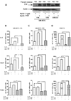
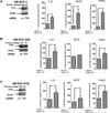
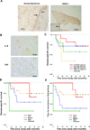
Results: TTP expression in HNSCC cell lines was found to be inversely correlated with the secretion of IL-6, vascular endothelial growth factor (VEGF), and prostaglandin E2 (PGE(2) )(.) Knockdown of TTP increased mRNA stability and the secretion of cytokines. Conversely, overexpression of TTP in HNSCC cells led to decreased secretion of IL-6, VEGF, and PGE(2) . Immunohistochemical staining of tissue microarrays for IL-6 demonstrated that staining intensity is prognostic for poor disease-specific survival (P = .023), tumor recurrence and development of second primary tumors (P = .014), and poor overall survival (P = .019).
Conclusions: The results of the current study demonstrated that down-regulation of TTP in HNSCC enhances mRNA stability and promotes secretion of IL-6, VEGF, and PGE(2) . Furthermore, high IL-6 secretion in HNSCC tissue is a biomarker for poor prognosis. In as much as enhanced cytokine secretion is associated with poor prognosis, TTP may be a therapeutic target to reduce multiple cytokines concurrently in patients with HNSCC.
Copyright © 2011 American Cancer Society.
Conflict of interest statement
The authors report no conflict of interest with this manuscript.
Figures


Similar articles
-
Inactivation or loss of TTP promotes invasion in head and neck cancer via transcript stabilization and secretion of MMP9, MMP2, and IL-6.Clin Cancer Res. 2013 Mar 1;19(5):1169-79. doi: 10.1158/1078-0432.CCR-12-2927. Epub 2013 Jan 24. Clin Cancer Res. 2013. PMID: 23349315 Free PMC article.
-
IL-6 antisense-mediated growth inhibition in a head and neck squamous cell carcinoma cell line.In Vivo. 2011 Jul-Aug;25(4):579-84. In Vivo. 2011. PMID: 21708999
-
Inflammatory mediators drive metastasis and drug resistance in head and neck squamous cell carcinoma.Laryngoscope. 2015 Mar;125 Suppl 3:S1-11. doi: 10.1002/lary.24998. Epub 2015 Feb 3. Laryngoscope. 2015. PMID: 25646683
-
Molecular and biological factors in the prognosis of head and neck squamous cell cancer.Mol Biol Rep. 2023 Sep;50(9):7839-7849. doi: 10.1007/s11033-023-08611-1. Epub 2023 Jul 26. Mol Biol Rep. 2023. PMID: 37493876 Review.
-
Vascular Endothelial Growth Factor Family and Head and Neck Squamous Cell Carcinoma.Anticancer Res. 2023 Oct;43(10):4315-4326. doi: 10.21873/anticanres.16626. Anticancer Res. 2023. PMID: 37772546 Review.
Cited by
-
The mRNA-binding Protein TTP/ZFP36 in Hepatocarcinogenesis and Hepatocellular Carcinoma.Cancers (Basel). 2019 Nov 8;11(11):1754. doi: 10.3390/cancers11111754. Cancers (Basel). 2019. PMID: 31717307 Free PMC article.
-
Important Cells and Factors from Tumor Microenvironment Participated in Perineural Invasion.Cancers (Basel). 2023 Feb 21;15(5):1360. doi: 10.3390/cancers15051360. Cancers (Basel). 2023. PMID: 36900158 Free PMC article. Review.
-
Expression of NF-κB and IL-6 in oral precancerous and cancerous lesions: An immunohistochemical study.Med Oral Patol Oral Cir Bucal. 2016 Jan 1;21(1):e6-13. doi: 10.4317/medoral.20570. Med Oral Patol Oral Cir Bucal. 2016. PMID: 26595830 Free PMC article.
-
Association Between Interleukin-6 and Head and Neck Squamous Cell Carcinoma: A Systematic Review.Clin Exp Otorhinolaryngol. 2021 Feb;14(1):50-60. doi: 10.21053/ceo.2019.00906. Epub 2021 Feb 1. Clin Exp Otorhinolaryngol. 2021. PMID: 33587847 Free PMC article. Review.
-
Effects of tristetraprolin on doxorubicin (adriamycin)-induced experimental kidney injury through inhibiting IL-13/STAT6 signal pathway.Am J Transl Res. 2020 Apr 15;12(4):1203-1221. eCollection 2020. Am J Transl Res. 2020. PMID: 32355536 Free PMC article.
References
-
- Dong G, Loukinova E, Smith CW, Chen Z, Van Waes C. Genes differentially expressed with malignant transformation and metastatic tumor progression of murine squamous cell carcinoma. J Cell Biochem Suppl. 1997;28–29:90–100. - PubMed
-
- Kanazawa T, Nishino H, Hasegawa M, et al. Interleukin-6 directly influences proliferation and invasion potential of head and neck cancer cells. Eur Arch Otorhinolaryngol. 2007;264:815–821. - PubMed
-
- Chen Z, Malhotra PS, Thomas GR, Ondrey FG, Duffey DC, Smith CW, et al. Expression of proinflammatory and proangiogenic cytokines in patients with head and neck cancer. Clin Cancer Res. 1999;5:1369–1379. - PubMed
-
- Pries R, Nitsch S, Wollenberg B. Role of cytokines in head and neck squamous cell carcinoma. Expert Rev Anticancer Ther. 2006;6:1195–1203. - PubMed
Publication types
MeSH terms
Substances
Grants and funding
- F32 DE021305/DE/NIDCR NIH HHS/United States
- P50-CA97248/CA/NCI NIH HHS/United States
- R21 DE019272/DE/NIDCR NIH HHS/United States
- R01-DE018512/DE/NIDCR NIH HHS/United States
- K02-DE019513/DE/NIDCR NIH HHS/United States
- 5F32 DE213052/DE/NIDCR NIH HHS/United States
- P20 RR017696/RR/NCRR NIH HHS/United States
- P50 CA097248/CA/NCI NIH HHS/United States
- R01 DE018290/DE/NIDCR NIH HHS/United States
- K02 DE019513/DE/NIDCR NIH HHS/United States
- R21 DE017966/DE/NIDCR NIH HHS/United States
- R01 DE018512/DE/NIDCR NIH HHS/United States
- T32-DE007057-33/DE/NIDCR NIH HHS/United States
- R21 DE017977/DE/NIDCR NIH HHS/United States
- R01-DE018290/DE/NIDCR NIH HHS/United States
- R21-DE017977/DE/NIDCR NIH HHS/United States
- R01 DE021423/DE/NIDCR NIH HHS/United States
- T32 DE007057/DE/NIDCR NIH HHS/United States
- 5R21 DE017966/DE/NIDCR NIH HHS/United States
LinkOut - more resources
Full Text Sources
Other Literature Sources

