. 2011 Aug 4;118(5):1255-63.
doi: 10.1182/blood-2011-02-337360. Epub 2011 Jun 7.

A transgene-encoded cell surface polypeptide for selection, in vivo tracking, and ablation of engineered cells

Affiliations

A transgene-encoded cell surface polypeptide for selection, in vivo tracking, and ablation of engineered cells

Xiuli Wang et al. Blood. .

Abstract

An unmet need in cell engineering is the availability of a single transgene encoded, functionally inert, human polypeptide that can serve multiple purposes, including ex vivo cell selection, in vivo cell tracking, and as a target for in vivo cell ablation. Here we describe a truncated human EGFR polypeptide (huEGFRt) that is devoid of extracellular N-terminal ligand binding domains and intracellular receptor tyrosine kinase activity but retains the native amino acid sequence, type I transmembrane cell surface localization, and a conformationally intact binding epitope for pharmaceutical-grade anti-EGFR monoclonal antibody, cetuximab (Erbitux). After lentiviral transduction of human T cells with vectors that coordinately express tumor-specific chimeric antigen receptors and huEGFRt, we show that huEGFRt serves as a highly efficient selection epitope for chimeric antigen receptor(+) T cells using biotinylated cetuximab in conjunction with current good manufacturing practices (cGMP)-grade anti-biotin immunomagnetic microbeads. Moreover, huEGFRt provides a cell surface marker for in vivo tracking of adoptively transferred T cells using both flow cytometry and immunohistochemistry, and a target for cetuximab-mediated antibody-dependent cellular cytotoxicity and in vivo elimination. The versatility of huEGFRt and the availability of pharmaceutical-grade reagents for its clinical application denote huEGFRt as a significant new tool for cellular engineering.


Figures
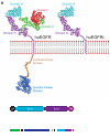
Figure 1
Figure 1
Generation of huEGFRt expressing construct. (A) Molecular model of human EGFR versus EGFRt proteins. Models were created using Accelrys Discovery Studio, Version 2.0 software and are based on the human EGFR crystal structure files 1YY9 (http://www.pdb.org/pdb/explore/explore.do?structureId = 1YY9), which provides the structure of the 4 extracellular domains (red, green, teal, and purple), and 3GOP (http://www.pdb.org/pdb/explore/explore.do?structureId = 3GOP), which provides the crystal structure of the intracellular juxtamembrane domain (brown) and the tyrosine kinase domain (blue). The transmembrane helix (magenta) is modeled on known helix structure. The structure of the last approximately 190 amino acids of the C-terminal intracellular domain is not known, so the placeholder for this is indicated (gray). (B) Schematic of the huEGFRt amino acid sequence. Amino acid ranges that were used from the GM-CSF receptor-α chain signal sequence (GMCSFRss, which directs surface expression) and the huEGFR sequence (domains III, IV and the transmembrane domain) are indicated. (C) Schematic of the CD19CAR-T2A-EGFRt construct contained in the lentiviral vector. Codon optimized sequence portions of the CD19-specific, CD28 costimulatory CAR (CD19CAR), followed by the self-cleavable T2A, and huEGFRt genes are indicated, along with the Elongation Factor 1 promoter sequences (EF-1p), the GM-CSF receptor-α chain signal sequences (GMCSFRss), and the 3 nucleotide stop codon.
Figure 2
Figure 2
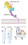
Generation and utility of a biotinylated cetuximab. (A) Model is based on the crystal structure file 1YY9 (http://www.pdb.org/pdb/explore/explore.do?structureId = 1YY9), which provides the structure of the 4 extracellular domains (red, green, teal, and purple), and the bound cetuximab Fab (yellow and orange). The transmembrane helix (magenta) is modeled on known helix structure. (B) Sodium dodecyl sulfate-polyacrylamide gel electrophoresis gels of nonreduced (left) and reduced (right) final biotinylated cetuximab product. (C) Titration of biotinylated cetuximab. A total of 106 EGFR+ cells were stained with 0 μg (black), 1.45 μg (red), 0.145 μg (orange), 14.5 ng (yellow), 1.45 ng (green), 0.145 ng (blue), or 14.5 pg (purple) of biotinylated cetuximab followed by 0.5 μg PE-conjugated streptavidin and analyzed by flow cytometry. A total of 14.5 ng of biotinylated cetuximab was deemed sufficient for future staining of 106 cells. (D) Biotinylated cetuximab can detect huEGFRt on transduced H9 cells. H9 cells were transduced with the CD19CAR-T2A-EGFRt containing lentivirus at a multiplicity of infection of 10 and analyzed 2 days later by flow cytometry using biotinylated cetuximab or biotinylated anti-Fc Ab (to detect the CD19CAR) followed by SA-PE (black histograms). Gray histograms represent nontransduced parental H9. Percentage positive staining is indicated in each histogram.
Figure 3
Figure 3
Selected EGFRt+ CD19CAR+ T cells can be expanded with maintenance of effector phenotype. (A) Schematic of the immunomagnetic huEGFRt selection procedure. (B) After 1 rapid expansion cycle, nonselected (Unslxd) and huEGFRt-selected (Slxd) T cells were phenotyped for surface EGFR (ie, EGFRt, with biotinylated cetuximab), Fc (ie, CAR), and effector T-cell markers TCRαβ, CD3, CD4, CD8, CD28, or intracellular granzyme A (black histogram) versus isotype control Ab (gray histogram) by flow cytometry. Percentage of positive staining is indicated in each histogram. (C) Fold expansion of nontransduced (EGFRt-) and transduced (EGFRt+) T cells in unselected cultures of 4 different transduced cell lines was determined after OKT3-mediated stimulation. (D) huEGFRt expression of selected T cells was determined by flow cytometry after each of 3 rounds of OKT3-mediated stimulation (Stim 1, 2, and 3). Percentage of cetuximab-mediated staining (black) compared with that of SA-PE alone (gray) is indicated in each histogram. (E) Nonselected (Unslxd) and huEGFRt-selected (Slxd) T cells were incubated for 4 hours with 51Cr-labled CD19+ LCL or SupB15 cells, or OKT3-epressing LCL cells as targets at the indicated effector to target ratios. Percentage cytotoxicity (mean ± SE) of triplicate wells is depicted. (F) Nonselected (Unslxd) and EGFRt-selected (Slxd) T cells were incubated with CD19+ LCL or SupB15 cells, or OKT3-epressing LCL cells and supernatants were analyzed by Bioplex. IFN-γ and TNF-α levels (mean ± SE) are shown.
Figure 4
Figure 4
The huEGFRt expressed on selected T cells is inert. (A) EGF does not bind to the surface of huEGFRt-expressing T cells. A431, EGFRt-selected T cells, and negative control T cells were analyzed by flow cytometry after incubation with PE-conjugated anti-EGFR, or either biotinylated cetuximab or biotinylated EGF followed by PE-conjugated streptavidin (black histogram) versus PE-conjugated isotype control antibody or streptavidin alone (gray histogram). Percentage positive staining is indicated in each histogram. (B) huEGFRt expressed on T cells is not phosphorylated on coincubation with EGF. Negative control T cells, huEGFRt-selected T cells, or A431 cells were incubated for 5 minutes with or without 100 ng/mL EGF and then lysed in the presence of phosphatase inhibitor. Lysates run on Western blots were then probed using antibodies specific for β-actin, the cytoplasmic domain of EGFR, or the phosphorylated tyrosine at position 1068 of EGFR.
Figure 5
Figure 5
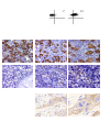
Use of EGFRt as a marker for in vivo detection of engineered cells. (A) Day 20 bone marrow cells harvested from mice that were engrafted with either unmodified huEGFRt-negative control T cells (Ctrl T cells) or huEGFRt-selected T cells (EGFRt+ T cells) were stained using peridinin chlorophyll protein-conjugated anti–human CD45 and biotinylated cetuximab followed by PE-conjugated streptavidin. Total percentage of CD45+ cells in bone marrow are indicated in the left-hand panels. Percentage of EGFRt+ cells within the CD45-gated population are indicated in the right-hand panels, using quadrants that were created based on isotype control staining. (B) Day 20 femurs were also analyzed by immunohistochemistry for CD45 versus EGFR expression. EGFR expression on paraffin-embedded breast tumor tissue was used as a positive control for EGFR staining. Representative ×40 and ×100 images are shown as indicated.
Figure 6
Figure 6
Use of EGFRt as a target for Erbitux-mediated ADCC of engineered cells. (A) 51Cr-labeled huEGFRt-selected T cells (inset depicts huEGFRt expression) were mixed with 1 μg/mL of Erbitux or Rituxan as a negative control before addition of GM-CSF–stimulated human PBMCs as effectors. The percentage 51Cr-release was then determined after a 4-hour incubation. Data are representative of 2 separate experiments. (B) NOD/scid mice were inoculated intravenously with ffLuc+huEGFRt+ CTLL-2 cells. On successful engraftment (day 0), the mice were divided into 2 groups (n = 5) and then treated with either Erbitux (Ctxmb) or Rituxan (Rtxn) intraperitoneally daily for 6 days. Data are ± SE; total flux levels of luciferase activity were measured by Xenogen imaging. *P = .0159, the flux of Erbitux versus Rituxan-treated mice at day 6 using the Mann-Whitney test. (C) Representative bioluminescence images of NOD/scid mice from (B) at day 6.

Similar articles

Cited by

References

    1. Cooper LJ, Kalos M, DiGiusto D, et al. T-cell genetic modification for re-directed tumor recognition. Cancer Chemother Biol Response Modif. 2005;22:293–324. - PubMed
    1. Berger C, Flowers ME, Warren EH, Riddell SR. Analysis of transgene-specific immune responses that limit the in vivo persistence of adoptively transferred HSV-TK-modified donor T cells after allogeneic hematopoietic cell transplantation. Blood. 2006;107(6):2294–2302. - PMC - PubMed
    1. Jensen MC, Popplewell L, Cooper LJ, et al. Antitransgene rejection responses contribute to attenuated persistence of adoptively transferred CD20/CD19-specific chimeric antigen receptor redirected T cells in humans. Biol Blood Marrow Transplant. 2010;16(9):1245–1256. - PMC - PubMed
    1. Fehse B, Richters A, Putimtseva-Scharf K, et al. CD34 splice variant: an attractive marker for selection of gene-modified cells. Mol Ther. 2000;1(5):448–456. - PubMed
    1. Fecci PE, Ochiai H, Mitchell DA, et al. Systemic CTLA-4 blockade ameliorates glioma-induced changes to the CD4+ T cell compartment without affecting regulatory T-cell function. Clin Cancer Res. 2007;13(7):2158–2167. - PubMed

Publication types

MeSH terms