. 2011 Jun 6:7:44.
doi: 10.1186/1744-8069-7-44.

Transcription factors Sp1 and Sp4 regulate TRPV1 gene expression in rat sensory neurons

Affiliations

Transcription factors Sp1 and Sp4 regulate TRPV1 gene expression in rat sensory neurons

Catherine Chu et al. Mol Pain. .

Abstract

Background: The capsaicin receptor, transient receptor potential vanilloid type -1 (TRPV1) directs complex roles in signal transduction including the detection of noxious stimuli arising from cellular injury and inflammation. Under pathophysiologic conditions, TRPV1 mRNA and receptor protein expression are elevated in dorsal root ganglion (DRG) neurons for weeks to months and is associated with hyperalgesia. Building on our previous isolation of a promoter system for the rat TRPV1 gene, we investigated the proximal TRPV1 P2-promoter by first identifying candidate Sp1-like transcription factors bound in vivo to the P2-promoter using chromatin immunoprecipitation (ChIP) assay. We then performed deletion analysis of GC-box binding sites, and quantified promoter activity under conditions of Sp1 / Sp4 over-expression versus inhibition/knockdown. mRNA encoding Sp1, Sp4 and TRPV1 were quantified by qRT-PCR under conditions of Sp1/Sp4 over-expression or siRNA mediated knockdown in cultured DRG neurons.

Results: Using ChIP analysis of DRG tissue, we demonstrated that Sp1 and Sp4 are bound to the candidate GC-box site region within the endogenous TRPV1 P2-promoter. Deletion of GC-box "a" or "a + b" within the P2- promoter resulted in a complete loss of transcriptional activity indicating that GC-box "a" was the critical site for promoter activation. Co-transfection of Sp1 increased P2-promoter activity in cultured DRG neurons whereas mithramycin-a, an inhibitor of Sp1-like function, dose dependently blocked NGF and Sp1-dependent promoter activity in PC12 cells. Co-transfection of siRNA directed against Sp1 or Sp4 decreased promoter activity in DRG neurons and NGF treated PC12 cells. Finally, electroporation of Sp1 or Sp4 cDNA into cultures of DRG neurons directed an increase in Sp1/Sp4 mRNA and importantly an increase in TRPV1 mRNA. Conversely, combined si-RNA directed knockdown of Sp1/Sp4 resulted in a decrease in TRPV1 mRNA.

Conclusion: Based on these studies, we now propose a model of TRPV1 expression that is dependent on Sp1-like transcription factors with Sp4 playing a predominant role in activating TRPV1 RNA transcription in DRG neurons. Given that increases of TRPV1 expression have been implicated in a wide range of pathophysiologic states including persistent painful conditions, blockade of Sp1-like transcription factors represents a novel direction in therapeutic strategies.


Figures
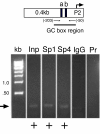
Figure 1
Figure 1
Transcription factors Sp1 and Sp4 are bound in vivo to the GC-box region of P2-promoter in DRG chromatin. Composite ethidium bromide-stained agarose gel showing evidence of PCR amplified products directed by oligodeoxynucleotide primers spanning TRPV1 GC-box "a" and "b" (top) using template DNA provided as: control input chromatin DNA without immunoprecipitation (Inp), chromatin immunoprecipitated with antisera against transcription factors (Sp1) or (Sp4), chromatin immunoprecipitated with non-immune (IgG), absence of chromatin template - primers only (Pr). Plus sign (+) denotes successful amplification of expected PCR product (arrow). There was no evidence in vivo of Sp3 binding (not shown). Each lane represents 1 of at least 3 independent ChIP assays. Molecular size ladder: kilobase (kb).
Figure 2
Figure 2
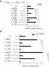
GC-box "a" is essential for TRPV1 P2-promoter activation in DRG neurons and NGF-treated PC12 cells. Comparison of P2-promoter activity in DRG neurons + NGF (A) or +/- NGF-treated PC12 cells (B) directed by: empty pGL3 reporter plasmid (pGL-E); control reporter plasmid (0.4 kb); 0.4 kb reporter with deletion of GC-box "a" (Del-a); 0.4 kb reporter with deletion of GC-box "b" (Del-b) or deletion of both GC-box "a & b". Deletion of GC-box "a" resulted in a complete loss of promoter activity when compared with the (0.4 kb) P2-promoter control in DRG neurons and NGF treated PC12 cells. Deletion of GC-box "b" directed a trend to decrease promoter activity in DRG neurons and NGF treated PC12 cells. Concurrent loss of GC-box a & b resulted in the lowest measurable promoter activity. When identical experiments were repeated under conditions of human Sp1 cDNA (A,B) over-expression, P2-promoter activity continued to be lost following deletion of GC-box "a" or GC-box "a & b". Loss of GC-box "b" under conditions of Sp1 (A,B) over-expression showed a small decrease of P2-promoter activity that attained significance in NGF treated PC12 cells. Diagram (left) indicates location of GC-box deletions and start site of transcription for P2-promoter expressing firefly luciferase (Luc). Error bars SEM (n = 3) quadruplicate measures. Significant differences: ANOVA (***) p <0.001; (*) p < 0.05.
Figure 3
Figure 3
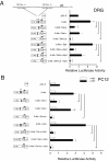
GC-box "b" modulates TRPV1 P2-promoter activity. Comparison of P2-promoter activity in DRG neurons + NGF (A) or +/- NGF-treated PC12 cells (B) directed by: empty pGL3 reporter plasmid (pGL-E); control reporter plasmid (0.4 kb); 0.4 kb reporter with deletion of GC-box "a" (Del-a); 0.4 kb reporter with deletion of GC-box "b" (Del-b) or deletion of both GC-box "a & b". When experiments were repeated under conditions of human Sp4 cDNA over-expression (A,B), P2-promoter activity continued to be lost following deletion of GC-box "a" or GC-box a & b. Loss of GC-box "b" under conditions of Sp4 (A,B) over-expression showed a small decrease of P2-promoter activity that was most evident in NGF treated PC12 cells. Diagram (left) indicates location of GC-box deletions and start site of transcription for P2-promoter expressing firefly luciferase (Luc). Error bars SEM (n = 3) quadruplicate measures. Significant differences: ANOVA (***) p < 0.001; (*) p < 0.05.
Figure 4
Figure 4
Transcription factors Sp1 and Sp3 increase P2-promoter activity in DRG neurons. P2-promoter reporter plasmid (0.4 kb) directs an ~8 fold increase in luciferase activity when compared with the empty reporter control (pGL-E). When construct 0.4 kb is co-transfected with a plasmid expressing Sp1, or Sp3, a significant increase in promoter activity was observed. However, co-transfection of Sp4 or a combination of Sp1/Sp3, Sp1/Sp4 or Sp3/Sp4 (equal ratios) failed to increase promoter activity beyond what was observed with the 0.4 kb alone. Error bars SEM (n = 3) triplicate measures. Significant differences: ANOVA (**) p < 0.01, (*) p < 0.05.
Figure 5
Figure 5
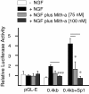
An inhibitor of Sp1 (mithramycin-a) dose-dependently blocks NGF and Sp1- dependent P2-promoter activity in PC12 cells. P2-promoter reporter plasmid (0.4 kb) directs an ~2 fold increase in luciferase activity when treated with NGF × 48 hours [14]. Treatment with an inhibitor (mithramycin-a) of Sp1 function that disrupts GC-box/transcription factor binding, blocked the NGF-dependent P2-promoter activity. When the experiment was repeated in the presence of co-transfected Sp1, the expected increase in activity directed by Sp1 was dose-dependently inhibited by mithramycin-a. Error bars SEM (n = 3) triplicate measures. Significant differences: ANOVA (***) p < 0.001, (*) p < 0.05.
Figure 6
Figure 6
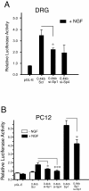
P2-promoter activity in DRG neurons is decreased by Sp1-siRNA and Sp1 and Sp4 siRNA also block P2 promoter activity in NGF treated PC12 cells. Co-transfection of Sp1-siRNA with the 0.4 kb P2-promoter construct resulted in a significant decrease in promoter activity when compared with co-transfection of the scrambled (scr) siRNA control whereas Sp4-siRNA co-transfection failed to show a decrease (A). In contrast, both Sp1-siRNA and Sp4-siRNA co-transfection experiments showed a significant decrease in NGF treated PC12 cells (B). Primary cultures of NGF-treated dorsal root ganglion (DRG) neurons were transfected with either (pGL-E) empty luciferase reporter plasmid; (0.4 kb + pBS) Luciferase reporter containing the P2-promoter plus empty siRNA vector pBS/U6; (0.4 kb + siRNA-Sp1) 0.4 kb plus siRNA construct containing the Sp1 directed hairpin encoding Sp1 nucleotides 881-901; (0.4 kb + siRNA-Sp4) 0.4 kb plus siRNA construct containing the Sp4 directed hairpin encoding Sp4 nucleotides 1551-1571. (siRNAs were a gift from G. Gill Lab, Tufts, Boston) [16] . The presence of the scrambled siRNA control plasmid reduced the expected promoter activity of the 0.4 kb reporter plasmid in DRG. Co-transfection of the Sp1 cDNA in PC12 cells (0.4 kb + Sp1) directed a further increase in P2-promoter activity that was significantly reversed by co-transfection of the Sp1-siRNA construct. Error bars SEM (n = 3) triplicate measures. Significant differences: ANOVA (*) p < 0.05; (***) p < 0.001.
Figure 7
Figure 7
Over-expression of transcription factor Sp1 and Sp4 mRNA increase endogenous TRPV1 mRNA in cultured DRG neurons. (A) Measurement of endogenous levels of rat Sp1 mRNA in cultured DRG neurons following transfection with empty/PN3 vector (left). Additional expression of an equivalent amount of human form of Sp1 mRNA was achieved following transfection with hSp1/PN3. (B) Endogenous TRPV1 mRNA levels were increased following over-expression of hSp1 in cultured DRG neurons (**) p < 0.005. (C) Measurement of control levels of rat Sp4 mRNA in cultured DRG neurons following transfection with empty/PN3 vector (left). Additional expression of an equivalent amount of human form of Sp4 mRNA was also achieved following transfection with hSp4/PN3. (D) Endogenous TRPV1 mRNA levels increased following over-expression of hSp4 in cultured DRG neurons (*). Error bars SEM (n = 3) triplicate measures. Two tailed unpaired t-test. Significance: (p < 0.05). Ct threshold values were derived from quantitative RT-PCR amplification of cultured rat DRG neuron RNA - see Methods.
Figure 8
Figure 8
Knock-down of transcription factors Sp1/Sp4 decrease endogenous TRPV1 mRNA in cultured DRG neurons. (A) Evidence of Sp1mRNA knockdown following transfection of siSp1 in cultured DRG neurons (***) p < 0.0001 (left). Apparent changes in Sp4 (middle) or TRPV1 mRNA (right) were not significant (ns). (B) Transfection of siSp4 resulted in an apparent knockdown of Sp1, Sp4 and TRPV1 mRNA. Combined knockdown of Sp1 + Sp4 using an equal ratio (1:1) of siSp1/siSp4 resulted in the most consistent knockdown of endogenous TRPV1 mRNA. Error bars SEM (n = 3) triplicates measures. Two tailed unpaired t-test. Significant differences: (*) p < 0.05; (***) p < 0.0001. RQ values of siRNA treated DRGs are compared relative to the RQ values of scrambled controls which represent baseline amounts of Sp1, Sp4 or TRPV1 mRNA following transfection of a scrambled siRNA or Spx control vector, see Methods for details.
Figure 9
Figure 9
Regulation of TRPV1 transcription at the rat P2-promoter. TRPV1 P2-promoter contains two tandem GC-box binding sites adjacent to the start site of TRPV1 transcription (arrow). GC-box "a" was found to be essential for transcriptional activation and appears to be the primary regulatory site in the P2-promoter and is co-regulated by factors Sp1 and Sp4. Depending on the cellular environment and potential state of transcription factor abundance/modification, this model proposes factor Sp4 playing a dominant role in the activation of TRPV1 transcription amongst the Sp1-like factors examined in this study. One type of transcription factor activation may arise from the activity of exogenous products of inflammation, such as NGF. Sp1, Sp4 and/or other members of the Sp1-like family (Spx) may also bind to GC-box region "b" providing additional modulation and full transcriptional activation. Classically transcriptional regulation is dynamic and rapidly responds to intrinsic and extrinsic changes of the cellular milieu. It is envisioned that transcriptional control is directed by a combination of protein modifications and/or formation of a multi-protein transcription factor complex to attract and activate RNA polymerase II (not shown). Differing 'sizes' of transcription factors represent their relative contribution to activation of TRPV1 transcription.

Similar articles

Cited by

References

    1. Caterina MJ, Schumacher MA, Tominaga M, Rosen TA, Levine JD, Julius D. The capsaicin receptor: a heat-activated ion channel in the pain pathway. Nature. 1997;389(6653):816–824. doi: 10.1038/39807. - DOI - PubMed
    1. Tominaga M, Caterina MJ, Malmberg AB, Rosen TA, Gilbert H, Skinner K, Raumann BE, Basbaum AI, Julius D. The cloned capsaicin receptor integrates multiple pain-producing stimuli. Neuron. 1998;21(3):531–543. doi: 10.1016/S0896-6273(00)80564-4. - DOI - PubMed
    1. Zygmunt PM, Petersson J, Andersson DA, Chuang H, Sorgard M, Di Marzo V, Julius D, Hogestatt ED. Vanilloid receptors on sensory nerves mediate the vasodilator action of anandamide. Nature. 1999;400(6743):452–457. doi: 10.1038/22761. - DOI - PubMed
    1. Premkumar LS, Ahern GP. Induction of vanilloid receptor channel activity by protein kinase C. Nature. 2000;408(6815):985–990. doi: 10.1038/35050121. - DOI - PubMed
    1. Shin J, Cho H, Hwang SW, Jung J, Shin CY, Lee SY, Kim SH, Lee MG, Choi YH, Kim J. et al.Bradykinin-12-lipoxygenase-VR1 signaling pathway for inflammatory hyperalgesia. Proc Natl Acad Sci USA. 2002;99(15):10150–10155. doi: 10.1073/pnas.152002699. - DOI - PMC - PubMed

Publication types

MeSH terms