. 2011 Jul 15;22(14):2646-58.
doi: 10.1091/mbc.E11-03-0239. Epub 2011 May 25.

Hierarchical regulation of mRNA partitioning between the cytoplasm and the endoplasmic reticulum of mammalian cells

Affiliations

Hierarchical regulation of mRNA partitioning between the cytoplasm and the endoplasmic reticulum of mammalian cells

Qiang Chen et al. Mol Biol Cell. .

Abstract

The mRNA transcriptome is currently thought to be partitioned between the cytosol and endoplasmic reticulum (ER) compartments by binary selection; mRNAs encoding cytosolic/nucleoplasmic proteins are translated on free ribosomes, and mRNAs encoding topogenic signal-bearing proteins are translated on ER-bound ribosomes, with ER localization being conferred by the signal-recognition particle pathway. In subgenomic and genomic analyses of subcellular mRNA partitioning, we report an overlapping subcellular distribution of cytosolic/nucleoplasmic and topogenic signal-encoding mRNAs, with mRNAs of both cohorts displaying noncanonical subcellular partitioning patterns. Unexpectedly, the topogenic signal-encoding mRNA transcriptome was observed to partition in a hierarchical, cohort-specific manner. mRNAs encoding resident proteins of the endomembrane system were clustered at high ER-enrichment values, whereas mRNAs encoding secretory pathway cargo were broadly represented on free and ER-bound ribosomes. Two distinct modes of mRNA association with the ER were identified. mRNAs encoding endomembrane-resident proteins were bound via direct, ribosome-independent interactions, whereas mRNAs encoding secretory cargo displayed predominantly ribosome-dependent modes of ER association. These data indicate that mRNAs are partitioned between the cytosol and ER compartments via a hierarchical system of intrinsic and encoded topogenic signals and identify mRNA cohort-restricted modes of mRNA association with the ER.


Figures

FIGURE 1:
FIGURE 1:
mRNAs display diverse patterns of subcellular partitioning. J558 murine plasmacytoma cells were fractionated by sequential detergent extraction and total cytosolic and ER membrane-associated RNA pools were isolated by Trizol extraction. The mRNA composition of the two pools was assayed by 96-gene qPCR arrays and compartmental enrichment values quantified using GPR software.
FIGURE 2:
FIGURE 2:
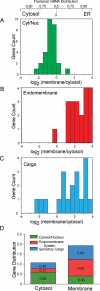
Subcellular mRNA distributions are marked by cohort-specific enrichment patterns. Gene product distributions (Figure 1) were grouped on the basis of predicted subcellular localization, and cohort-specific subcellular distributions patterns were analyzed. Predicted subcellular distributions were determined by manual interrogation of existing databases. (A) mRNAs encoding cytosolic and nucleoplasmic proteins (Cyt/Nuc). (B) mRNAs encoding resident proteins of the endomembrane system. (C) mRNAs encoding secretory pathway cargo. (D) Cohort-specific population distributions of mRNAs in the cytosol and ER membrane RNA pools. Total gene distributions were determined, and the overall and relative pool compositions were determined as a function of a three-cohort model.
FIGURE 3:
FIGURE 3:
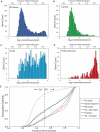
Genome-scale analysis of subcellular mRNA distribution reveals topogenic signal-independent partitioning patterns. Publicly available subcellular gene production distribution data were analyzed by a three-cohort model. The genome database for the K-562 (human myelogenous leukemia) cell line was used. Gene product cohorts were identified by algorithmic sorting, using gene ontology (GO) criteria. (A) All genes. (B) Genes encoding cytosolic and nucleoplasmic proteins, selected via the GO category “cytoplasm” (GO: 0005737) and filtered to remove topogenic signal-encoding genes. (C) Secretory pathway cargo. The K-562 gene set was sorted using the GO categories “extracellular” (GO: 0005615) and “plasma membrane” (GO: 0005886). (D) Endomembrane system. The K-562 gene set was sorted using a custom GO category “endomembrane” to include genes whose translation products reside in the ER membrane, the ER lumen, or the Golgi apparatus or lysosomes. (E) Subcellular mRNA distributions were analyzed by cumulative density distribution, using a six-cohort model: no topogenic signal, signal sequence-encoding, single transmembrane domain-encoding (monotopic), multiple transmembrane domain-encoding (polytopic), and single/multi-transmembrane domain plus signal sequence.
FIGURE 4:
FIGURE 4:
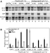
mRNAs display cohort-specific modes of interaction with the ER membrane. RM were purified from J558 murine plasmacytoma and H929 human myeloma cells by equilibrium density gradient centrifugation and mRNA–ER membrane interactions were determined by biochemical fractionation. RM suspensions were diluted in physiological salts buffer (Control), 0.5 M KCl (KCl), 20 mM EDTA (EDTA), or 0.5 M KCl/20 mM EDTA (K/E) and incubated on ice. Membrane-bound (P) and released (S) fractions were separated by ultracentrifugation and total RNA was isolated. (A) rRNA (28S, 18S) distributions were determined by dye staining, and mRNAs encoding the ER-resident proteins GRP94 (J558, H929), BiP (J558), calreticulin (CRT)(H929), λ light chain (J558), and κ light (chain (H929) were determined by Northern blot analysis. (B) Digital images for rRNA and mRNA distributions are depicted. rRNA and mRNA distributions were quantified by ImageJ analysis of SYBR Safe stained RNA gels or phosphorimager scans of Northern blots. Data represent mean ± SD of seven (J558) or three (H929) individual experiments.
FIGURE 5:
FIGURE 5:
mRNA–ER binding interactions of mRNAs are distinguished by protonated amine extraction. RM were purified from J558 murine plasmacytoma cells by equilibrium density gradient centrifugation and mRNA–ER membrane interactions were examined by extraction with the protonated amine buffers (neutral Tris or neutral imidazole). J558 RM suspensions were diluted in physiological salts buffer (Control), neutral Tris/HCl (0.1, 0.25, 0.5 M), or neutral imidazole/HCl (0.1, 0.25, 0.5 M), and incubated on ice, and the membrane-associated and -released fractions were isolated by ultracentrifugation. Samples were processed as described in the Figure 4 caption. (A) Digital images of rRNA gels and phosphorimager data (BiP, λ light chain). (B) Data from three independent experiments are summarized, with mean ± SD values indicated.
FIGURE 6:
FIGURE 6:
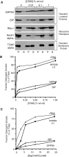
mRNAs encoding endomembrane-resident proteins are bound to the ER via ribosome-independent interactions. The ribosome dependence of mRNA–ER interactions for mRNAs encoding secretory pathway cargo (λ light chain) and endomembrane-resident proteins (GRP94, BiP) was determined by detergent solubilization profiling. (A) J558 plasmacytoma RM were diluted into buffers containing 0.3 M KCl and the indicated concentrations of the detergent DDM. The detergent-soluble and -resistant fractions were separated by ultracentrifugation, and the protein composition of the two fractions was determined by immunoblot analysis of the ER-resident lumenal proteins GRP94 and BiP, the ribosome-interacting proteins ribophorin I and Sec61α, and the ER-resident membrane protein TRAPα. The detergent solubilization profiles for ribosomes (rRNA), mRNAs encoding the secretory pathway cargo protein λ light chain (λ LC), and the resident endomembrane (ER) proteins BiP and GRP94 are depicted in (B and C), for the detergents DDM and BigCHAPS, respectively. rRNA levels were determined by SYBR Green Supermix staining of denaturing RNA gels; mRNA levels were determined by Northern blot and phosphorimager analysis.
FIGURE 7:
FIGURE 7:
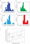
mRNAs display cohort-restricted modes of interaction with the ER membrane. Three independent J558 RM preparations were isolated and extracted with 0.5 M KCl/20 mM EDTA, as described in the Figure 4 caption. Total RNA was isolated from all fractions by Trizol extraction, and the mRNA composition was analyzed by qPCR array analysis, using Lonza's StellARray qPCR Array. Two Lonza gene arrays were examined: 1) murine antigen-processing and presentation, and 2) growth and development. Together these provide a diverse representation of genes encoding prominent cytosolic, nucleoplasmic, secretory pathway cargo, and resident endomembrane proteins. qPCR data were analyzed as described in the Figure 1 caption. (A) All genes. (B) Genes encoding cytosolic and nucleoplasmic proteins. (C) Genes encoding secretory pathway proteins. (D) Genes encoding resident proteins of the endomembrane system. In (E), the gene product distribution data for genes common to the experiments depicted in Figures 1 and 2 and (A–D) were examined for correlation. Statistical analysis was performed by Spearman's correlation. The 80% and 95% confidence intervals for the data correlation analysis are depicted by the solid and dashed lines, respectively.

Similar articles

Cited by

References

    1. Adelman MR, Sabatini DD, Blobel G. Ribosome-membrane interaction: non-destructive disassembly of rat liver rough microsomes into ribosomal and membranous components. J Cell Biol. 1973;56:206–229. - PMC - PubMed
    1. Adesnik M, Lande M, Martin T, Sabatini DD. Retention of mRNA on the endoplasmic reticulum membranes after in vivo disassembly of polysomes by an inhibitor of initiation. J Cell Biol. 1976;71:307–313. - PMC - PubMed
    1. Aragón T, van Anken E, Pincus D, Serafimova IM, Korennykh AV, Rubio CA, Walter P. Messenger RNA targeting to endoplasmic reticulum stress signalling sites. Nature. 2009;457:736–740. - PMC - PubMed
    1. Bendtsen JD, Nielsen H, von Heijne G, Brunak S. Improved prediction of signal peptides: SignalP 3.0. J Mol Biol. 2004;340:783–795. - PubMed
    1. Blobel G. Intracellular protein topogenesis. Proc Natl Acad Sci USA. 1980;77:1496–1500. - PMC - PubMed

Publication types

LinkOut - more resources